• 검색 결과가 없습니다.

The effect of casein phosphopeptide amorphous calcium phosphate on the shear bond strength of orthodontic brackets

N/A
N/A
Protected

Academic year: 2022

Share "The effect of casein phosphopeptide amorphous calcium phosphate on the shear bond strength of orthodontic brackets"

Copied!
6
0
0

로드 중.... (전체 텍스트 보기)

전체 글

(1)

The effect of casein phosphopeptide amorphous calcium phosphate on the in vitro shear bond strength of orthodontic brackets

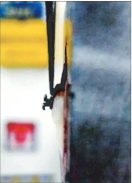
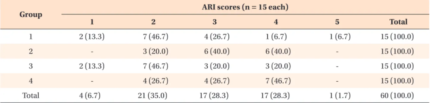
Objective: The purpose of this study was to evaluate the effect of casein phosphopeptide amorphous calcium phosphate (CPP-ACP) on the shear bond strength (SBS) of brackets bonded to non-demineralized teeth with either phosphoric acid etching or self-etching primer. Methods: Sixty human premolars were randomly assigned to 1 of 4 treatment groups (n = 15 each): phosphoric acid etching (group 1); self-etching primer (group 2); CPP-ACP for 2 weeks + phosphoric acid etching (group 3), and CPP-ACP for 2 weeks + self-etching primer (group 4). After bonding of the maxillary premolar metal brackets, specimens were subjected to shear forces in a testing machine. Scanning electron microscopy was used to observe etching patterns on the enamel surfaces of all teeth. A 2-way analysis of variance was used to test for effects of CPP-ACP and etching system on SBS. Results: Significantly higher mean SBSs were observed in groups subjected to phosphoric acid etching (i.e., groups 1 and 3; p < 0.05).

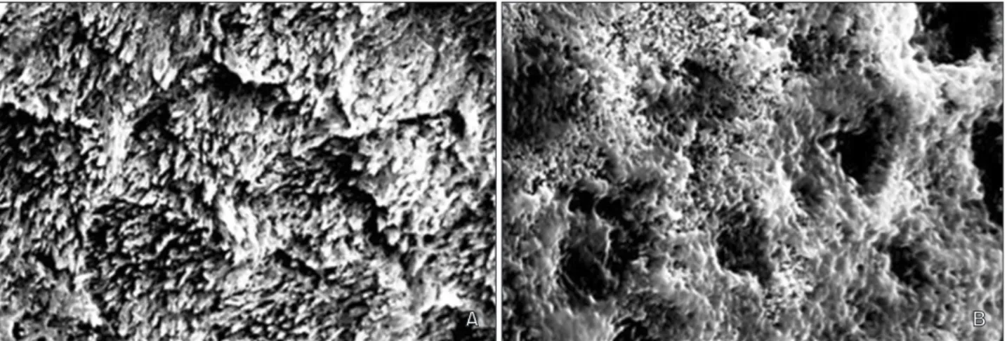
On the other hand, SBSs did not appear to be influenced by CPP-ACP (i.e., groups 3 and 4; p > 0.05). We observed a uniform and clear etched pattern on the enamel surface of the phosphoric acid etching groups. Conclusions: CPP- ACP does not significantly affect the SBS of orthodontic brackets bonded to non-demineralized teeth, regardless of which adhesive method is used to bond the brackets.

[Korean J Orthod 2013;43(1):23-28]

Key words: CPP-ACP, Shear bond strength, Self-etching primer, Bracket Sun-Youn Park

a

Jung-Yul Cha

c

Kyoung-Nam Kim

b

Chung-Ju Hwang

c

a

Department of Orthodontics, College of Dentistry, Yonsei University, Seoul, Korea

b

Research Institute of Dental

Biomaterials and Bioengineering, BK21 Project, College of Dentistry, Yonsei University, Seoul, Korea

c

Department of Orthodontics, College of Dentistry and Institute of Cranio- facial Deformity, Yonsei University, Seoul, Korea

Received April 24, 2012; Revised August 15, 2012; Accepted August 18, 2012.

Corresponding author: Chung-Ju Hwang.

Professor, Department of Orthodontics, College of Dentistry, Yonsei University, 50 Yonsei- ro, Seodaemun-gu, Seoul 120-752, Korea.

Tel +82-2-2228-3106 e-mail [email protected]

© 2013 The Korean Association of Orthodontists.

The authors report no commercial, proprietary, or fi nancial interest in the products or companies described in this article.

This is an Open Access article distributed under the terms of the Creative Commons Attribution Non-Commercial License (http://creativecommons.org/licenses/by-nc/3.0) which permits unrestricted non-commercial use, distribution, and reproduction in any medium, provided the original work is properly cited.

pISSN 2234-7518 • eISSN 2005-372X

http://dx.doi.org/10.4041/kjod.2013.43.1.23

(2)

INTRODUCTION

Casein phosphopeptide amorphous calcium phosphate (CPP-ACP) is a dental product that strengthens and remi neralizes teeth while also acting as an anticariogenic agent.

1-3

CPP-ACP can dramatically decrease lesion depth and increase both remineralization and mineral content of enamel, thereby improving ratios of both calcium and phosphate.

4-7

Both of these minerals form a complex with CPP-ACP on the dental plaque of tooth surfaces, creating a supersaturated state that resists acid challenges and prevents demineralization; the calcium and phosphate ions themselves also enhance remineralization by pene trating the demineralized enamel structure.

2,4

Further, enamel surfaces remine- ralized by CPP-ACP are more resistant to acid challenges with hydroxyapatite of high concentrations of calcium and phosphate ions.

8

CPP-ACP application is one of many techniques that have been proposed for simultaneously enhancing remineralization and reducing the occurrence of dental caries and white spot lesions (i.e., the decalcification of enamel that often occurs adjacent to orthodontic brackets).

9-11

Such lesions are a significant clinical problem for orthodontists; in one study population, an increase in the number of white spots was observed in 50% of patients who underwent fixed orthodontic treatments.

12

There is much interest in using CPP-ACP as a prophy- lactic for preventing decalcification during orthodontic treatments. However, this will not be feasible if the compound negatively impacts bracket adhesion. Of the 3 phases of the orthodontic adhesion process (i.e., etching, priming, and bonding), the first seems most likely to be impacted by CPP-ACP, which increases mineral content and acid resistance of enamel. Such interference could ultimately reduce the shear bond strength (SBS) of bracket bonding, though the extent of this effect might be expected to vary depending on the type of etching technique employed - for example, phosphoric acid or a self-etching primer can be used, the latter of which combines the etching and priming events into a single step.

We developed the current study to evaluate the effects of both CPP-ACP and different etching processes on the SBS of brackets attached to non-demineralized teeth. We also explored whether SBS was impacted by interactions between these 2 factors.

MATERIALS AND METHODS

Our sample was comprised of 60 human maxillary and mandibular premolars that had been extracted for orthodontic reasons from patients aged between 18 and

28 years; all individuals provided informed consent prior to the study. Teeth were only included in the study if they had an intact buccal surface and were free of caries, cracks, and enamel defects. The teeth were cleaned after extraction and any residual soft tissues were removed.

They were then stored in distilled water according to ISO standards 11405/2003. All teeth were tested within 1 month of extraction after being embedded in cold acrylic resin with a mounting jig, such that the labial surface was perpendicular to the bottom of the mold.

Teeth were randomly assigned to 1 of 4 treatment groups (n = 15 each): 35% phosphoric acid + Transbond XT primer + Transbond XT resin (group 1); Transbond Plus Self-etching Primer (TPSEP) + Transbond XT resin (group 2); CPP-ACP paste applied for 2 weeks + 35%

phosphoric acid + Transbond XT (group 3); and CPP- ACP paste applied for 2 weeks + TPSEP + Transbond XT resin (group 4).

Teeth in group 1 were etched with 35% phosphoric acid for 15 seconds, washed for 10 seconds, and air- dried. Transbond XT primer was applied and light-cured for 10 seconds. Brackets were bonded with Transbond XT resin. Teeth in Group 2 were treated with TPSEP, which was rubbed on the enamel surfaces for 5 seconds and gently air-dried for 5 seconds. Brackets were bonded with Transbond XT resin. Maxillary premolar metal brackets (Victory Series

TM

brackets; 3M Unitek, Monrovia, CA, USA) were positioned parallel to the base of the acrylic block. In groups 3 and 4, the teeth were pre-treated with CPP-ACP paste (Toothmousse; GC Co., Tokyo, Japan) for 2 weeks before the brackets were bonded. This protocol was selected because previous work has shown that the remineralizing effects of CPP-

Figure 1. Shear bond strength test with the Instron

machine showing where the debonding force was applied.

(3)

ACP last for at least 2 weeks after its application.

5,7,13

CPP-ACP was applied twice a day according to the manufacturer’s instructions. Three minutes after appli- cation, the specimens were stored in artificial saliva in a 37

o

C incubator; the saliva was changed daily, and the enamel surface was cleaned with distilled water prior to application of the CPP-ACP paste.

All specimens were stored in artificial saliva for 24 hours in a 37

o

C incubator prior to undergoing shear force testing in a universal testing machine with a crosshead speed of 0.5 mm/minute; each trial lasted until bracket failure occurred (Figure 1). Once the debon ding was complete, we examined the enamel surface of the tooth under a stereomicroscope at 50

× magnification. We used the modified adhesive rem- nant index (ARI) to evaluate the amount of residual adhesive on each tooth surface. Classifications were as follows: 1, the entire composite, with an impression of the bracket base, remained on the tooth; 2, > 90%

of the composite remained; 3, > 10% but < 90%

of the composite remained on the tooth; 4, < 10% of composite remained on the tooth surface; 5, no com- posite remained on the tooth. We also examined the acid-etched enamel surfaces with scanning electron microscopy (SEM) in order to compare the differences between group1/ group2 (no CPP-ACP) and group 3/

group 4 (CPP-ACP).

We used Levene variance homogeneity and Shapiro- Wilk’s tests to investigate the homogeneity of variance and distribution, respectively, of bond strength data.

Two-way analysis of variance (ANOVA) tests were used to test for differences between groups and to explore any interactions between CPP-ACP pre-treatment and etching type. A Fisher’s exact test was used to evaluate differences in ARI scores between groups. Significance was defined as p < 0.05.

RESULTS

Mean SBS values and standard deviations are shown in Table 1. Bond strength was not significantly affected by pre-treatment with CPP-ACP (Table 2). However, SBS was impacted by etching system. Bond strengths were significantly higher for teeth prepared with phosphoric acid etching (group 1: 18.66 ± 2.31 megapascal [MPa];

group 3: 18.48 ± 2.19 MPa) than with the self-etch adhesive system (group 2: 15.75 ± 1.77 MPa; group 4: 15.51 ± 1.71 MPa) (p < 0.05). CPP-ACP and type of adhesive system did not interact to affect SBS (p > 0.05)

Table 1. Shear bond strength values (MPa) of the 4 treatment groups

Group

(n = 15 each) Mean SD Minimum Maximum

1 18.66 2.31 13.68 20.95

2 15.75 1.77 13.06 18.31

3 18.48 2.19 14.71 20.91

4 15.51 1.71 12.65 18.18

MPa, Megapascal; SD, standard deviation.

Table 2. Results from two-way ANOVA test investigating the effects of our explanatory variables (presence/absence of CPP-ACP pre-treatment, type of etching system, and the interaction between these) on SBS

Sum of

squares F p-value

CPP-ACP 0.70 0.17 0.68

ES 129.60 31.93 < 0.001

CPP-ACP × ES 0.01 0.00 0.95

Anova, Analysis of variance; CPP-ACP, casein phospho- peptide amorphous calcium phosphate; SBS, shear bond strength; ES, etching system.

Table 3. Frequency distribution of ARI scores for each of the 4 treatment groups

Group ARI scores (n = 15 each)

1 2 3 4 5 Total

1 2 (13.3) 7 (46.7) 4 (26.7) 1 (6.7) 1 (6.7) 15 (100.0)

2 - 3 (20.0) 6 (40.0) 6 (40.0) - 15 (100.0)

3 2 (13.3) 7 (46.7) 3 (20.0) 3 (20.0) - 15 (100.0)

4 - 4 (26.7) 4 (26.7) 7 (46.7) - 15 (100.0)

Total 4 (6.7) 21 (35.0) 17 (28.3) 17 (28.3) 1 (1.7) 60 (100.0)

Data are presented as number (% of total). Fisher’s exact test; p > 0.05.

Adhesive remnant index (ARI) scores: 1, the entire composite, with an impression of the bracket base, remained on the tooth;

2, > 90% of the composite remained on the tooth; 3, > 10% but < 90% of the composite remained on the tooth; 4, < 10% of

composite remained on the tooth; 5, no composite remained on the tooth.

(4)

(Table 2).

ARI values were similar across all groups (Table 3), though we did notice that the self-etch adhesive system group (group 2 and group 4) tended to display adhesive failure at the enamel/adhesive interface.

Our SEM analysis revealed no significant difference associated with CPP-ACP preparation. We only noticed different enamel surfaces between the 35% phosphoric acid treatment groups (group 1 and 3) and the TPSEP groups (group 2 and 4) that etching was much more pronounced, uniform, and clear on enamel etched with 35% phosphoric acid, while enamel prepared with TPSEP was characterized by much shallower etching (Figures 2 and 3).

DISCUSSION

Our results indicate that SBS is impacted more by etching technique than by use of CPP-ACP. This suggests that there will not be negative orthodontic side effects of employing CPP-ACP to reduce the likelihood of decalcification and/or prevent formation of dental caries. This is true regardless of whether teeth are prepared with a phosphoric acid or self-etching primer system, the latter of which appears to be associated with lower SBS.

Our CPP-ACP results are in agreement with previous findings demonstrating that CPP-ACP does not com- promise bracket bond strength when a 3-step bonding procedure is used.

14-17

In fact, the authors of these studies all suggested that pre-treatment with CPP-ACP Figure 3. Scanning electron microscopy images of acid-etched enamel surfaces after 2 weeks of pre-treatment with casein phosphopeptide amorphous calcium phosphate. A, Surface of a tooth etched with 35% phosphoric acid (×5,000). B, Surface of a tooth etched with Transbond Plus Self-Etching Primer (×5,000).

Figure 2. Scanning electron microscopy images of acid-etched enamel tooth surfaces. A, Surface of a tooth etched with

35% phosphoric acid (×5,000). B, Surface of a tooth etched with Transbond Plus Self-Etching Primer (×5,000).

(5)

before bonding actually increased bracket SBS.

14-16

As in the current study, CPP-ACP was used in these instances as a prophylactic prior to bonding. However, because the previous authors stored specimens in distilled water, they were unable to consider the impacts of saliva, which enhances the effectiveness of CPP-ACP.

Demand for a simpler and more effective bonding technique has led to the development of self-etching primers, which are comprised of phosphoric acid and a methacrylate group. When these primers are applied to enamel, the phosphate group dissolves and removes the calcium from the hydroxyapatite, forming a complex that is incorporated into the network during primer polymerization: etching and monomer diffusion occur simultaneously. By decreasing the number of steps re- quired during the adhesion process (e.g., etching, rin- sing, and drying), these primers potentially reduce the likelihood of contamination - an important factor deter mining SBS.

18

While there is no question that self- etching primers are effective,

19-21

SEM examinations of etched enamel have revealed primer-dependent variations in etching patterns. Surfaces etched with 35%

phosphoric acid tend to be very porous, characterized by long resin tags and numerous enamel prisms that form a typical honeycomb pattern. Surfaces prepared with TPSEP, on the other hand, have less prominent etching patterns, with short resin tags and very thin hybrid layers.

20,22,23

Thus, a combination technique of CPP-ACP pre- treat ment (which increases enamel acid resistance and mineral content) and TPSEP etching seemed likely to reduce bracket SBS.

Surprisingly, however, we did not find any evidence that a CPP-ACP pre-treatment reduced SBS for teeth prepared with the self-etch adhesive system (p > 0.05 for a comparison of groups 2 and 4). Additionally, our SEM analysis revealed similar etching patterns in groups that did and did not receive the CPP-ACP pre-treatment (i.e., groups 1 vs. 3/groups 2 vs. 4). These results add to the growing body of literature suggesting that CPP- ACP does not compromise SBS.

24-26

However, we did find that mean SBS was higher in the phosphoric acid etching groups (groups 1 and 3) than in the self-etch adhesive system groups (groups 2 and 4; p < 0.05) - an observation also consistent with previous study.

27

The higher SBS values reported here were primarily a result of the location at which the debonding force was applied. Klocke and Kahl-Nieke

28

noted that SBS dropped by 41.5% when debonding forces were moved from a position close to the enamel to the bracket wing. In vitro conditions are difficult to reproduce in a laboratory set ting because forces would need to be applied from multiple directions at once. Under clinical conditions, bond failures are usually caused by the

application of force loads to the wings. To standardize the test as much as possible and to exert a pure shear force, we chose to apply a debonding force to the interface of the bracket and the tooth surface.

CONCLUSION

1. The use of CPP-ACP did not affect mean SBS, regard less of the adhesive system used to bond brac- kets to the teeth in our study sample (p > 0.05). All SBS values were clinically acceptable.

2. Significantly lower SBS values were observed in the groups prepared with the self-etch adhesive system (Group 2: 15.75 ± 1.77 MPa; Group 4: 15.51 ± 1.71 MPa) than in those bonded with phosphoric acid etching (Group 1: 18.66 ± 2.31 MPa; Group 3: 18.48

± 2.19 MPa) (p < 0.05).

3. There were no significant differences in the ARI values of the 4 treatment groups. However, the self-etch adhesive system group tended to display adhesive failure at the enamel/adhesive interface.

4. Enamel etching patterns were similar on teeth that were and were not pre-treated with CPP-ACP.

Based on these results, we conclude that pre- treatment with CPP-ACP does not significantly affect the SBS of orthodontic brackets. Additionally, given the values of SBS observed for the self-etching primer groups, we advocate the use of both this method and 35% phosphoric acid etching for bonding orthodontic brackets.

REFERENCES

1. Reynolds EC, del Rio A. Effect of casein and whey- protein solutions on caries experience and feeding patterns of the rat. Arch Oral Biol 1984;29:927-33.

2. Reynolds EC, Cain CJ, Webber FL, Black CL, Riley PF, Johnson IH, et al. Anticariogenicity of calcium phosphate complexes of tryptic casein pho sphopep- tides in the rat. J Dent Res 1995;74:1272-9.

3. Reynolds EC. Anticariogenic complexes of amor- phous calcium phosphate stabilized by casein pho- sphopeptides: a review. Spec Care Dentist 1998;18:

8-16.

4. Choi HJ, Choi YC, Kim KC, Choi SC. Remineralization depth of CPP- ACP on demineralization human enamel in vitro. J Korean Acad Pediatr Dent 2008;35:278-86.

5. Shen P, Cai F, Nowicki A, Vincent J, Reynolds EC.

Remineralization of enamel subsurface lesions by

sugar-free chewing gum containing casein phospho-

peptide-amorphous calcium phosphate. J Dent Res

2001;80:2066-70.

(6)

6. Kumar VL, Itthagarun A, King NM. The effect of casein phosphopeptide-amorphous calcium phosphate on remineralization of artificial caries-like lesions: an in vitro study. Aust Dent J 2008;53:34- 40.

7. Zabokova-Bilbilova E, Stafilov T, Sortirovska- Ivkovska A, Sokolovska F. Prevention of enamel deminera lization during orthodontic treatment:

an in vitro study using GC tooth mousse. Balkan J Stomatol 2008;12:133-7.

8. Iijima Y, Cai F, Shen P, Walker G, Reynolds C, Rey- nolds EC. Acid resistance of enamel subsurface lesions remineralized by a sugar-free chewing gum containing casein phosphopeptide-amorphous cal- cium phos phate. Caries Res 2004;38:551-6.

9. White JM, Eakle WS. Rationale and treatment ap- proach in minimally invasive dentistry. J Am Dent Assoc 2000;131(Suppl):13S-9S.

10. Zero DT, Fontana M, Martínez-Mier EA, Ferreira- Zandoná A, Ando M, González-Cabezas C, et al.

The biology, prevention, diagnosis and treatment of dental caries: scientific advances in the United States. J Am Dent Assoc 2009;140 (Suppl 1):25S-34S.

11. Sudjalim TR, Woods MG, Manton DJ. Prevention of white spot lesions in orthodontic practice: a contemporary review. Aust Dent J 2006;51:284-9.

12. Gorelick L, Geiger AM, Gwinnett AJ. Incidence of white spot formation after bonding and banding.

Am J Orthod 1982;81:93-8.

13. Reynolds EC. Remineralization of enamel subsurface lesions by casein phosphopeptide-stabilized calcium phosphate solutions. J Dent Res 1997;76:1587-95.

14. Xiaojun D, Jing L, Xuehua G, Hong R, Youcheng Y, Zhangyu G, et al. Effects of CPP-ACP paste on the shear bond strength of orthodontic brackets. Angle Orthod 2009;79:945-50.

15. Keçik D, Cehreli SB, Sar C, Unver B. Effect of acidu- lated phosphate fluoride and casein phospho- peptide-amorphous calcium phosphate application on shear bond strength of orthodontic brackets.

Angle Orthod 2008;78:129-33.

16. Tabrizi A, Cakirer B. A comparative evaluation of ca- sein phosphopeptide-amorphous calcium pho sphate and fluoride on the shear bond strength of ortho- dontic brackets. Eur J Orthod 2011;33:282-7.

17. Uysal T, Baysal A, Uysal B, Aydınbelge M, Al- Qunaian T. Do fluoride and casein phosphopeptide- amorphous calcium phosphate affect shear bond

strength of orthodontic brackets bonded to a demi neralized enamel surface? Angle Orthod 2011;

81:490-5.

18. Buyukyilmaz T, Usumez S, Karaman AI. Effect of self-etching primers on bond strength--are they reliable? Angle Orthod 2003;73:64-70.

19. Arnold RW, Combe EC, Warford JH Jr. Bonding of stainless steel brackets to enamel with a new self- etching primer. Am J Orthod Dentofacial Orthop 2002;122:274-6.

20. Bishara SE, VonWald L, Laffoon JF, Warren JJ.

Effect of a self-etch primer/adhesive on the shear bond strength of orthodontic brackets. Am J Orthod Dentofacial Orthop 2001;119:621-4.

21. Grubisa HS, Heo G, Raboud D, Glover KE, Major PW. An evaluation and comparison of orthodontic bracket bond strengths achieved with self-et ching primer. Am J Orthod Dentofacial Orthop 2004;126:

213-9.

22. Iijima M, Ito S, Yuasa T, Muguruma T, Saito T, Mizo- guchi I. Bond strength comparison and scanning electron microscopic evaluation of three orthodontic bonding systems. Dent Mater J 2008;27:392-9.

23. Triolo PT Jr, Swift EJ Jr, Mudgil A, Levine A. Effects of etching time on enamel bond strengths. Am J Dent 1993;6:302-4.

24. Al-Twaijri S, Viana G, Bedran-Russo AK. Effect of prophylactic pastes containing active ingredients on the enamel-bracket bond strength of etch- and-rinse and self-etching systems. Angle Orthod 2011;81:788-93.

25. Cehreli SB, Sar C, Polat-Özsoy O, Unver B, Ozsoy S.

Effects of a fluoride-containing casein phospho- peptide-amorphous calcium phosphate complex on the shear bond strength of orthodontic brackets. Eur J Orthod 2012;34:193-7.

26. Adebayo OA, Burrow MF, Tyas MJ. An SEM eva- lua tion of conditioned and bonded enamel fol- low ing carbamide peroxide bleaching and casein phosphopeptide-amorphous calcium phosphate (CPP-ACP) treatment. J Dent 2009;37:297-306.

27. Holzmeier M, Schaubmayr M, Dasch W, Hirschfelder U. A new generation of self-etching adhesives:

com parison with traditional acid etch technique. J Orofac Orthop 2008;69:78-93.

28. Klocke A, Kahl-Nieke B. Influence of force location

in orthodontic shear bond strength testing. Dent

Mater 2005;21:391-6.

수치

Figure 1. Shear bond strength test with the Instron  machine showing where the debonding force was applied
Table 3. Frequency distribution of ARI scores for each of the 4 treatment groups
Figure 2. Scanning electron microscopy images of acid-etched enamel tooth surfaces. A, Surface of a tooth etched with  35% phosphoric acid (×5,000)

참조

관련 문서

This study aimed to investigate whether dentin surface preparation with diamond rotary instruments of different grit sizes affects the shear bond strength of

Purpose: This study examined the effects of glass infiltration treatments on the shear bond strength (SBS) between zirconia core and ultra low-fusing porcelain veneer.. SBS

Objective: We compared the shear bond strength (SBS) of lingual retainers bonded to bovine enamel with three different resins using direct and indirect methods.. Methods: Both ends

compared the shear bond strength (SBS) and the sites of bond failure for brackets bonded to teeth between the direct bonding technique (Transbond XT) and

The aims of this study were to perform an in-vitro evaluation to compare: 1) the shear bond strength (SBS), adhesive remnant index (ARI), and color change between self-etched

Objective: The purpose of this in vitro study was to examine the effects of fluoridated, casein phosphopeptide–amorphous calcium phosphate complex (CPP-ACP)-containing, and

The objectives of this in vitro study were to measure the shear bond strength (SBS) of metal brackets on gold alloys (treated by using a silica coating) at different time

Objective: To study and compare the effects of different demineralization- inhibition methods on the shear bond strength (SBS) and fracture mode of an adhesive used to bond