Effects of silanation time on shear bond strength between a gold alloy surface and metal bracket
Objective: We aimed to investigate the effects of silanation time on the shear bond strength (SBS) of metal brackets on gold alloy in a silicoating procedure and compare the SBS of metal brackets on gold alloy and enamel. Methods:
Type III gold alloy plates were sandblasted with 30-μm silicon dioxide.
Excess particles were removed with gentle air after silica coating, and silane was applied. Maxillary central-incisor metal brackets were bonded to each conditioned alloy surface with a light curing resin adhesive for 1 s, 30 s, 60 s, or 120 s after applying silane. The brackets were also bonded to 36 upper central incisors with the same adhesive. All samples were cured for 40 s with a light emitting diode curing light. The SBS was tested after 1 h and after 24 h. The adhesive remnant index (ARI) of the samples was also compared. Results: The 60-s and 120-s silanation time groups showed a higher SBS than the other groups (p < 0.05). Samples tested after 24 h showed a significantly higher SBS than did the samples tested after 1 h (p < 0.05). The 1-s group showed higher ARI scores. The one-way analysis of variance and Student–Newman–Keuls test showed that the SBS values of the 60-s and 120-s silanation time groups were not significantly different from the SBS values of enamel. Conclusions:
Adequate silanation time is required to produce sufficient bond strength during silicoating.
[Korean J Orthod 2013;43(3):127-133]
Key words: Bonding, Biomaterial science Min-Ho Jung
aWon-Jun Shon
bYoung-Seok Park
cShin-Hye Chung
ba
Department of Orthodontics, School of Dentistry and Dental Research Institute, Seoul National University, Seoul, Korea
b
Department of Conservative Dentistry, School of Dentistry and Dental Research Institute, Seoul National University, Seoul, Korea
c
Department of Oral Anatomy, School of Dentistry and Dental Research Institute, Seoul National University, Seoul, Korea
Received October 24, 2012; Revised December 7, 2012; Accepted December 8, 2012.
Corresponding author: Min-Ho Jung.
Adjunct Associate Professor, Department of Orthodontics, School of Dentistry and Dental Research Institute, Seoul National University; SNU HONORS Orthodontic Clinic, 3rd Fl, Taenam Bldg, 72-3 Chamwon-dong, Seocho-gu, Seoul 137-909, Korea.
Tel +82-2-599-4001 e-mail [email protected]
© 2013 The Korean Association of Orthodontists.
The authors report no commercial, proprietary, or financial interest in the products or companies described in this article.
This is an Open Access article distributed under the terms of the Creative Commons Attribution Non-Commercial License (http://creativecommons.org/licenses/by-nc/3.0) which permits unrestricted non-commercial use, distribution, and reproduction in any medium, provided the original work is properly cited.
pISSN 2234-7518 • eISSN 2005-372X
http://dx.doi.org/10.4041/kjod.2013.43.3.127
INTRODUCTION
Clinicians are often faced with the problem of bon- ding orthodontic brackets to teeth that already have undergone different types of restorations, especially in adult patients. The prevalence of malocclusion in adults is equal to or greater than the prevalence in children and adolescents;
1therefore, the percentage of adult orthodontic patients is high.
2Stress with a bonding procedure such as posterior tooth banding (which is more prone to gingival inflammation and attachment loss
3) has become a matter of concern to orthodontists.
Metallic surfaces can be encountered on the labial sur- faces of the molars and premolars and the lingual sur- faces of the anterior or posterior teeth.
Conventional acid etching is ineffective for preparing metal surfaces for mechanically retaining orthodontic attachments. Surface roughening before bracket bonding is therefore a prerequisite for sufficient bracket-to-alloy bonding.
4,5Sandblasting, the most common method for surface preparation, creates scratch-like irregularities that increase the surface area, thereby enhancing me- cha nical bond strength.
6A recently introduced silica coating method facili- tates mechanical retention by sandblasting and chemi- cophysical bonding between a composite resin and an alloy surface with a silane.
6By using an intraoral sandblaster, alloy surfaces are treated with aluminum oxide particles that are modified with silicic acid. The particles form a reactive silica layer on the substrate.
Silane must thereafter be applied to allow chemical bonding with a resin-based system such as composite adhesives.
7Silica coating has been used in many dental appli- cations such as repairing intraoral fractured ceramic surfaces,
8,9treating ceramic bracket bases for rebon- ding,
10and repairing resin-bonded prostheses.
11Several studies were recently published in regard to bracket bonding on metal surfaces.
12-14In silicoating procedures, the ideal timing of silane appli cation before bracket bonding remains equivocal.
Manufacturer recommendations for silanation are typi- cally 5 min for extraoral use (3M ESPE Sil; 3M ESPE Dental Products. St. Paul, MN, USA).
15However, 5 min is excessively long in clinical situations. For that reason, manufacturers also recommend 30 s (3M ESPE Sil)
15or 1 min (Pore-Etch and porcelain conditioner; Reliance Orthodontic Products Inc., Itasca, IL, USA) in intraoral use.
16However, there has been no data concerning bond strength differences in regard to these time differences.
The objectives of this in vitro study were to measure the shear bond strength (SBS) of metal brackets on gold alloys (treated by using a silica coating) at different time intervals after applying silane and compare these SBS
values with the SBS values on an enamel surface.
MATERIALS AND METHODS
Thirty-two 1.5-mm thick and 10-mm long gold alloy square plates (type III gold consisting of 50% gold, 5%
palladium, 32.5% silver, and 11.45% copper; Argen Co., San Diego, CA, USA) and 36 human central incisors, which were extracted for periodontal purposes and stored in thymol solution (0.1% wt/vol), were used in this study. The criterion for tooth selection was that a tooth had to be free of restoration, cracks, caries, attrition, or white spot lesion. The upper central incisors were randomly divided into 2 groups: 1 h group and 24 h group. Institutional Review Board of the Seoul National University Dental Hospital authorized the authors to proceed with this experimental study.
All plates and teeth were embedded in a cold-curing acrylic resin (Leocryl; Leone, Sesto Fiorentino, Italy) with acrylic rings (Taejin Acrylic, Seoul, Korea). Each plate was oriented so that its surface was parallel to the force applied during the shear bond tests.
The silica coating process was performed on all alloy plates after embedding. A sandblasting device (Air- Flow Handy II; EMS Corp., Dallas, TX, USA) was filled with 30-mm aluminum oxide grain that was modified with silicic acid (Cojet-Sand; 3M ESPE, Seefeld, Ger- many). The grain was used for surface roughening.
In accordance with the manufacturer’s instructions, the abrasive was applied perpendicularly to the metal surface at a distance of 10 mm under 2.5 bar pressure for 15 seconds. Excess particles were removed with gen tle air after the silica coating procedure. A silane coupling agent (ESPE-Sil; 3M ESPE) was then applied to the plate surface and air-dried with oil-free air.
A power analysis (alpha level, p < 0.05; beta level, < 0.20;
2-tailed) was performed in this study by using the mean and standard deviation, on the basis of the results of a previously conducted pilot study. As a result of this analysis, 18 samples were used for each group.
Because 2 factors (i.e., silanation time and test timing)
had to be evaluated, we assigned alloy plates to 8
groups. In the 1-s silanation time group, brackets were
bonded immediately after the silane application. We
tried to immediately bond the brackets, but bracket
positioning and removal of excessive adhesive takes
time; therefore, we named the group the “1-s silanation
time group,” for the sake of convenience. In the 30-s,
60-s, and 120-s silanation time groups, the specimens
were air-dried for 30 s, 60 s, and 120 s, respectively,
after the silane application to allow chemical adhesion
between the silane and the silica-coated surface and to
acquire a dry field before bracket bonding. To evaluate
the effect of test timing, specimens were stored in water
in a thermostatic chamber at 37
oC for 1 h or for 24 h before SBS testing.
Immediately after surface conditioning and air drying, maxillary central-incisor metal brackets (item number 017-875; 3M Unitek, Monrovia, CA, USA) were bonded to each conditioned alloy surface with a light-cured composite adhesive (Transbond XT; 3M Unitek). To maximize the bond strength, a thin uniform coat of adhesive primer was applied to the bracket base and was light cured for 10 s with light emitting diode (LED) curing lights (Ortholux LED; 3M Unitek), as demonstrated in previous research.
12Resin adhesives were then applied to the bracket bases. According to the manufacturer, the average surface area of each bracket base is 10.56 mm
2.
Each bracket was positioned on the prepared alloy sur- face with sufficient pressure to expel excess adhesive, which was then carefully removed. Previous study suggests that 40 s of curing time is required to obtain proper bond strength of metal brackets on metal plates when using LED curing lights.
14Therefore, the brackets were light cured for 40 s. As the manufacturer recommends, the light source was held 1 - 2 mm above the bracket, and the mesial and distal edges were cured for an equal amount of time on each side. A minimum light intensity of at least 2,000 mW/cm
2was verified by using a handheld curing radiometer (Demetron 100;
Demetron Research, Danbury, CT, USA).
After photopolymerization, the plates were stored in water in a thermostatic chamber at 37
oC for 1 h or for 24 h. A universal testing device was used to determine the bracket SBS (LF Plus; Ametek, Albany, NY, USA).
For this test, acrylic rings were mounted in a jig with the brackets positioned vertically. Shear force at a cross-head speed of 1 mm/min was transmitted to the bracket by means of a square plate of the same size. The force required to shear the bracket was recorded and converted into units of stress (MPa), based on a known bracket area.
Bond strength testing was performed on 4 alloy plates in each group. The tested alloy surfaces were polished with a Shofu Gold Polishing Kit (HP 0303; Shofu Inc., Tokyo, Japan) and were cleaned for 10 min in an ultrasonic bath (Bransonic; Ultrasonic Cleaner, Shel- ton, CT, USA) containing ethylacetate. The plates were then air dried with oil-free air before reusing them for the next experiment. These procedures followed the methods used in previous studies.
13,14The polished plates were randomly reassigned to a group for the next experiment. This procedure was repeated for the alloy plates until the whole test was completed.
To compare the SBS of the brackets on a gold surface and on an enamel surface, the brackets were bonded to the tooth surface of each group by using the same
adhesive after 30 s of etching with 37% phosphoric acid; the brackets were light cured for 40 s. After 1-h and 24-h storage in a thermostatic chamber, SBS testing was performed.
After bond strength testing, the gold alloy plates were examined under 10× magnification to detect adhesive remnants on the alloy surfaces, on the basis of ARI system.
17The ARI scale ranges from 1 to 5 to define the sites of bond failure. The ARI assessment was performed with a Zeiss OPMI 111 microscope (Mednet Locator Inc., Memphis, TN, USA).
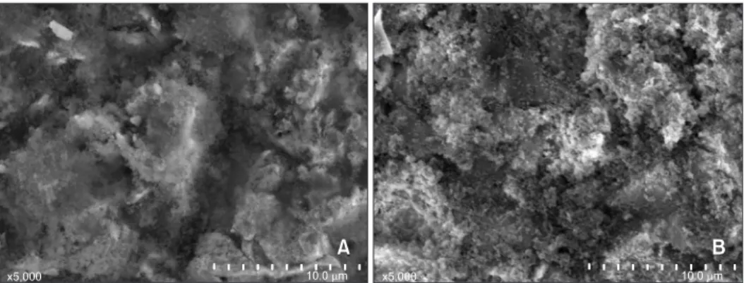
After applying a CoJet silica coating (3M ESPE) and silane, the specimens were sputter-coated with carbon evaporation (SCD-005; Leica Microsystems, Wetzlar, Germany) and examined at 5,000× magnification with a scanning electron microscope (SEM) (JSM-6380; JEOL, Akishima, Japan).
Statistical analysis
The programming language R was used for all stati- stical analyses. Descriptive statistics (i.e., mean, standard deviation) and inferential statistical analyses were per- formed.
After checking the normality assumption and the equa- lity of variance, two-way analysis of variance (ANOVA) was implemented to deduce the significant influential factors on the SBS of the alloy plates with regard to two variables: silanation time and testing timing. Differences between the groups were assessed by using the Student–
Newman–Keuls (S-N-K) multiple comparisons test with a level of significance at p less than 0.05. One-way ANOVA and S-N-K test were used to compare the SBS of enamel and gold surfaces after 24 h.
The Kruskal–Wallis test for the silanation time and the Wilcoxon rank sum test for the test time were used to determine whether differences in ARIs existed between the groups. For the silanation time, a pairwise comparison test using the Wilcoxon rank sum test with Bonferoni correction was performed to determine whether there were differences between 1 s and 30 s, 30 s and 60 s, and 60 s and 120 s.
RESULTS
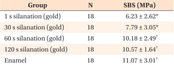
Table 1 shows the means, standard deviations, and range of SBS values, based on silanation time and test timing. The results of the two-way ANOVA test revealed significant differences between the groups (p < 0.05).
A significantly greater SBS was observed in samples
subjected to 60 s and 120 s silanation time that were
examined after 24 h, compared to the SBS of the other
groups (p < 0.05). Figure 1 shows the distribution of the
SBS values in relation to silanation time and test timing
factor.
Table 1. Shear bond strength of the specimens after 1 hour and after 24 hours Test timing (hour) Silanation time
(second) N Shear bond strength (MPa) Significance
1 1 18 6.23 ± 2.62* (1.63 - 11.24)
1 30 18 7.79 ± 3.05* (1.80 - 13.34) Test timing (hour)
§1 60 18 10.18 ± 2.49
†(5.85 - 16.99) 1 < 24
1 120 18 10.57 ± 1.64
†(8.19 - 14.35)
24 1 18 10.34 ± 2.41
†(6.58 - 15.11)
24 30 18 10.43 ± 2.68
†(5.46 - 16.52) Silanation time (second)
§24 60 18 11.86 ± 1.81
‡(9.48 - 15.97) 1 = 30 < 60 = 120
24 120 18 12.59 ± 2.52
‡(8.23 - 16.38)
Values are presented as number only or mean ± standard deviation (range).
*
−‡Items with the same superscripts indicate a homogenous subset, after performing the Student−Newman−Keuls multiple comparisons test (p < 0.05).
§
p < 0.05 indicates a significant result after two-way analysis of variance.
Figure 1. A, Scanning electron micrographs of a specimen after sandblasting with silica (5,000× magnification) and B, after silane application.
Table 2. Frequency distribution of the ARI scores Silanation time
(second) Test timing
(hour) N ARI scores*
Significance
1 2 3 4 5
1 1 18 0 2 4 9 3 Test timing (hour)
24 18 0 2 8 7 1 1 = 24
30 1 18 0 3 9 5 1
24 18 4 3 8 3 0 Silanation time (second)
60 1 18 4 4 8 2 0 1 > 30
†, 30 = 60, 60 = 120
24 18 3 6 7 2 0
120 1 18 4 8 6 0 0
24 18 6 8 4 0 0
*Adhesive remnant index (ARI) scores: 5, no composite remains on the specimen; 4, less than 10% of the composite remains on the specimen surface; 3, more than 10% but less than 90% of the composite remains on the specimen; 2, more than 90%
of the composite remains on the specimen; 1, all of the composite, with an impression of the bracket base, remains on the specimen.
†