황반하출혈의 망막하 Tenecteplase 주입술
Subretinal Tenecteplase Injection for Submacular Hemorrhage in Age-related Macular Degeneration
김양재, 김주영, 성산, 유용성, 권오웅, 김순현
Yang Jae Kim, Ju Young Kim, San Seong, Yong Sung You, Oh Woong Kwon, Soon Hyun Kim
누네안과병원 망막센터
Retina Center, Nune Eye Hospital, Seoul, Korea
Purpose: To investigate the efficacy and safety of a surgical procedure with subretinal tenecteplase (TNK) injection for displacement of thick submacular hemorrhage in patients with age-related macular degeneration (AMD).
Methods: A retrospective, non-comparative, interventional case study was conducted. Medical records of patients with submacular hemorrhage secondary to AMD who underwent subretinal TNK injection were reviewed. Best-corrected visual acuity (BCVA) and spectral domain optical coherence tomography (SD-OCT) at baseline, 1 month, 3 months, and final follow-up after initial treatment were mea- sured. Postoperative complications were identified.
Results: Eleven eyes of eleven patients who underwent subretinal TNK injection for submacular hemorrhage were included. Mean BCVA at baseline was 1.85 ± 0.90 logarithm of the minimum angle of resolution (logMAR), which improved to 1.17 ± 0.63 logMAR at 3 month follow-up and 0.94 ± 0.58 logMAR at final follow-up (p = 0.011). SD-OCT results, including central foveal thickness (495.7 ± 410.7 μm → 230.5 ± 166.4 μm) and submacular hemorrhage thickness (263.3 ± 196.0 μm → 0 μm), significantly improved (p = 0.028). Complications included retinal detachment (n = 2), epiretinal membrane (n = 1), and vitreous hemorrhage (n = 1).
Conclusions: Vitrectomy with subretinal TNK injection may be a good treatment option for displacement of submacular hemorrhage leading to functional and anatomical improvement.
Keywords: Age-related macular degeneration; Submacular hemorrhage; Subretinal injection; Tenecteplase (TNK)
Introduction
The natural prognosis of submacular hemorrhage resulting from age-related macular degeneration (AMD) is generally poor [1-4]. Submacular hemorrhage interferes with retinal function through several mechanisms. Iron released from hemoglobin is toxic to retinal microcirculation and chorio- capillaris. Shearing forces exerted by the contracting clot are
involved in photoreceptor damage. Thick hemorrhages are physical barriers to the exchange of nutrients, oxygen, and metabolites between photoreceptors and the choroid [5,6].
Several procedures have been proposed to displace subma- cular hemorrhage away from the fovea, including pneumatic displacement with or without tissue plasminogen activator (tPA) [7-9], pars plana vitrectomy (PPV) with subretinal tPA [10-12], and intravitreal anti-vascular endothelial growth Address reprint requests to Soon Hyun Kim, MD, PhD
Retina Center, Nune Eye Hospital, #404 Seolleung-ro, Gangnam-gu, Seoul 06198, Korea Tel: 82-2-1661-1175, Fax: 82-2-2086-7779
E-mail: [email protected]
Received: 2017. 3. 23 Revised: 2017. 7. 4 Accepted: 2017. 7. 19
factor (VEGF) injection with or without pneumatic displace- ment [13,14]. However, there remains no gold standard treat- ment for submacular hemorrhage in AMD.
tPA is a recombinant plasminogen activator that binds to the fibrin component of thrombus. It converts bound plas- minogen to plasmin to initiate local fibrinolysis. As men- tioned earlier, tPA has been used to facilitate clearance of submacular hemorrhage [8-14]. However, dose-dependent retinal toxicity in animal and human eyes have been report- ed with tPA [15-18].
More recently, a new third-generation thrombolytic agent, tenecteplase (TNK, Metalyse; Boehringer Ingelheim, Ingel- heim, Germany), has been developed to overcome the lim- itations of tPA. TNK has a 6-fold prolonged plasma half-life, 15-fold higher fibrin specificity, and 80-fold reduced binding affinity to physiological plasminogen activator inhibitor (PAI-1) [19]. Another advantage of TNK is that its vehicle has less than one-third of the potentially toxic L-arginine content than the equivalent dose of tPA [20]. The penetration and toxicity of TNK has been reported in previous studies [20-23]. We recently reported a case of thick submacular hemorrhage resulting from polypoidal choroidal vasculop- athy (PCV) that was successfully treated with subretinal TNK injection [24].
We previousely reported the result of intravitreal TNK in- jection for acute submacular hemorrhage [25]. Even though it is an effective and safe treatment strategy, submacular hemorrhage is not removed by intravitreal pharmacological injection with gas tamponade followed by additional intrav- itreal injections in some patients. The objective of this study was to evaluate the efficacy and safety of PPV with subret- inal TNK injection for submacular hemorrhage resulting from AMD in these patients.
Materials and Methods
We performed a retrospective case series study for eleven eyes from eleven patients who underwent PPV with subreti- nal TNK injection for submacular hemorrhage secondary to AMD at Nune Eye Hospital (Seoul, Korea) from November 2010 to August 2015. This study adhered to the ethical stan- dards in the Declaration of Helsinki. Subretinal TNK injec- tion for submacular hemorrhage secondary to AMD was ap- proved by the Nune Eye Hospital Institutional Review Board
(IRB No. 2010-NEH-001). Informed consent was obtained from all individual participants included in the study.
The inclusion criteria were as follows: 1) patients without displacement of subretinal hemorrhage in macula 1-2 weeks after previous intravitreal TNK injection with pneumatic displacement for submacular hemorrhage and/or previous anti-vascular endothelial growth factor (VEGF) agent in- jection for AMD, 2) patients who had significant vitreous hemorrhage and submacular hemorrhage in AMD diagnosis, and 3) patients who were followed up for at least 5 months after PPV with subretinal TNK injection. Patients with sub- macular hemorrhage secondary to any condition other than AMD were excluded from this study. Patients with evidence of end-stage AMD with severe scarring or atrophy at initial presentation and those with duration of symptoms resulting from submacular hemorrhage longer than one month or who had eyes with an old, whitish discolored submacular hem- orrhage were also excluded. In addition, patients who had previously undergone ocular trauma, PPV, or ocular surgery except cataract surgery were also excluded.
All patients received comprehensive ophthalmic examina- tions, including anterior segment examination and dilated biomicroscopic fundus examination. Best-corrected visual acuity (BCVA) was determined using a Snellen visual acu- ity (VA) chart. Patients who could not visualize the largest Snellen line were shown a 10/400 eye chart at a shorter test distance and then successively asked to count fingers or de- termine hand movements under bright illumination. BCVA was converted to the logarithm of the minimum angle of resolution (logMAR) units for statistical analysis. According to the Holladay method, hand movement was set at 3.0, and counting fingers at 2.0 [26]. Color fundus photography and fluorescein angiography (FA) with indocyanine green angi- ography (ICGA) (Spectralis HRA; Heidelberg Engineering;
Heidelberg, Germany) were performed before vitrectomy with subretinal TNK injection. Spectral-domain optical co- herence tomography (SD-OCT, Spectralis OCT; Heidelberg Engineering, Heidelberg, Germany) was performed before surgery and during follow-up visit. In eyes without a view of the retina because of vitreous hemorrhage, B-scan ultraso- nography was used to identify the extent of hemorrhage.
Diagnosis for neovascular AMD and submacular hem- orrhage were based on the results obtained from FA and ICGA at either initial presentation or after the resolution of submacular hemorrhage. Patients were classified as PCV or
typical exudative AMD. PCV was diagnosed based on ICGA findings, the presence of a branched vascular network, and evidence of terminal polypoidal lesions. Other cases were classified as typical exudative AMD.
Operations were performed by experienced vitreoretinal surgeons (Y.S.Y, O.W.K, and S.H.K). Patients with a visu- ally significant cataract precluding adequate intraoperative visualization underwent phacoemulsification. PPV and pos- terior vitreous detachment formation were performed using an Alcon Accurus or Constellation 23-gauge system (Alcon Laboratories, Fort Worth, TX, USA). TNK was diluted with buffered saline solution (100 μg/0.1 mL) (concentration was based on previous reports by Rowley et al. [20] and McAllis- ter et al. [27] and our previous report [24]) and injected into the subretinal space via a 40-gauge needle to create bullous retinal detachment of the posterior pole. Retinal detachment induced by subretinal injections of TNK involved the fovea.
Partial or total air/fluid exchange was performed. C3F8 gas or silicone oil was used for tamponade. Thereafter, the face- down position was maintained for 48 hours.
The main outcome measures included BCVA, central fo- veal thickness (CFT), thickness of submacular hemorrhage, and the extent of submacular hemorrhage at baseline, 1 month, 3 months, and final follow-up after initial operation.
CFT was defined as the distance between the internal limit- ing membrane and the Bruch membrane at the foveal center.
The thickness of submacular hemorrhage was defined as the distance between the inner segment–outer segment line and the inner border of RPE. The extent of hemorrhage was measured in square millimeters on a reference scan image obtained with SD-OCT using built-in caliper function. All measurements were conducted by a single retinal specialist (J.Y.K).
Statistical analyses were performed with a commercially available software package (PASW Statistics v. 18.0; IBM Corp., Armonk, NY, USA). Wilcoxon’s matched pairs signed ranks test was used to compare BCVA between baseline and postoperative time points (1 month, 3 months, and final fol- low-up). Additional analysis was conducted for six patients whose submacular hemorrhage size and thickness could be identified preoperatively. A Wilcoxon signed rank test was performed to determine changes in the size and the thick- ness of submacular hemorrhage. Statistical significance was defined as a p-value less than 0.05.
Table 1. Summary of patient characteristics Case No./ Age/SexDxVHSMH duration (day)Amount of TNK (mL)TamponadePreop TxPreop VA (logMAR) Postop 3 months VA (logMAR)
Final VA (logMAR)F/U duration (months)Postop anti-VEGF injection 1/62/MPCV(+)270.05SO(-)321.452 2/68/FCNV(-)100.05C3F8A + TNK + gas 1.31.31.4474 3/65/MPCV(+)190.05C3F8(-)321.4306 4/62/MCNV(-)130.05C3F8L + TNK + gas1.410.52137 5/58/MPCV(+)100.05C3F8TNK + gas31.31140 6/57/FPCV(-)210.05C3F8A0.70.102710 7/69/FCNV(-)300.05C3F8A + TNK + gas1.60.520.522317 8/66/MPCV(-)200.05C3F8A + TNK + gas0.40.70.450 9/52/FPCV(+)220.05SO(-)222100 10/76/FCNV(-)280.05C3F8L + TNK + gas210.770 11/69/FCNV(+)30.05C3F8(-)21179 Dx = diagnosis; VH = vitreous hemorrhage; SMH = submacular hemorrhage; TNK = tenecteplase; Preop = preoperative; Tx = treatment; VA = visual acuity; Postop = postoperative; F/U = follow up; VEGF = vascular endothelial growth factor; M = male; F = female; PCV = polypoidal choroidal vasculopathy; CNV = choroidal neovascularization; SO = silicone oil; A = Avastin (bevacizumab); L = Lucentis (ranibizumab).
Results
Eleven eyes of 11 patients who underwent surgery for sub- macular hemorrhage were included. There were 5 men (45.5%) and 6 women (54.5%). They had a median age of 65 years, ranging from 52 to 76 years. Three eyes (27.3%) had been previously treated with intravitreal anti-VEGF injec- tions for neovascular AMD. The median number of previous injections in these eyes was 20, ranging from 2 to 42. There were nine phakic eyes and two pseudophakic eyes.
The median time between onset of symptoms and sur- gery was 20 days, ranging from 3 to 30 days. Six eyes had been previously treated with intravitreal TNK injection (50 μg/0.05 mL) with C3F8 gas injection (0.3 mL) for submacu- lar hemorrhage. There were five eyes with vitreous hemor- rhage. Procedures performed during initial surgery included subretinal injection of TNK (n = 11), C3F8 gas tamponade (n
= 9), silicone oil tamponade (n = 2), intravitreal bevacizumab injection (n = 5), and cataract extraction (n = 8).
Median follow-up time was 13 months, ranging from 5 to 47 months. During postoperative follow-up, 8 (72.7%) pa- tients received additional intravitreal anti-VEGF injections.
The median number of additional postoperative injections in these patients was 4, ranging from 2 to 17. Five eyes (45.5%) were diagnosed with typical neovascular AMD and six eyes (54.5%) were diagnosed with polypoidal choroidal vascu- lopathy (PCV) (Table 1). Mean BCVA of all eyes at baseline was 1.85 ± 0.90 logMAR, ranging from 0.4 to 3.0 logMAR.
Mean BCVA at 1 month after initial surgery was 1.49 ± 0.68 logMAR, ranging from 0.15 to 2 logMAR. Mean BCVA at 3 months after surgery was 1.17 ± 0.63 logMAR, ranging 0.1 to 2.0 logMAR. At final follow-up, mean BCVA was 0.94 ± 0.58 logMAR, ranging from 0 to 2.0 logMAR. BCVA was significantly improved at 1 month (p = 0.049), 3 months (p = 0.010), and final follow-up (p = 0.011) after the initial opera- tion. Eight of 11 eyes had improved BCVA.
In six eyes that were available for SD-OCT preoperatively, mean CFT, mean thickness of submacular hemorrhage, and mean extent of submacular hemorrhage were 495.7 ± 410.7 μm, 263.3 ± 196.0 μm, and 28.7 ± 24.8 mm2,respectively. At one month after the initial operation, mean CFT, mean thick- ness of submacular hemorrhage, and mean extend of subma- cular hemorrhage were 210.2 ± 123.5 μm, 52 ± 66.7 μm, and 3.81 ± 6.99 mm2, respectively. At 3 months after the initial operation, mean CFT was 230.5 ± 166.4 μm, with clearly re- Table 2
. Summary of OCT data of submacular hemorrhage without vitreous hemorrhage cases Case No.Extend of Preop SMH (mm2 )Preop SRHT (μm)Preop CFT (μm) Extend of SMH at Postop 1 month (mm2 )
SRHT at Postop 1 month (μm)CFT at Postop 1 month (μm) Extend of SMH at Postop 3 months (mm2 )
SRHT at Postop 3 months (μm)CFT at Postop 3 months (μm) 231.395938643.7818044100481 43.742312600.64471240073 635.594001,1590021100217 74.251012340.64521270097 871.111142060012100132 1026.091412517.783323700383 OCT = optical coherence tomography; Preop = preoperative; SMH = submacular hemorrhage; SRHT = thickness of subretinal hemorrhage; CFT = central foveal thickness; Postop = post- operative.
solved submacular hemorrhage. At the final follow-up, mean CFT was 193.7 ± 86.4 μm. CFT was decreased significantly at 1 month (p = 0.028), and 3 months (p = 0.028) after the initial operation. However, it did not change significantly at final follow-up (p = 0.116). The thickness of submacular
hemorrhage and extend of submacular hemorrhage were significantly decreased at 1 month (both p = 0.028), and 3 months (p = 0.028, p = 0.027, respectively) after the initial operation (Table 2, Fig. 1).
Following surgery, VA remained stable or deteriorated in 3 (27.3%) patients and was improved by at least 1 line in 8 (72.7%) patients. Complications in the study included retinal detachment (n = 2, 18%), epiretinal membrane (n = 1, 9%), vitreous hemorrhage (n = 1, 9%), and recurrent submacular hemorrhage (n = 1, 9%) (Table 3).
Discussion
tPA has been used to treat submacular hemorrhage [8-12].
However, tPA has not been shown to penetrate the retina in experimental studies, and there are reports of tPA dose-relat- ed retinal toxicity [15-18]. Johnson and colleagues reported dose-related retinal toxicity of tPA in rabbits [15]. They suggested that the L-arginine vehicle in the commercially available solution rather than tPA itself was responsible for the retinotoxicity. It has been reported that tPA can increase N-Methyl-D-aspartate (NMDA)-induced retinal cell damage [28,29]. Chen et al. reported that the toxicity of tPA in human eyes can induce diffuse RPE perturbations, poor recovery of visual acuity, and decreased photopic and scotopic a-waves and b-waves in the electroretinogram (ERG) [17].
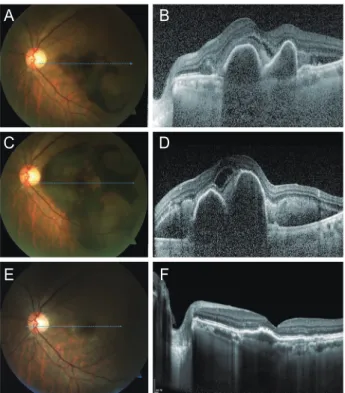
TNK is a variant of tPA that was produced by recombi- nant DNA technology after tPA underwent multiple point mutations. This new thrombolytic agent was developed to overcome several tPA limitations for treating myocardial in- farctions. Compared to tPA, TNK has a longer half-life and greater fibrin specificity with greater efficacy in thrombus Figure 1. Illustrative case. Fundus photography (A) and spectral-do-
main optical coherence tomography (SD-OCT) (B) of submacular hemorrhage in case 7 at first visit before intravitreal tenecteplase (TNK) and avastin injection with gas tamponade. One week after the primary treatment, fundus photography (C) and SD-OCT (D) of the left eye of case 7 showed aggravated submacular hemorrhage. Post- operative fundus photography (E) and SD-OCT (F) taken 3 months after vitrectomy with subretinal TNK injection showed that the sub- macular hemorrhage had mostly disappeared.
A
C
E
B
D
F
Table 3. Summary of postoperative complications
Case No. Cx Secondary procedure Term between primary operation and
secondary procedure
2 VH TPPV 6 weeks
5 RD SB + gas 2 months
6 ERM TPPV + membranectomy + ILM 8 months
7 Submacular hemo Intravitreal TNK + gas 7 months
9 RD TPPV + SE + SO exchange + endolaser 5 weeks
Cx = complications; VH = vitreous hemorrhage; TPPV = trans pars plana vitrectomy; RD = retinal detachment; SB = sclearal buckling; ERM = epiretinal membrane; ILM = internal limiting membrane; TNK = tenecteplase; SE = scleral encircling; SO = silicone oil.
dissolution [19]. Furthermore, the vehicle of TNK has less than one-third of the L-arginine content used for tPA. These characteristics could provide significant advantages for treat- ment of submacular hemorrhage [20]. In human eyes, McAl- lister et al. reported that intravitreal TNK injection had a safe and favorable effect on submacular hemorrhage [22,23].
We recently reported a case of subretinal TNK injection in a submacular hemorrhage from PCV [24]. In this case, we identified the efficacy and safety of direct subretinal TNK injection for submacular hemorrhage secondary to PCV.
The patient’s vision fully recovered without retinal pigment epithelium change. Scotopic and photopic ERG showed no prolongation of implicit time with slightly decreased a-wave and b-wave amplitude.
Several studies have recently shown that anti-VEGF treat- ment with or without gas tamponade for submacular hem- orrhage from neovascular AMD had positive results. Kim et al. reported that anti-VEGF monotherapy was a useful treatment option for exudative AMD accompanied by sub- macular hemorrhage [13]. Shin and colleagues have shown that anti-VEGF therapy with gas tamponade may yield better treatment outcomes than anti-VEGF monotherapy for eyes with thick subretinal hemorrhage [14]. However, surgery is unavoidable if there is accompanying vitreous hemorrhage or if pharmacological treatment with or without gas tampon- ade has failed.
In our clinic, the primary treatment for submacular hem- orrhage from neovascular AMD is intravitreal TNK injec- tion with C3F8 gas tamponade. Intravitreal TNK injection with gas tamponade may be an effective treatment for sub- macular hemorrhage [25]. However, submacular hemorrhage can remain in some patients at the macula or more severe submacular hemorrhage can be found even after primary intravitreal TNK and/or anti-VEGF injection with gas tam- ponade. In this study, 6 of 11 eyes received intravitreal TNK injection with C3F8 gas tamponade preoperatively. However, the results of intravitreal TNK injection in these cases were unsatisfactory. When intravitreal TNK injection with gas tamponade failed, surgery including pars plana vitrectomy, subretinal TNK injection, and gas tamponade may have good results. Therefore, in our clinic, indications for subret- inal TNK injection for submacular hemorrhage include ac- companying vitreous hemorrhage and unsatisfactory results from intravitreal TNK injection with gas tamponade.
A direct comparison between subretinal TNK and tPA
injection is difficult because different procedures are used in different cases. However, results of subretinal TNK in- jection in this study may not be inferior to previous results of subretinal tPA injection for submacular hemorrhage. In previous studies, improved visual acuity was observed in 45- 82% of eyes after subretinal tPA injection [10,11,30]. In this study, 9 of 12 (75%) eyes had improved BCVA. In addition, submacular hemorrhage of all eyes improved 3 months after subretinal TNK injection. Complications in the study includ- ed retinal detachment (n = 2, 18%), epiretinal membrane (n = 1, 9%), vitreous hemorrhage (n = 1, 9%), and recurrent sub- macular hemorrhage (n = 1, 9%). Complications after subret- inal TNK injection were similar to subretinal tPA injection complications reported in the literature. Surgical removal of submacular hemorrhage with tPA has been associated with significant complications, including retinal detachment (4.0- 19.3%), vitreous hemorrhage (1.9-40%), and glaucoma (6%) [10,11,12,30]. Recurrence of submacular hemorrhage (5.9- 27%) has also been reported [10,11,30].
This study has several limitations. First, it was a small, retrospective case series study without a control group.
Second, although visual acuities were best corrected, Early Treatment Diabetic Retinopathy Study (ETDRS) visual acu- ities were not available. Third, unlike our previous case re- port, we did not perform both preoperative and postoperative ERG for all cases. Therefore, we could not exactly evaluate the retinal toxicity of TNK. It was also difficult to discrimi- nate the causes of complications between TNK and surgical technique. In addition, data obtained in this study were not from a single surgeon and that could be a confounding fac- tor.
In conclusion, vitrectomy with subretinal TNK injection may be a good treatment option for submacular hemorrhage secondary to neovascular AMD if submacular hemorrhage has accompanying vitreous hemorrhage or if primary in- travitreal pharmacological injection and/or gas tamponade is unsatisfactory. To our knowledge, this is the first study on the efficacy and safety of subretinal TNK injection for submacular hemorrhage. Large, long-term, prospective, and comparative clinical trials are needed to evaluate the efficacy and safety of this procedure in the future.
Conflicts of Interest
The authors have no conflicts to disclose.
References
1. Bennett SR, Folk JC, Blodi CF, Klugman M. Factors prognostic of visual outcome in patients with subretinal hemorrhage. Am J Ophthalmol 1990;109:33-7.
2. Avery RL, Fekrat S, Hawkins BS, Bressler NM. Natural history of subfoveal subretinal hemorrhage in age-related macular de- generation. Retina 1996;16:183-9.
3. Berrocal MH, Lewis ML, Flynn HW Jr. Variations in the clin- ical course of submacular hemorrhage. Am J Ophthalmol 1996;122:486-93.
4. Scupola A, Coscas G, Soubrane G, Balestrazzi E. Natural history of macular subretinal hemorrhage in age-related macular de- generation. Ophthalmologica 1999;213:97-102.
5. Glatt H, Machemer R. Experimental subretinal hemorrhage in rabbits. Am J Ophthalmol 1982;94:762-73.
6. Toth CA, Morse LS, Hjelmeland LM, Landers MB 3rd. Fibrin di- rects early retinal damage after experimental subretinal hemor- rhage. Arch Ophthalmol 1991;109:723-9.
7. Ohji M, Saito Y, Hayashi A, et al. Pneumatic displacement of sub- retinal hemorrhage without tissue plasminogen activator. Arch Ophthalmol 1998;116:1326-32
8. Hassan AS, Johnson MW, Schneiderman TE, et al. Management of submacular hemorrhage with intravitreous tissue plasmino- gen activator injection and pneumatic displacement. Ophthal- mology 1999;106:1900-6; discussion 1906-7.
9. Hesse L, Schmidt J, Kroll P. Management of acute submacular hemorrhage using recombinant tissue plasminogen activator and gas. Graefes Arch Clin Exp Ophthalmol 1999;237: 273-7.
10. Haupert CL, McCuen BW 2nd, Jaffe GJ, et al. Pars plana vitrec- tomy, subretinal injection of tissue plasminogen activator, and fluid-gas exchange for displacement of thick submacular hem- orrhage in age-related macular degeneration. Am J Ophthalmol 2001;131:208-15.
11. Olivier S, Chow DR, Packo KH, et al. Subretinal recombinant tissue plasminogen activator injection and pneumatic displace- ment of thick submacular hemorrhage in age-related macular degeneration. Ophthalmology 2004;111:1201-8.
12. Fine HF, Iranmanesh R, Del Priore LV, et al. Surgical outcomes after massive subretinal hemorrhage secondary to age-related macular degeneration. Retina 2010;30:1588-94.
13. Kim JH, Chang YS, Kim JW, et al. Intravitreal anti-vascular endo- thelial growth factor for submacular hemorrhage from choroi- dal neovascularization. Ophthalmology 2014;121:926-35.
14. Shin JY, Lee JM, Byeon SH. Anti-vascular endothelial growth
factor with or without pneumatic displacement for submacular hemorrhage. Am J Ophthalmol 2015;159:904-14.e1.
15. Johnson MW, Olsen KR, Hernandez E, et al. Retinal toxicity of recombinant tissue plasminogen activator in the rabbit. Arch Ophthalmol 1990;108:259-63.
16. Hrach CJ, Johnson MW, Hassan AS, et al. Retinal toxicity of com- mercial intravitreal tissue plasminogen activator solution in cat eyes. Arch Ophthalmol 2000;118:659-63.
17. Chen SN, Yang TC, Ho CL, et al. Retinal toxicity of intravitreal tissue plasminogen activator: case report and literature review.
Ophthalmology 2003;110:704-8.
18. Jung JJ, Cho SW. Retinal toxicity of intravitreal tissue plasmino- gen activator on submacular hemorrhage. J Korean Ophthalmol Soc 2009;50:800-3.
19. Tanswell P, Modi N, Combs D, Danays T. Pharmacokinetics and pharmacodynamics of tenecteplase in fibrinolytic therapy of acute myocardial infarction. Clin Pharmacokinet 2002;41:1229- 45.
20. Rowley SA, Vijayasekaran S, Yu PK, et al. Retinal toxicity of intrav- itreal tenecteplase in the rabbit. Br J Ophthalmol 2004;88:573-8.
21. Kwan AS, Vijayasekaran S, McAllister IL, et al. A study of retinal penetration of intravitreal tenecteplase in pigs. Invest Ophthal- mol Vis Sci 2006;47:2662-7.
22. McAllister IL, Vijayasekaran S, Khong CH, Yu DY. Investigation of the safety of tenecteplase to the outer retina. Clin Exp Ophthal- mol 2006;34:787-93.
23. McAllister IL, Chen SD, Patel JI, et al. Management of submacular haemorrhage in age-related macular degeneration with intrav- itreal tenecteplase. Br J Ophthalmol 2010;94:260-1.
24. Kwon YH, Lim SJ, Jeung WJ, et al. Subretinal tenecteplase injec- tion in a submacular hemorrhage from polypoidal choroidal vasculopathy: a case report. Retin Cases Brief Rep 2012;6:400-5.
25. Lee JP, Park JS, Kwon OW, et al. Management of acute subma- cular hemorrhage with intravitreal injection of tenecteplase, anti-vascular endothelial growth factor and gas. Korean J Oph- thalmol 2016;30:192-7.
26. Holladay JT. Proper method for calculating average visual acuity.
J Refract Surg 1997;13:388-91.
27. McAllister IL, Vijayasekaran S, Yu DY. Intravitreal tenecteplase (metalyse) for acute management of retinal vein occlusions.
Invest Ophthalmol Vis Sci 2013;54:4910-8.
28. Kumada M, Niwa M, Hara A, et al. Tissue type plasminogen activator facilitates NMDA-receptor-mediated retinal apoptosis through an independent fibrinolytic cascade. Invest Ophthal- mol Vis Sci 2005;46:1504-7.
29. Mali RS, Cheng M, Chintala SK. Plasminogen activators promote excitotoxicity-induced retinal damage. FASEB J 2005;19:1280-9.
30. Moisseiev E, Ben Ami T, Barak A. Vitrectomy and subretinal injec-
tion of tissue plasminogen activator for large submacular hem- orrhage secondary to AMD. Eur J Ophthalmol 2014;24:925-31.