CASE REPORT
ISSN 2508-1926Copyright © 2016 The korean retina society
This is an Open Access article distributed under the terms of the Creative Commons Attribution Non-Commercial License (http://creativecommons.org/licenses/by-nc/3.0/) which permits unrestricted non-commercial use, distribution, and reproduction in any medium, provided the original work is properly cited.
Journal of Retina 2016;1(2):105-107 http://dx.doi.org/10.21561/jor.2016.1.2.105
유리체 출혈 제거를 위한 유리체 절제술 후 이차적 황반 원공 발생
Secondary Macular Hole Formation after Pars Plana Vitrectomy for Vitreous Hemorrhage
김정희, 장철원, 황덕진
Jeong Hee Kim, Cheol Won Jang, Duck Jin Hwang
한길안과병원
Hangil Eye Hospital, Incheon, Korea
Purpose: To describe a case of a secondary macular hole (MH) after pars plana vitrectomy for vitreous hemorrhage.
Case summary: A 79-year-old woman with decreased visual acuity in her right eye was diagnosed with a grade 4 vitreous hemorrhage and underwent pars plana vitrectomy. After 1 week, an MH was detected using a fundus examination and optical coherence tomogra- phy (OCT). One month after surgery, the MH remained stationary with a best corrected visual acuity (BCVA) of 20/1,000. She underwent a second partial vitrectomy, and closure of the MH was demonstrated by OCT. BCVA was 20/63 at 1 month after the second operation.
Conclusions: We report a case of secondary full-thickness macular hole formation without vitreofoveal traction after vitrectomy for vit- reous hemorrhage.
Keywords: Secondary macular hole; Vitrectomy; Vitreous hemorrhage
Introduction
Although the prevalence of secondary macular hole (MH) formation is relatively low, its clinical findings and prognosis have been widely reported. Tangential macular traction has long been proposed to be a cause of MH formation [1-3].
However, some reported cases appear to have occurred with- out such traction, raising the possibility that traction is not the only precipitating factor in MH formation [3]. We report such a case after vitrectomy with no significant intraopera- tive complications.
Case Report
A 79-year-old woman presented with a 4-day history of de- creased visual acuity in the right eye. She had pseudophakia in the right eye and an artificial left eye due to an unknown cause. There was no underlying disease. Her best corrected visual acuity (BCVA) was counting fingers at 20 cm. The initial fundus examination revealed a grade 4 vitreous hem- orrhage, and a B-scan showed no evidence of retinal detach- ment. Pars plana vitrectomy was performed without internal limiting membrane (ILM) peeling. Neither an epiretinal membrane nor an MH was observed. Triamcinolone ace- tonide was used for vitreous visualization. No air or gas tamponade was applied, and there were no intraoperative
Address reprint requests to Duck Jin Hwang, MD
Hangil Eye Hospital, #35 Bupyeong-daero, Bupyeong-gu, Incheon 21388, Korea Tel: 82- 32-503-3322, Fax: 82-32-504-3322
E-mail: [email protected]
Received: 2016. 10. 14 Revised: 2016. 11. 1 Accepted: 2016. 11. 1
106
JOURNAL OF RETINA
http://dx.doi.org/10.21561/jor.2016.1.2.105 complications. An MH was detected at 1 week postoperative
using a fundus examination and optical coherence tomog- raphy (OCT) (Fig. 1A). We waited 4 weeks for spontaneous MH closure. One month after surgery, the MH remained stationary (Fig. 1B) with a BCVA of 20/1,000, and a second partial vitrectomy with ILM peeling and perfluoropropane (C3F8) gas tamponade were performed. Triamcinolone ace- tonide was used for vitreous visualization and ILM peeling.
OCT demonstrated closure of the MH, and BCVA was 20/63 at 1 month after the second operation (Fig. 2)
Discussion
Vitreofoveal traction is the major mechanism of MH forma- tion, as supported by imaging and histopathological data [1- 6]. However, certain cases with underlying conditions, such as intraretinal exudation in diabetic retinopathy and epireti- nal membranes, have been reported in association with sec- ondary MH formation [1,2].
In the present case, the reason for secondary MH develop- ment with no remarkable traction to the fovea was unclear.
There was no retinal vein occlusion or diabetic retinopathy.
Considering the patient’s age, degenerative factors, such
as subtle defects or breaks in the ILM (traction- or senes- cence-induced), might have allowed hydration of the fovea and distorted the tissue to form a full-thickness MH [3,5]
Lipham and Smiddy’s explanation that spontaneous foveal dehiscence may be the result of a degenerative process also supports the alternate mechanism seen in our case [5]. An- other possible but less likely explanation for secondary MH formation is that the traction component could have been below the resolution of the OCT, and a possible mediator might be the outer wall of a vitreoschisis [3]. Although we did not take OCT images before the initial vitrectomy due to vitreous hemorrhage, it is possible that the vitreous hemor- rhage might have masked spontaneous vitreofoveal traction, which might have resulted in spontaneous fine vitreofoveal dehiscence. Iatrogenic trauma during the initial vitrectomy is another possible mechanism of secondary MH formation in our case. The latter two mechanisms, however, are less likely than the first.
In summary, we report a case of secondary full-thickness MH formation without vitreofoveal traction after vitrectomy.
Although the pathogenesis of MH formation is still uncer- tain, it may involve degenerative and tractional factors. In the present case, the MH closed after ILM peeling and gas tamponade. Most current studies indicate that ILM peeling is the preferred treatment for both idiopathic and secondary MHs. MH repair surgery with ILM peeling achieved favor- able anatomic and functional outcomes in the present case.
Conflicts of interest
The authors report no conflicts of interest. The authors alone are responsible for the content and writing of the paper.
References
1. Tomita Y, Noda K, Shinoda H, et al. Secondary macular hole for- mation with presumed evulsion of foveal hard exudates in a pa- tient with diabetic retinopathy. Jpn J Ophthalmol 2010;54:366-8.
2. Kumagai K, Ogino N, Furukawa M, et al. Surgical outcomes for patients who develop macular holes after pars plana vitrecto- my. Am J Ophthalmol 2008;145:1077-80.
3. Smiddy WE. Macular hole formation without vitreofoveal trac- tion. Arch Ophthalmol 2008;126:737-8.
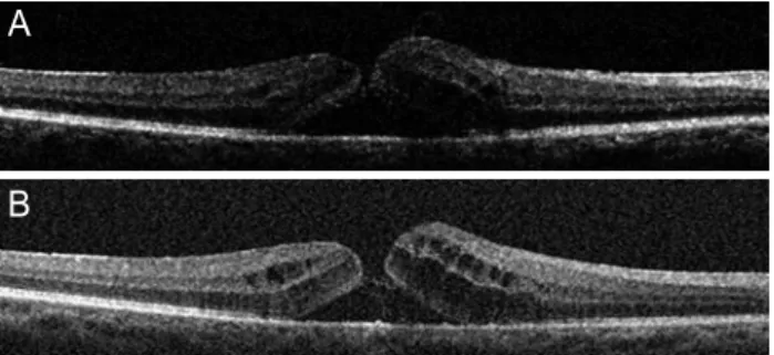
4. Lee SH, Park KH, Kim JH, et al. Secondary macular hole forma- Figure 1. Optical coherence tomography images. Full-thickness
macular hole formation detected at postoperative week 1 (A). Spon- taneous closure was not observed until postoperative week 4 (B).
Internal limiting membrane peeling and gas tamponade were not performed in the first operation.
Figure 2. Optical coherence tomography image. One month after the second partial vitrectomy with internal limiting membrane peel- ing and gas tamponade, the macular hole was closed.
A
B
107
Kim JH, et al. Secondary MH after PPV
http://dx.doi.org/10.21561/jor.2016.1.2.105 tion after vitrectomy. Retina 2010;30:1072-7.
5. Lipham WJ, Smiddy WE. Idiopathic macular hole following vit- rectomy: implications for pathogenesis. Ophthalmic Surg Lasers
1997;28: 633-9.
6. Kimura H, Kuroda S, Nagata M. Macular hole formation in post- vitrectomized eyes. Retina 2005;25:521-3.