Tuberc Respir Dis 2009;67:462-466
CopyrightⒸ2009. The Korean Academy of Tuberculosis and Respiratory Diseases. All rights reserved.
전종격동에서 발생한 악성 횡문근양 종양 1예
연세대학교 의과대학
1내과학교실,
2진단병리학교실
오경진1, 이기병1, 홍순원2, 정교태1, 최홍규1, 김형중1, 안철민1, 장윤수1
A Case of Malignant Rhabdoid Tumor in the Anterior Mediastinum
Kyung Jin Oh, M.D.1, Ki Byung Lee, M.D.1, Soon Won Hong, M.D., Ph.D.2, Kyo Tae Jung, M.D.1, Hong Kyu Choi, M.D.1, Hyung Jung Kim, M.D., Ph.D.1, Chul Min Ahn, M.D., Ph.D.1, Yoon Soo Chang, M.D., Ph.D.1 Departments of 1Internal Medicine, 2Pathology, Yonsei University College of Medicine, Seoul, Korea
Malignant rhabdoid tumors arise primarily from the kidney. Extrarenal malignant rhabdoid tumors are rare, with the liver, central nervous system, and skin reported as the primary sites. Malignant rhabdoid tumors of the mediastinum are extremely rare among extrarenal malignant rhadoid tumors; only 3 cases have been reported to date, all characterized by aggressive clinical behavior. We experienced a 35-year-old woman diagnosed with malignant rhabdoid tumor in the anterior mediastinum with multiple metastases. The tumor was surgically unresectable, and treated with palliative radiation therapy. Three-month after radiation treatment, she died from dissemination of the malignant rabdoid tumor.
Key Words: Rhabdoid Tumor; Mediastinum
Address for correspondence: Yoon Soo Chang, M.D., Ph.D.
Department of Internal Medicine, Yonsei University College of Medicine, 712, Eonju-ro, Gangnam-gu, Seoul 135-720, Korea
Phone: 82-2-2019-3309, Fax: 82-2-3463-3882 E-mail: [email protected]
Received: Sep. 1, 2009 Accepted: Sep. 28, 2009
서 론
악성 횡문근양 종양(malignant rhabdoid tumor)은 주 로 유소아의 신장이나 중추신경계에서 발생하는 질병으 로1, 1978년 신장에서 발생한 예가 처음 기술되었고 초기 의 보고 당시 조직학적으로 횡문근 종양과 유사한 소견을 보여 윌름즈 종양의 횡문근 종양 변이로 생각되었으나2, 이후의 연구에서 면역조직화학 검사상 횡문근 종양과는 연관성이 없는 것으로 밝혀져 1981년 Haas 등3에 의해 악 성 횡문근양 종양으로 명명되었다. 발생 부위에 따라 신 장에 발생한 경우는 악성 신성 횡문근양 종양(malignant renal rhabdoid tumor), 신장 이외의 조직에서 발생한 경 우를 악성 신외성 횡문근양 종양(malignant extrarenal rhabdoid tumor)으로 구분되었다4.
신장 이외의 간, 뇌, 피부, 연조직 등에서 발생한 악성 횡문근양 종양에 대한 증례 보고들이 있었으나1,4,5, 악성 횡문근양 종양이 종격동에서 발생하는 경우는 극히 드물 어 선천성 종양을 포함하여 전세계적으로 3예만이 보고되 었으며5,6, 국내 보고는 예가 없는 실정이다. 저자들은 전 종격동에서 발생한 악성 횡문근양 종양 1예를 경험하여 이를 보고하는 바이다.
증 례
환 자: 박○○, 여자, 35세
주 소: 흉부 단순촬영에서 발견된 흉부 종괴 현병력: 상기 환자는 내원 2개월 전부터 발생한 기침과 객담이 있었고 내원 1개월 전부터는 전신 쇠약감, 식욕 부진 및 5 kg의 체중 감소가 있었다. 지역 의원에서 경과 관찰 및 투약하였으나 증상 호전 없어 시행한 흉부 X-선 검사 및 흉부 전산화 단층촬영에서 폐장 및 전종격동의 종괴 발견되어 정밀 검사를 위해 본원 호흡기 내과에 내원 하였다.
과거력: 특이 소견은 없었다.
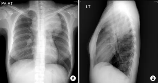
Figure 1. Chest X-ray sug- gests huge mass lesion on left upper lung fileds (A, B).
Figure 2. Chest computed tomography shows 8.5 cm sized anterior mediastinal mass with contrast window (A), with 가족력: 특이 소견은 없었다.
진찰 소견: 내원 당시 신체 활력 징후는 정상이었고, 좌 측 쇄골 상부에서 고정된 다발성 임파절이 촉진되었으며, 액와부 및 서혜부의 임파절은 촉지되지 않았다. 청진상 심음, 호흡음은 정상이었으며 수포음은 들리지 않았다.
복부 진찰상 배는 부드럽고 종괴나 장기가 촉지 되지 않았 으며 장음은 정상이었고 압통이나 반발통은 없었다.
검사 소견: 말초 혈액 검사에서 백혈구 25,580/mm3 (다 핵구 84.5%, 림프구 9.7%, 단핵구 3.5%, 호산구 0.4%), 혈색소 11.5 g/dL, 혈소판 778,000/mm3이었다. 혈청 생화 학 검사에서 AST 37 IU/L, ALT 58 IU/L였고 ALP 331 IU/L, LDH 695 IU/L으로 증가되어 있었으며 BUN 2.6 mg/dL, Creatinine 0.4로 측정되었다.
방사선 소견: 단순 흉부 촬영에서 좌상부폐야를 침윤하 는 종괴가 의심되었고(Figure 1), 흉부 전산화 단층촬영에 서 전종격동 및 좌측쇄골상부 림프의 다발성 종대가 관찰 되었으며 가장 큰 종괴의 직경은 8.5 cm으로 전종격동에 있었다(Figure 2). 전신 뼈 스캔에서 좌측 후방 쇄골에 전 이가 의심되고(Figure 3A), 양전자 방출 단층촬영에서 전 종격동과 양측 폐문부, 좌측 쇄골상부 임파절 외에도 양측 부신 및 양측 갈비사이근육에 전이 소견 보이는 상태로 수 술적인 제거는 불가능한 상태였다(Figure 3B).
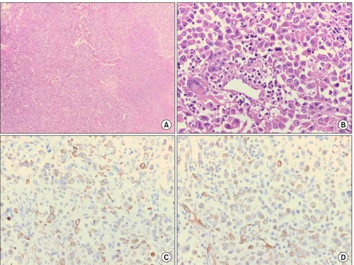
조직 소견: 폐 및 경부 림프절 조직검사에서 주변조직 으로 광범하게 침윤되는 원형의 종양세포가 호산성으로 유리질의 봉합체를 포함하는 호산성 세포질과 뚜렷한 핵 소체가 관찰되고 핵이 한쪽으로 치우쳐 있는 양상 보였으
Figure 3. Whole body bone scan suggests metastasis to left lower posterior ribs (A), and PET-CT shows mul- tiple metastasis to lymph node, both adrenal glands (B).
Figure 4. Histopathologic findings shows round tumor cells infiltrating surrounding tissues with eosinophilic cytoplasm containing amorphous inclusion body (H&E stain; A, ×40, B, ×400), positive staining in cytoplasm (Immunohistochemical staining; C, cytokeratin, ×400, D, vimentin, ×400).
며(Figure 4A, B), 면역 염색상 세포각질항원(cytokeratin) 과 비멘틴(vimentin)에 양성 소견 보여 전종격동의 악성 횡문근양 종양으로 진단되었다(Figure 4C, D).
치료 및 경과: 환자의 전신상태는 ECOG 수행상태 3점 으로 항암화학 요법과 동시에 방사선 치료를 시행하기는 어렵다고 판단하여 방사선 치료를 먼저 시행하기로 하였 다. 총 15회, 총 3,000 cGy 방사선치료 받았으나 종괴의 크기는 줄어들지 않았다. 환자 전신 상태 호전되지 않았 고 치료에도 반응 없어 더 이상의 치료는 의미 없을 것으 로 판단하고 보존적 치료를 하며 경과를 관찰하던 중 진단 3개월 후 방사선 치료 후 생긴 폐렴 및 이에 속발된 패혈 증으로 사망하였다.
고 찰
악성 횡문근양 종양은 주로 유소아에서 발생하는 질병 이지만 성인에서도 발생할 수 있어 평균 발병연령은 20개 월이지만 생후 3주에서 50세까지 다양한 연령층에서 발생 이 보고되었다1,7,8. 발병 후 생존기간은 평균 6개월로 5년 생존율이 20% 미만인1,7 매우 침습적인 질병으로 조기 진 단과 수술적 절제 여부가 중요하다.
악성 횡문근양 종양은 면역 염색에서 비멘틴에 75∼
100%가 양성을 보이며 세포각질항원과 EMA에서도 각각 59∼100%, 63∼100%가 양성을 보이며 S-100 단백이나 데스민(desmin), 미오글로빈(myoglobin) 등의 골격근 표 지자들에서는 음성을 보인다. 본 증례에서도 비멘틴과 세 포각질항원에 양성을 보이는 전형적인 조직병리학적 소 견을 보였다.
치료로는 가능한 완전 절제하는 것이 목표이며 방사선 치료와 유육종증이나 콩팥모세포종에서 시도되는 ifosfa- mide, carboplatin, etoposide 등의 병합 항암 화학요법이 생존 기간 연장에 도움이 된다는 제안이 있으나5, 재발 및 전이가 잘 되어 예후가 불량한 것으로 알려져 있다. 특히 방사선 치료나 병합 항암 화학요법이 생존기간을 연장시 킨 몇몇 보고는 소아가 대부분이며 성인의 경우라 하더라 도 전이가 없고 원발 종괴의 완전절제나 부분절제가 가능 한 경우에 국한된 경우가 많았다9,10.
악성 신외성 횡문근양 종양은 폐, 간, 림프절로 조기 전 이를 보이며 특히 전종격동에서 발생한 두 예6는 종괴가 큰 혈관 및 주요 장기들과 인접해 있어 완전 절제가 불가 능하였고, 방사선 치료 및 항암화학요법으로는 완전 관해
으나 한 환자는 증례보고 당시까지 생존하고 있었다. 본 증례는 기존 보고와 유사하게 진단 당시 폐와 림프절 뿐만 아니라 뼈와 부신 전이도 동반되어 있고 방사선 치료에 효과가 없었으며 진단 3개월 만에 사망하였다. 종격동에 발생한 악성 횡문근양 종양은 아직까지 보고된 예가 적어 정확한 생존기간이나 예후에 대한 정확한 보고가 없고 치 료법에 대해서도 표준화되어 있지 않은 실정으로 현재까 지는 다른 악성 신외성 횡문근양 종양의 치료법을 시도해 보고 있다. 최근에는 유방암과 악성흑색종의 치료에서 이 용되는 전초림프절생검이 악성 신외성 횡문근양 종양의 조기 진단과 국소 병변의 치료에 도움이 될 것으로 제안되 기도 하나11 아직까지 효과적으로 시도되고 있지는 않다.
참 고 문 헌
1. Oda Y, Tsuneyoshi M. Extrarenal rhabdoid tumors of soft tissue: clinicopathological and molecular genetic review and distinction from other soft-tissue sarcomas with rhabdoid features. Pathol Int 2006;56:287-95.
2. Beckwith JB, Palmer NF. Histopathology and prognosis of Wilms tumors: results from the First National Wilms' Tumor Study. Cancer 1978;41:1937-48.
3. Haas JE, Palmer NF, Weinberg AG, Beckwith JB.
Ultrastructure of malignant rhabdoid tumor of the kid- ney. A distinctive renal tumor of children. Hum Pathol 1981;12:646-57.
4. Maschek H, Werner M, Busche G, Weinel P. Congeni- tal rhabdoid tumor in the mediastinum and liver. Case report and review of the literature. Pathologe 1992;13:
172-8.
5. Wick MR, Ritter JH, Dehner LP. Malignant rhabdoid tu- mors: a clinicopathologic review and conceptual dis- cussion. Semin Diagn Pathol 1995;12:233-48.
6. Falconieri G, Moran CA, Pizzolitto S, Zidar A, Angione V, Wakely PE Jr. Intrathoracic rhabdoid carcinoma: a clinicopathological, immunohistochemical, and ultrastr- uctural study of 6 cases. Ann Diagn Pathol 2005;9:279- 83.
7. Kodet R, Newton WA Jr, Sachs N, Hamoudi AB, Raney RB, Asmar L, et al. Rhabdoid tumors of soft tissues:
a clinicopathologic study of 26 cases enrolled on the Intergroup Rhabdomyosarcoma Study. Hum Pathol 1991;22:674-84.
8. Parham D, Weeks D, Beckwith JB. The clinicopatho- logic spectrum of putative extrarenal rhabdoid tumors.
An analysis of 42 cases studied with immunohistoch-
1994;18:1010-29.
9. Puri DR, Meyers PA, Kraus DH, Laquaglia MP, Wexler LH, Wolden SL. Radiotherapy in the multimodal treat- ment of extrarenal extracranial malignant rhabdoid tumors. Pediatr Blood Cancer 2008;50:167-9.
10. Walterhouse D, Watson A. Optimal management strat-
egies for rhabdomyosarcoma in children. Paediatr Dru- gs 2007;9:391-400.
11. Mazzocchi M, Chiummariello S, Bistoni G, Marchetti F, Alfano C. Extrarenal malignant rhabdoid tumour of the heel: a case report. Anticancer Res 2005;25:4573-6.