DOI 10.17480/psk.2015.59.5.201
Human Liver Microsomes과 HepG2 세포를 이용한 약물유래 간독성 평가 방법의 최적화
최종민 · 전장수 · 김상겸# 충남대학교 약학대학
(Received April 24, 2015; Revised July 16, 2015; Accepted July 17, 2015)
The Optimization of Method for Prediction of Drug-Induced Liver Injury Using HepG2 Cells Cultured with Human Liver Microsomes
Jong Min Choi, Jang Su Jeon and Sang Kyum Kim# College of Pharmacy, Chungnam National University, Daejeon 305-764, Korea
Abstract — The aim of the present study was to optimize in vitro method for the prediction of drug-induced liver injury using human liver microsomes (HLM). Cytotoxicity test of cyclophosphamide and acetaminophen in HepG2 cells cultured with HLM showed that the newly established condition using 0.375 mg/ml HLM for 24 hr incubation was comparable or more sensitive than the previously established condition using 0.75 mg/ml HLM for 12 hr incubation. Although the cytotoxic effect of troglitazone was completely attenuated by 0.75 mg/ml HLM, it was augmented by 0.375 mg/ml HLM in the pres- ence of the NADPH-generating system. The cytotoxic effect of chlormezanone, a withdrawn drug due to hepatotoxicity in human, was increased by HLM in the presence of the NADPH-generating system. In contrast, the cytotoxic effect of metha- pyrilene, a withdrawn drug due to hepatotoxicity in rats, was decreased by HLM in the presence of the NADPH-generating system. The present study suggests that the optimized in vitro method using HLM can be useful for the prediction of drug- induced hepatotoxicity.
Keywords □ drug toxicity, cytochrome P450, microsomes, in vitro assay, optimization
신약 개발에서 약물유래 간독성에 대한 연구는 약물의 안전성 을 확보하기 위한 필수적인 과정으로 인식되고 있다.1,2)약물유 래 간독성의 기전은 다양하지만 대사 활성화는 약물유래 간독성 의 주요한 원인 중 하나이다.3)
신약 개발에서 약물유래 간독성을 예측하기 위해 다양한 in vitro assay가 개발되어 왔다. 일차 배양 인간 간세포는 phase I 효소와 phase II 효소가 발현되어 약물유래 간독성의 평가에서 흔히 사용하는 in vitro 세포 모델이다.4)하지만, 일차 배양 인간 간세포는 불안정한 in vitro 표현형, 개체차, 조작의 어려움, 고비 용 등의 이유로 그 사용에 한계를 가진다.5)이러한 한계를 극복
하기 위해 일차 배양 인간 간세포를 대체할 수 있는 여러 가지 세포주와 유전자 조작된 세포 모델들이 연구되었다.4,6-9)이 모델 들은 안정한 표현형, 실험의 재현성 등의 장점이 있지만, 대체로 대사 활성화를 매개하는 주요한 효소인 cytochrome P450(CYP) 의 활성이 낮고, in vivo 실험과의 불일치, 특허 침해 등으로 사 용에 제한이 된다.
선행 연구에서 약물유래 간독성을 예측하는 새로운 모델로서 human liver microsomes(HLM)과 함께 배양한 HepG2 세포 모 델을 개발하였다.10) HLM의 활용은 CYP 등의 대사능이 낮은 HepG2 세포 모델에 대사효소를 간접적으로 부여함으로써 약물 의 독성 평가에서 대사 활성화와 무독화 그리고 HLM 단백질 결 합 등의 독성 기전을 규명하기 위해 사용되었다. 하지만, HLM과 독성물질의 반응시간이 12시간으로 실험 수행에서 불편함이 있고 자체 독성의 발현에도 한계가 있었다. 이러한 문제점을 보완하고 자 본 연구에서는 실험의 편의와 자체 독성 발현의 가능성을 증 진하기 위해 HLM의 반응시간과 농도 조건을 최적화하는 실험을
#
Corresponding Author Sang Kyum Kim
College of Pharmacy, Chungnam National University, Daejeon 305-764, Korea
Tel.: 042-821-5930 Fax.: 042-823-6566 E-mail: [email protected]
Short Report
종설수행하였다. 이를 위하여 cyclophosphamide와 acetaminophen을 대표 물질로 선정하고 선행 연구에서 보고된 조건과 새로운 조 건을 비교하여 연구 방법을 최적화하였다. 추가적으로 최적화된 조건에서 간독성이 관찰되어 시장에서 퇴출된 약물인 troglitazone, chlormezanone과 methapyrilene의 독성평가를 수행하였다.
재료 및 방법
시약
Cyclophosphamide, acetaminophen, troglitazone, dimethyl sulfoxide(DMSO), glucose 6-phosphate(G6P), glucose 6-phos- phate dehydrogenase(G6PDH), 3-[4,5-dimethylthiazol-2-yl]- 2,5-diphenyltetrazoliumbromide(MTT), NADP+, phenacetin, coumarin, dextromethorphan, bupropion, carbamazepine, tolbu- tamide, chlorzoxazone, testosterone은 Sigma-Aldrich(St. Louis, MO, USA)에서 구입하였다. Dulbecco’s modified Eagle’s medium (DMEM), PenStrep, trypsin-EDTA은 GIBCO(Grand Island, NY, USA)에서 구입하였다. Fetal bovine serum(FBS)은 HyClone (Logan, UT, USA)에서 구입하였다. Pooled human liver microsomes(BD UltraPool HLM 150), S-mephenytoin은 BD Gentest(Woburn, MA, USA)에서 구입하였다. Chlormezanone과 methapyrilene은 Santa Cruz Biotechnology(Santa Cruz, CA, USA)에서 구입하였다. Midazolam은 부광 약품(서울)에서 구입 하였다. 모든 시약은 분석용 또는 HPLC용 등급을 사용하였다.
세포배양 및 세포생존율 측정
간암세포주인 HepG2는 한국 세포주은행(Korea Cell Line Bank, Seoul, Korea)에서 구입하여 사용하였다. HepG2 세포는 10% FBS와 항생제(100 U/ml penicillin, 100 µg/ml strepto- mycin)가 포함된 high-glucose DMEM 배지에서 5% CO2, 37oC 의 환경을 유지시켜 배양하였다. 세포생존율을 측정하기 위하여 MTT assay를 사용하였다.11)배지를 제거한 후, 각 well에 100 µl MTT용액(0.5 mg/ml)을 첨가하였다. 세포생존율은 임의대로 100%
로 표시한 대조군에 대하여 백분율로 나타내었다.
HLM을 이용한 세포독성 평가
HLM을 serum-free DMEM에 5배 희석하고 0.2-µm syringe filter로 여과하여 사용하였다. HLM mixture(최종부피: 1 ml)는 여과된 HLM 210 또는 420 µl(0.375 또는 0.75 mg/ml), 100 mM NADP+ 20µl, 1 M G6P 12.7 µl, G6PDH(100 U/ml) 20 µl, 80 mM MgCl2 50µl, 150 mM KCl 41 µl, supplemented DMEM 646.3 또는 436.3 µl로 구성되었다. HepG2 세포는 96-well plate (1.5×10⁴세포/well)에서 24시간 동안 배양하였다. 각 well의 배 양 배지를 제거하고 시험물질이 희석된 DMEM 배지 50 µl를 세
포에 처리하였고, 즉시 HLM mixture 50 µl를 첨가하였다. 12 또 는 24시간 동안 배양한 후, 배지는 serum-free DMEM 200 µl로 교환하였고, 36 또는 24시간 동안 배양하였다. 세포생존율 측정 은 MTT assay를 사용하였다. 열에 의해 불활성화된 HLM은 45oC에서 30분간 반응하고 0.2-µm syringe filter로 여과하여 사 용하였다.
열에 의해 불활성화된 HLM의 CYP 활성 평가
열에 의해 불활성화된 HLM의 CYP 활성은 이전에 보고된 방 법을 토대로 측정되었다.12)반응 용액은 최종 농도 0.2 mg/ml human microsomal protein, 0.1 M phosphate buffer(pH 7.4), 1 mM NADPH와 다양한 CYP isoform의 CYP isoform-specific 개별 기질의 혼합액(A set: phenacetin, coumarin, S-mephenytoin, dextromethorphan, 및 midazolam; B set: bupropion, tolbu- tamide, chlorzoxazone, 및 testosterone)에 의해 최종 부피 200 µl 로 구성되었다. 기질은 각각의 Michaelis-Menten constant(Km) 값에 맞게 다음과 같이 농도를 설정하였다: phenacetin 50 µM, coumarin 10µM, bupropion 50 µM, tolbutamide 100 µM, S- mephenytoin 100µM, dextromethorphan 5 µM, chlorzoxazone 50µM, midazolam 5 µM, 및 testosterone 50 µM. 모든 실험에 사용된 기질은 acetonitrile에 녹여 계열 희석하였다. 반응에 사 용된 기질에 포함된 acetonitrile의 조성은 최종 1.0%(A set) 및 0.5%(B set)이 되도록 설정하였다. 45oC에서 30분간 HLM을 불 활성화한 후에, 최종농도 1 mM NADPH를 가하여 반응을 시작 하였다. 반응 용액은 10분간 37oC 진탕배양기를 이용하여 반응 하였다. 200 µl 부피의 ice-cold 반응 종결액(내부 표준물질로 100 nM carbamazepine을 포함한 acetonitrile)을 가하여 반응을 종결 하였다. 각 반응액을 4oC, 3,000 rpm에서 20분간 원심분리한 후, 각 샘플 반응액(A, B set)을 96 well에 1 : 1로 희석하고, LC-MS/
MS로 분석하였다.
통계 분석
실험결과에서 얻은 모든 값은 평균±표준편차 또는 평균±%CV 로 나타내었다. 통계적 유의적 차이의 정도는 Student’s t-test 또 는 Dunnett’s test for multiple comparison를 사용하여 P <
0.05, P < 0.01, P < 0.001인 값에 대해 유의적인 것으로 처리하 였다.
실험결과 및 고찰
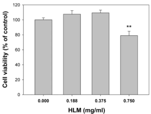
본 연구에서는 선행 연구에서 구축된 HLM을 이용한 약물유 래 간독성 예측 평가방법에서 실험의 편의성과 독성발현 가능성 증진을 위해 HLM 반응시간 및 농도 조건을 최적화하는 실험을 수행하였다.10) HepG2 세포에 0.75 mg/ml의 HLM을 가하고 24
시간 반응하였을 때, 세포생존율이 20% 감소하였다(Fig. 1). 반 면 0.188 또는 0.375 mg/ml의 HLM을 가한 반응한 조건에서는 세포 독성이 유발되지 않았다(Fig. 1). 대사체의 형성을 최대화하 고 HLM 자체의 독성이 없는 조건을 최적으로 조건으로 판단하 여 이후 연구에서는 0.375 mg/ml의 HLM으로 24시간 반응 또는 0.75 mg/ml의 HLM으로 12시간 반응을 선택하여 실험하였다. 대 사 독성을 유발하는 물질인 cyclophosphamide와 acetaminophen 을 대표 물질로 선정하고 HLM을 0.375 mg/ml 농도에서 24시간 반응하는 조건과 선행 연구에서 구축한 조건인 HLM 0.75 mg/
ml 농도에서 12시간을 반응하는 조건에서 이들 물질의 세포독성 을 평가하였다(Fig. 2). HLM에 존재하는 NADPH 의존성 약물 대사효소의 반응을 유발하기 위해 NADPH-generating system 이 있는 조건에서 HLM을 세포 배양액에 추가하였다. Cyclo- phosmamide의 독성은 0.75 또는 0.375 mg/ml의 HLM 첨가에 의해 현격히 증가하였다. Acetaminophen의 독성은 40 mM의 농 도에서만 0.75 mg/ml의 HLM에 의해 증가하였으며 반면 0.375
Fig. 1 − Evaluation of cytotoxicity of HLM in HepG2 cells. HepG2
cells were treated with the NADPH-generating system in the presence of 0, 0.188, 0.375 or 0.75 mg/ml HLM. After incubation for 24 hr, cell viability was determined by the MTT assay. Each value represents the mean±SD for four separate samples. **Significantly different from the control at P<0.01 (Student’s t-test).
Fig. 2 − Evaluation of cyclophosphamide- and acetaminophen-induced cytotoxicity in HepG2 cells cultured with HLM. HepG2 cells were treated with cyclophosphamide (A) or acetaminophen (C) in the presence of the NADPH-generating system and 0.75 mg/ml HLM.
After incubation for 12 hr, the medium was changed to serum-free DMEM without the test compounds and HLM, and cells were
incubated for 36 hr. HepG2 cells were treated with cyclophosphamide (B) or acetaminophen (D) in the presence of the NADPH-
generating system and 0.375 mg/ml HLM. After incubation for 24 hr, the medium was changed to serum-free DMEM without the test
compounds and HLM, and cells were incubated for 24 hr. Cell viability was determined by the MTT assay. Each value represents the
mean±SD for four separate samples. *,**,***Significantly different between two groups treated with the same concentration of test
compound (P<0.05, P<0.01, or P<0.001, Student’s t-test).
mg/ml의 HLM을 가하였을 때에는 20, 30 그리고 40 mM의 acetaminophen의 독성이 증가하였다. 이 결과는 HLM의 농도를 50%로 감소시키고 반면 반응시간을 2배로 증가시켰을 때 대사 활성화에 의한 세포독성이 이전 조건과 유사하거나 더욱 현격하 게 관찰됨을 시사한다.
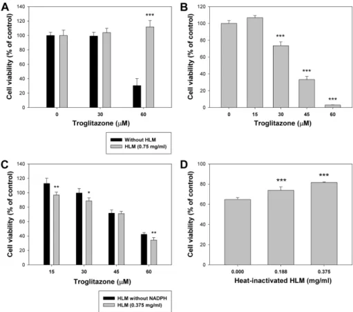
본 연구에서 최적화된 HLM의 반응조건을 활용하여 간독성으 로 시장에서 퇴출된 약물 중 하나인 troglitazone의 독성 평가를 수행하였다. 이전에 구축한 조건에서는 HLM에 의해 troglitazone 의 독성이 완전히 억제되었다(Fig. 3A). HepG2 세포와 troglitazone 의 배양시간을 24시간으로 증가시켰을 때 세포 독성은 12시간 배 양했을 때에 비하여 증가하였으며(Fig. 3B), 0.375 mg/ml의 HLM 과 NADPH-generating system이 있는 조건에서 troglotazone의 독성은 유의적으로 증가하였다(Fig. 3C). NADPH-generating system 유무에 따른 troglitazone의 독성 비교를 통해 tro- glitazone의 독성이 적어도 부분적으로 대사 활성화에 의해 매개 될 가능성을 시사한다. Troglitazone의 독성 양상을 정확히 규명
하기 위해 열을 가하여 불활성화된 HLM을 이용하여 독성 평가 를 수행하였다. HLM을 45oC에서 30분간 가열하였을 때,
Fig. 3 − Evaluation of troglitazone-induced cytotoxicity in HepG2 cells cultured with HLM. (A) HepG2 cells were treated with troglitazone in the presence of the NADPH-generating system and 0.75 mg/ml HLM. After incubation for 12 hr, the medium was changed to serum- free DMEM without the test compounds and HLM, and cells were incubated for 36 hr. (B) HepG2 cells were treated with troglitazone for 24 hr. (C) HepG2 cells cultured with 0.375 mg/ml HLM were treated with troglitazone in the presence or absence of the NADPH- generating system. (D) HepG2 cells were treated with troglitazone 30 µM, the NADPH-generating system, and heat-inactivated HLM. After incubation for 24 hr, the medium was changed to serum-free DMEM without the test compounds and HLM, and cells were incubated for 24 hr. Cell viability was determined by the MTT assay. Each value represents the mean±SD for four separate samples. *,**,***Significantly different from the control at P<0.05, P<0.01, or P<0.001, respectively (Student’s t test for two-group comparison and one-way ANOVA followed by Dunnett’s test for multiple comparison).
Table I − Determination of CYP activities in heat-inactivated HLM using LC-MS/MS
CYP enzyme Heat-inactivated HLM (% of control)
1A2 8.2±1.5
2A6 3.1±0.2
2B6 2.3±0.1
2C8 2.1±1.3
2C9 1.2±0.7
2C19 1.3±0.0
2D6 1.1±0.4
2E1 5.7±5.0
3A4(M)* 0.6±0.5
3A4(T)** 0.5±0.2
CYP activities in heat-inactivated HLM were expressed as a
percentage of control value obtained from active HLM. Each data
represents the mean value±%CV of duplicate incubations. *M-
midazolam as a substrate, **T-testosterone as a substrate.
CYP1A2, 2A6, 2B6, 2C8, 2C9, 2C19, 2D6, 2E1과 3A4의 활성 이 정상적인 HLM에서의 활성에 비하여 10% 미만으로 감소하 였다(Table I). Troglitazone의 독성은 열에 의해 불활성화된 HLM을 가하였을 때 HLM의 농도에 의존적으로 약화되었다(Fig.
3D). 이러한 결과는 HLM에 의한 troglitazone의 무독화가 troglitazone과 HLM의 비선택적인 단백결합에 기인함을 시사한 다. 2형 당뇨 치료제로 사용되었던 troglitazone의 간독성 기전은 친전자성 대사체 형성, 미토콘드리아독성, bile salt export pump (BSEP) 억제 등 매우 다양하며 정확히 밝혀지지 않았다.13,14) HLM을 이용한 독성 평가 방법을 통해 troglitazone의 간독성에 대사 활성화가 부분적으로 관여함을 확인할 수 있었다.
시장에서 퇴출된 약물들인 chlormezanone과 methapyrilene의 간독성에서 대사 활성화의 관여 가능성을 알아보고자 새로 구축 된 조건에서 독성 평가를 수행하였다(Fig. 4). Chlormezanone의 세포독성은 HLM이 없는 조건에서 2000 µM 이상의 농도에서 관 찰되었다(Fig. 4A). HLM이 가해진 조건에서 NADPH-generating system 유무에 따른 chlormezanone의 LC50(95% 신뢰구간)은
각각 7724 µM(3874-15399)과 3781 µM(1571-9102) 이었다(Fig.
4A, B). 이 결과는 HLM이 있는 조건에서 chlormezanone의 독 성이 NADPH에 의존적으로 증가함을 시사한다. 근이완제로 사 용되었던 chlormezanone의 간독성 기전은 명확하지 않으나 본 연구를 통해 그 원인이 대사 활성화에 기인할 가능성을 제시한 다. NADPH-generating system 유무에 따른 methapyrilene의 LC50은 각각 515 µM(185-1434), 604 µM(316-1157)이었으며, HLM에 의한 methapyrilene의 독성이 NADPH-generating system에 의존적으로 감소하였다(Fig. 4C, D). 본 연구결과는 methapyrilene의 NADPH 의존성 대사가 무독화일 가능성을 시 사한다. Methapyrilene은 랫트에서 간암 및 간독성이 발생하여 시장에서 철수된 약물로서 인간에서는 간독성이 보고되지 않았 다. 이전 연구에서 랫트의 CYP 매개성 대사가 대사 활성화이며 결과적으로 독성대사체에 의한 간독성의 가능성을 제시하였다.15,16) 향후 종차를 확인하기 위한 CYP 매개성 methapyrilene의 대사 와 간독성의 상관성 연구가 필요할 것이다. 종합적으로 본 연구 에서 확립된 HLM을 이용한 간독성 예측 평가 모델은 대사 활
Fig. 4 − Evaluation of chlormezanone- and methapyrilene-induced cytotoxicity in HepG2 cells cultured with HLM. HepG2 cells were treated
with chlormezanone (A) or methapyrilene (C) for 24 hr. HepG2 cells cultured with 0.375 mg/m l HLM were treated with
chlormezanone (B) or methapyrilene (D) in the presence or absence of the NADPH-generating system. After incubation for 24 hr,
the medium was changed to serum-free DMEM without the test compounds and HLM, and cells were incubated for 24 hr. Cell
viability was determined by the MTT assay. Each value represents the mean±SD for four separate samples. *,**,***Significantly
different from the control at P<0.05, P<0.01, or P<0.001, respectively (Student’s t test for two-group comparison and one-way
ANOVA followed by Dunnett’s test for multiple comparison).
성화 및 무독화를 모두 확인할 수 있는 방법으로써 신약 개발에 서 간독성 예측에 유용하게 활용될 것이다.
결 론
본 연구에서는 선행 연구에서 구축된 HLM을 이용한 독성평 가 방법을 최적화하는 실험을 수행하였다. Cyclophosphamide와 acetaminophen, troglitazone의 독성 평가를 통해 기존의 HLM 반응 조건보다 새로 확립된 조건이 독성에 더 민감함을 확인하 였고, 시험물질과 HLM의 24시간 반응은 실험의 편의성을 증가 시키고 HLM의 사용량을 감소시켰다. 새로 확립된 HLM 조건에 서 chlormezanone과 methapyrilene의 독성 평가를 통해 퇴출된 약물의 간독성에서 대사 활성화의 관여 가능성을 확인할 수 있 었다. Chlormezanone의 간독성은 NADPH 의존성 대사 활성화 그리고 methapyrilene은 NADPH 의존성 무독화의 가능성을 시 사한다. 최적화된 HLM 이용 in vitro 독성 평가 모델은 실험의 편의성과 간독성 예측력이 증진된 방법으로서 신약 개발에서 in vitro 독성 스크리닝 시스템에 유용할 것으로 예상된다.
감사의 말씀
이 연구는 2014년 충남대학교 학술연구비에 의해 지원되었음.
References
1) Kaplowitz, N. : Idiosyncratic drug hepatotoxicity. Nat. Rev.
Drug Discov. 4, 489 (2005).
2) Guengerich, F. P. and MacDonald, J. S. : Applying mechanisms of chemical toxicity to predict drug safety. Chem. Res. Toxicol.
20, 344 (2007).
3) Park, B. K., Kitteringham, N. R., Maggs, J. L., Pirmohamed, M.
and Williams, D. P. : The role of metabolic activation in drug- induced hepatotoxicity. Annu. Rev. Pharmacol. Toxicol. 45, 177 (2005).
4) Donato, M. T., Lahoz, A., Castell, J. V. and Gomez-Lechon, M. J. : Cell lines: a tool for in vitro drug metabolism studies.
Curr. Drug Metab. 9, 1 (2008).
5) Guillouzo, A. : Liver cell models in in vitro toxicology. Environ.
Health Perspect. 106 Suppl. 2, 511 (1998).
6) Aninat, C., Piton, A., Glaise, D., Le Charpentier, T., Langouet, S., Morel, F., Guguen-Guillouzo, C. and Guillouzo, A. :
Expression of cytochromes P450, conjugating enzymes and nuclear receptors in human hepatoma HepaRG cells. Drug Metab. Dispos. 34, 75 (2006).
7) Doehmer, J., Dogra, S., Friedberg, T., Monier, S., Adesnik, M., Glatt, H. and Oesch, F. : Stable expression of rat cytochrome P-450IIB1 cDNA in Chinese hamster cells (V79) and metabolic activation of aflatoxin B1. Proc. Natl. Acad. Sci. U.S.A. 85, 5769 (1988).
8) Gomez-Lechon, M. J., Donato, T., Jover, R., Rodriguez, C., Ponsoda, X., Glaise, D., Castell, J. V. and Guguen-Guillouzo, C. : Expression and induction of a large set of drug-metabolizing enzymes by the highly differentiated human hepatoma cell line BC2. Eur. J. Biochem. 268, 1448 (2001).
9) Hosomi, H., Fukami, T., Iwamura, A., Nakajima, M. and Yokoi, T. : Development of a highly sensitive cytotoxicity assay system for CYP3A4-mediated metabolic activation. Drug Metab. Dispos. 39, 1388 (2011).
10) Choi, J. M., Oh, S. J., Lee, J. Y., Jeon, J. S., Ryu, C. S., Kim, Y. M., Lee, K. H. and Kim, S. K. : Prediction of drug-induced liver injury in HepG2 cells cultured with human liver mic- rosomes. Chem. Res. Toxicol. 28, 872 (2015).
11) Mosmann, T. : Rapid colorimetric assay for cellular growth and survival: application to proliferation and cytotoxicity assays. J.
Immunol. Methods 65, 55 (1983).
12) Lee, K. S. and Kim, S. K. : Direct and metabolism-dependent cytochrome P450 inhibition assays for evaluating drug-drug interactions. J. Appl. Toxicol. 33, 100 (2013).
13) Smith, M. T. : Mechanisms of troglitazone hepatotoxicity.
Chem. Res. Toxicol. 16, 679 (2003).
14) Ikeda, T. : Drug-induced idiosyncratic hepatotoxicity: prevention strategy developed after the troglitazone case. Drug Metab.
Pharmacokinet. 26, 60 (2011).
15) Graham, E. E., Walsh, R. J., Hirst, C. M., Maggs, J. L., Martin, S., Wild, M. J., Wilson, I. D., Harding, J. R., Kenna, J. G., Peter, R. M., Williams, D. P. and Park, B. K. : Identification of the thiophene ring of methapyrilene as a novel bioactivation- dependent hepatic toxicophore. J. Pharmacol. Exp. Ther. 326, 657 (2008).
16) Howell, B. A., Yang, Y., Kumar, R., Woodhead, J. L., Harrill, A. H., Clewell, H. J., 3rd, Andersen, M. E., Siler, S. Q. and Watkins, P. B. : In vitro to in vivo extrapolation and species response comparisons for drug-induced liver injury (DILI) using DILIsym: a mechanistic, mathematical model of DILI. J.
Pharmacokinet. Pharmacodyn. 39, 527 (2012).