A Trial of Apolipoprotein E Genotyping in Noninsulin Dependent Diabetes Mellitus by Polymerase Chain Reaction Method
Dept. of Internal Medicine, Endocrine Research Institute, Kyung Hee University Hospital.
Paeng, Jung Ryung. ·Kim, Deog Yoon. • Woo, Jung Taek.
Kim, Sung Woon. • Yang, In Myung. • Kim, Jin Woo.
Kim, Young Seol. • Kim, K wang Won. • Choi, Y ong Kil.
Key words : Genotyping. Apolipoprotein E. isoforms. Polymerase chain reaction.
I . INTRODUCTION
Apo E is one of various factors which regulate the lipid metabolism. It is a constituent of plasma lipoproteins such as chylomicrons, very low density lipoproteins(VLDL) and high density lipoproteins.
The major physiological function of apo E plays a central role in cholesterol transport!), and it serves as a ligand for apo B, E receptor2l. The isoforms, designated E2, E3, and E4, differ by single amino acid residue with substitution of cysteine for argi- nine3·4·5l. In addition to this major isoforms, several other rare apo E isoproteins have recently been de- scribed6·7l. The E4 isoform(112 arg and 158 arg) is associated with increased levels of plasma total cho- lesterol and betalipoprotein8l and increased suscepti- bility to coronary heart disease. Hyperlipidemia, a risk for development of atherosclerosis occurs fre- quently in non-insulin dependent diabetic mellitus (NIDDM). Eto et al. have been reported that apo E polymorphism may be a cause of hyperlipidemia in type
ll
(NIDDM) diabetes. In their study, the E2 isoform is associated with decreased levels of choles- terol and betalipoprotein8l. And there are reports concerned with relationship between typeV
hyper- lipoproteinemia and apo E4/E49•10l.A number of isoforms of apo E are separable by one dimentional isoelectric focusing(IEF) method as
well as by two dimentional gel IEF method11•12·13l,
followed by SDS-polyacrylamide gel electrophoresis (PAGE) 14l.
Because this method is based on charge differenc- es of apo E isoproteins, it is not possible to identify neutral amino acid substitution, nor to locate the site of the mutation.
These methods has been well known for time-con- suming as well as there has no clear discrimination of IEF band. Recently, apo E typing at the DNA level was introduced by PCR method15l. In addition, It is now possible to define mutation site of apo E gene by application of DNA typing method16·17).
However, clinical study from large population is not reported until recently. In this report, we have tried restriction fragment analysis of apo E gene as a simpler and faster method for typing the common apo E isoforms. to elucidate the relationship be- tween diabetes accompanied by hyperlipidemia, apo E genotype and apo E allele frequencies of diabetics were studied.
ll • Materials and Methods 1. Amplification of apo E gene
Blood samples were obtained from the patients with NIDDM. DNA was extracted from the periph-
*~ :c~.g.. 1992\1 7-%! 26~-¥-El 31~7/}Al Ireland DublinO!l''l 7~~:!fl ~120 -*} IMALT ~~l~~
OjlJ,i
~lE:!fl ~ qJ.eral leucocyte as described previously18l. And ampli- fied by PCR in a DNA thermal cycler(Hybaid) using oligonecleotide primers F4(5 '-ACAGAATTC GCC CCGGCCTGGT ACAG-3') and F6 ( 5 '-T AAG C TTGGCACGGCTGTCCAAGA-3') as described by Emi et aP3•19l.
Oligonucleotide primers were synthesized by the phosphoramidite method using a DNA synthesizer (Applied Biosystems Inc.). Taq DNA polymerase was obtained from KIST, Korea. Each amplification reagents contained 1 pg of DNA, 100 pmol of each primer, 10% dimethylsulfoxide, and 2.5 unit of Taq polymerase in a final volume of 100 p R and then added few drops of mineral oil to prevent evapora- tion. At first, reaction mixture was heated at 95oC for 5 min for denaturation, and then subjected to 30 cycles of amplification by primer annealing(68°C for 50 sec). Extention(70°C for 50 sec), and denatu- ration(950C for 50 sec).
2. Electrophoresis and restriction fragments isotyping
After PCR amplification, the amplified DNA was recovered by ethanol precipitation, 5 units of Hha I in recovered DNA were added to each reaction mix- ture for digestion of PCR products(1.5h at 37°C).
Each reaction mixture was loaded on 8% polyacryl- amide gel(l mm thick x 12 em long) and electro-
E4 TAAGCTTG GCA CGG CTG
GCG CAG GCC CGG CTG
E2 T
E3 T 112
E4 G1G CGC GGC CGC CTG GTG CAG GCC ATG CTC CTG CGG GTG CGC CTC CTG CGT AAG CGG CTC
E2 T
E3
c
158E4 CTG CAG AAG CGC CTG GCGAATTCTGT 244
phoresed for 3h under constant current ( 45 rnA).
After electrophoresis, the gel was stained with ethidium bromide for 15 min and DNA fragments were visualized by UV illuminator. The sizes of Hha I fragments were estimated by comparision with DNA molecular-weight marker
V
(Boehringer ma- nnheim). Apo E genotyping relies on cleavage at Hha I sites to distinguish E2, E3, and E4 sequences.PCR product was amplified a specific region of the exon 4(Fig. 1, 2).
plasma apo E amino acids 299
precursor 317
protein amino acids
1163 mRNA
nucleotides exon 1 exon 2 44
• -66 3597 bp exon 3 193 exon 4 860
Fig. 1. Structure of the human apo E gene.
A schematic outline of the human apo E gene is shown in relation to the mRNA which it encodes, the primary translation product of the mRNA, and the mature plasma protein product of the gene.
TCC AAG GAG CTG CAG GCG GGC GCG GAC ATG GAG GAC
GTG CAG TAC CGC GGC GAG GGC CAG AGC ACC GAG GAG GCC TCC CAC CTG CGC AAG CTC CGC GAT GCC GAT GAC
GCA GTG TAC CAG GCC GGG
Rg. 2. DNA sequences of apo E isoforms and location of Hha I cleavage sites.
(substitution site at codons 112 and 158 are boxed)
The second step is cleavage of amplified products with Hha I for PAGE. Each of the isoforms is dis- tinguished by a unique combination of Hha I frag- ment sizes. According to Hha I fragment sizes, six apo E genotypes were determined as shown in Fig.
3. The cut sites are shown 6 places in E4, 5 places in E3, 4 places in E2(Fig. 3).
112 158
48 35
£3----~+---~---+----91
91 83
Ez----~r---++--- Fig. 3. Hha I cleavage maps are given for ampli-
fied sequences. The distances(in bp) be- tween polymorphic Hha I sites that distin- guish isoforms are shown for each cleavage map.
ill. RESULTS
PCR products(224 bp) electrophoresed on 1.5%
agarose gel(Fig. 4).
244 bp
Fig. 4. Agarose gel electrophoresis after PCR amplification.
lane 1, size marker.
lane 2-8, PCR products ( 244 bp)
PCR product is digested with Hha I restriction enzyme and electrophoresed on polyacrylamide gel.
Each genotype posessed unique combinations of Hha I fragments sizes(Fig. 5). The E3/E3 homozygote contained the 91 bp fragment(112 cys), as well as 48 bp and 35 bp fragments from cleavage at the Hha I site at 158 arg. The E4/E4 homozygote also contained these 48 and 35 bp sizes(l58 arg), as well as a unique 72 bp size from cleavage at 112 arg. E3 /E2 heterozygote contained both sets of fragments from each apo E allele, 91 and 83 bp(Fig. 5).
Fig. 5. 8% polyacrylamide gel electrophoresis of Hha I fragments after PCR amplification.
91 bp 83 72 48 35
lane 1, E4/E4(72 bp). lane 2,4-6, E3/E3(91 bp).
lane 3, E3/E2(91.83 bp). lane 7, size marker.
lane 8, PCR product ( 244 bp)
Apo E genotype frequency in normal subjects was 0% for E2/E2, 9.6% for E3/E2, 72.6% for E3/E3, 0% for E4/E2, 15.1% for E4/E3, 2.7% for E4/E4.
In the patients with NIDDMs, the gene frequency of apo E gene was 0% for E2/E2, 12.7% for E3/E2, 64.6% for E3/E3, 0% for E4/E2, 20.2% for E4/E3, 2.5% for E4/E4.
As shown in table 1, the apo E allele frequencies in normal subjects showed 4.8% for E2, 84.9% for E3, 10.3% for E4. And in NIDDMs showed 6.3%
for E2, 81.0% for E3, 12.7% for E4. The frequency of apo E3/E3 was most prevalent in both NIDDM and normal groups, whereas apo E2/E2 homozygote was not found in both NIDDM and normal groups.
The distribution of apo E isoforms was no signifi- cant difference between normal and NIDDMs.
N. DISCUSSION
The human apo E gone spans 3.59 kb including 4 exons and 3 introns20·21). Genetic variation at the apo E locus is an important determination of lipid levels and regard as a risk for developement of atherosclerosis22>. Until recently most methods for detection of allelic variation have relied on IEF method. It require time-consuming and necessary ultracentrifugation for the separation of lipoproteins.
Recently much attention has been focused on the relationship between apo E alleles and hyperlipo- proteinemia. Utermann et al. found that the apo E2 /E2 homozygote is related to type
ill
hyperlipopro- teinemia23>. And other apo E2 allele has been report- ed to be associated with higher levels of plasma tri- glyceride and VLDU'>. PCR technology has allowed genotyping of apo E isof orms by direct detection of nucleotide substitutions in genomic DNA rather than IEF of isoproteins. Our results clearly show that typing of the three major apo E isoforms from minute quantities of DNA can be done using the PCR methodology(Fig. 3).As shown in Table 1, apo E3/E3 were a prevalent genotype, and a lower frequency of genotype E3/E2 were found in the Korean than those reported for the Black et. al from the caucasian. But apo E4/E3 and E2/E2 showed a similar patterns. Black et al.
suggested that the frequency of apo E3 was signifi- cantly less in diabetic than in normal subjects25>.
Table 2 shows distribution of apo E allele f requen- cies in our study and a patients with NIDDM previ- ously repoted. The frequencies of apo E genotype in our population agree almost with those reported by previous authors although their method based on protein characterization26· 27 · 28>.
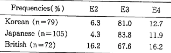
The apo E allele frequencies in diabetic subjects (E2, 6.3% ; E3, 81.0% ; E4, 12.7%) are similar to those in Japanese diabetic subjects(E2, 4.3% ; E3, 83.8% ; E4, 11.9%). Besides Eto et al. have been
Table 1. Apo E genotypes and phenotypes in Korean and previous report Apo E
2/2 3/2 3/3 4/2 4/3 4/4 Genotype(%)
NIDDM (n=79) 0.0 12.7 64.6 0.0 20.2 2.5 Normal (n=73) 0.0 9.6 72.6 0.0 15.1 2.7 Black et al. 0.0 26.0 43.0 4.0 19.0 7.0 Phenotype(NIDDM, n=72)
Apo E allele frequencies(%) E2 E3 E4
NIDDM (n=79) 6.3 81.0 12.7
Normal (n = 73) 4.8 84.9 10.3
Table 2. Apo E Allele Frequencies in NIDDM Frequencies ( % ) E2 E3 E4 Korean (n=79) 6.3 81.0 12.7 Japanese (n= 105) 4.3 83.8 11.9 British (n=72) 16.2 67.6 16.2
proposed that the apo E3/E2 and apo E4/E3 diabet- ic subjects is closely related to hyperlipemia26>. In particular, they suggested that E4 allele is closely related to higher levels of plasma cholesterol in NIDDM, although some investigator did not find any difference29·30·31>.
In our present study it was not clear which type of hyperlipoproteinemia is associated with the apo E allele because detailed lipoprotein analysis was not included. Table 3 shows a comparison between our present study(genotyping) and previously reported (phenotyping) in normal groups. Both Korean and Japanese have also a similar pattern as shown in NIDDMs.
Table 3. Apo E Genotype in Korean and Apo E Phenotype Frequencies in Other Races ApoE
2/2 3/2 3/3 4/2 4/3 4/4 Genotype (% )
NIDDM (n=79) 0.0 12.7 64.6 0.0 20.2 2.5 Normal (n=73) 0.0 9.6 72.6 0.0 15.1 2.7 Apo E Phenotype(%) : Nondiabetic subjects Japanese (n=68) 0.0 10.3 76.4 1.5 10.3 1.5 Finnish (n=490) 1.0 13.1 58.9 0.0 24.3 2.7
Interestingly, our results showed a great differ- ence in the apo E allele frequencies when compared with those of the Europians(British, Finnish). Fur- ther studies will be required for setting the relation- ship between the apo E4 allele and hyperlipopro- teinemia in NIDDM.
Futhermore, Hha I apo E genotyping could not detect rare variants, unless substituions alter Hha I sites within the amplified region using F4 and F6 primer. 'And a possibility of methylation may have prevent~d Hha I cleavage of PCR products. Howev- er, sequencing or single-strand conformation poly- morphism analysis of apo E genes may be useful to detect rare variants. PCR technique could be a use- ful for diagnosis of genetic desease, or for detection of unknown polymorphisms using survey of many different restriction enzymes.
V •
CONCLUSIONThere was no significant difference of apo E isoforms distribution between diabetic and normal groups in apo E genotypig. The apo E allele frequen- cies of Korean and Japanese diabetics were similar, but there was a great difference as compared with those of caucasian. In order to prove this difference between isoelectric focusing, we suggest tha:t fur- ther studies should be subdivided by type of lipop- roteinemia. Apo E genotyping by PCR method may be identified in conjunction with apo E phenotyping.
ACKNOWLEGEMENTS
This study was supported by Kyung Hee University Medical Center. We also thank Dr, Dae-sil Lee for kindly providing Taq polymerase.
~~~-~~%·~~~·~~~·~~~
~~~·~~1l·~llt~·~~~
75:§i~li~
Lll.i!l-
1.11~e1 ~.:r~'cfh ~.g. JI;•q ~ -'6'-0l ~ill %lfl-£1 JJ:l qoJ~ ~ cj o1 -B-a=Pel.r;}. Apolipoprotein E(apo E) B-~ 1I~
~ 01 ~ JI.Al ~ -'6'- _g_ ~A~ .i!} -B-~£1 <>1 JI.Al ~ -'6'-~
~ cj ~ it~ 5l.A} apo E isoform.Ql ~~ cl Al15] Q :iL 'V_r;}. ~-2- apo E _g_~.A}~% 1€-fi>l~r1l polymerase chain reaction(PCR) BJ!:fj o1 o1 %£1 J1. 'V_r;}. Apo E~ 2997~~ o}olx{!-.9...£. £1<>1 'V_.:il 37~.Ql isoform% ?!":il. 'V_~r11 112Jti.ll} 158Jt)_~~ o}olx{}
A~~~ ~}ol1J oJl u:}e.} E2, E3, E4.£. -T-~~1:}.
~ ~-ToJlA~~ apo E gene% PCR!:fj.£..£. -'6'-~Al
7'i
HhaI
~1 ~ E~.£. ~§}Al ~4%
polyacryl- amide gel electrophoresis i>} 0:1n
J?1 ti} J1. ~ 4j-ti} lll _g_ ~ 7-} ~ % ~78
ti} ~ 1:}. Hha I .g. arginine% encod- ing t~}~ GCGC site~ cj ~ ~ B-t>l-~ K~ o l o:l 112 arg(E4).i!} 158 arg(E3, E4)~ ~ B-t>l-.Al tfr112 cys(E2, E3).2} 158 cys(E2)~ GTGC.£. ~ B-
ti)-.Al *~1:}. 0lc:j~ ~l~E~~ ~A~% 01-§-ti}O:l Hha I restriction fragment length~ 3:-"l-til-~ 1:}.
'cfh~ ~.A} 79t§oJlA~ ztzt~ _g_~7-}~% ~*
~ ~:i!} E2(6.3%), E3(81.0%), E4(12.7%)£.. t..l-E}
Jit.£.o:l 78AJcj 73l:§oJlA~~ %~7-}~.g. E2(4.8%), E3(84.9%), E4(10.3%).£. 1-}E}J;tl:}.
0lAJ~ ~.ll}oJlA~Ji!.~ 'cfh~ ~7.}_2} t:ll.::f:~.J!}~
_g_ ~.A} ~ .£ oJlA~ ~ .18-lJl ~ _g_ ~ A~ _g. itt ~ 1:} • .::Z. 2.-l 1-} _g_~.A} ~.£oJl 'V_ o:j A~~ 4-i:!.l ~.ll}-2} ~ ~~
~.ll}~ 0~4- _g_.A}ti}.Allfr _g_~ .Al9:1.Ql ~.i!}-2}~ ~1- 0l~ Ji!.~ 1:}. 01 c:j ~ ~.i!}.Ql ~1-0l-i=- cj~;Q-.9.1 ~l-01
~ _g_~.A}~% ~78t>l-~ PCR!:fj.i!} B-~1J% ~~
til-~ isoelectric focusing
n
.9.1 ~1-0 1.£. ;.~zt
£1 D=l i;J=4- PCR~ol _g_~7-}~% ~78ti}JI ~B-t>}-i=-r1l
*
..B..~ ~0]:71- ~ -3!.£..£. -"1-li~l:}.
REFERENCES
1. Mahley, R. W. Apolipoprotein E :Cholesterol transport protein with expanding role in cell bi- ology. Science 240 : 622-630, 1988.
2. Mahley, R. W., T. L. Innerarity, S. C. Rail, Jr., and K. H. Weisgraber. Plasma apolipoprotein structure and function. J. Lipid Res. 24 : 1277
-1294, 1984.
3. Weisgraber, K. H., S. C. Rail. Jr., and R. W.
Mahley. Human E apoprotein heterogneity.
Cystein-arginine interchanges in the amino acid sequence of the apo E isoforms. J. Bio. Chern.
256 : 9077-9083, 1981.
4. Zannis, V.I., and J. L. Breslow. Human VLDL apo E isoprotein polymorphism is explained by genetic variation and post-translational modifi- cation. Biochemistry 20: 1033-1041, 1981.
5. Utermann, G., U. Langenback, U. Beisiegel, and W. Weber. Genetics of the apolipoprotein E system in man. Am. J. Hum. Genet. 32 : 339- 347, 1980.
6. McLean, J. W., N. A. Elshourbagy, D. J. Chang, R. W. Mahley, and J. M. Taylor. Human apolipoprotein E mRNA. eDNA cloning and nu- cleotide sequencing of a new variant. J. Bioi.
Chern. 259 : 6498-6504, 1984.
7. Breslow, J. L. Human apolipoprotein molecular biology and genetic variation. Annu. Rev.
Biochem. 54 : 699-727, 1985.
8. Boerwinkle, E., S. Visvikis, D. Welsh, J. Stein- metz, S.M. Hanash, and C.F. Sing. The use of measured genotype information in the analysis of quantitative phenotypes in man. ll . The role of the apolipoprotein E polymorphism in deter- mining levels, variability, and covariability of cholesterol, betalipoprotein, and triglycerides in a sample of unrelated individuals. Am. J. Med.
Genet. 27 : 567-582, 1989.
9. Ghiselli, G., E. J. Schaefer, L. A. Zech, L. E.
Gregg, and H. B. Brewer, Jr. Increased preva- lence of apolipoprotein E4 in type V hyperli- poproteinemia. J. Clin. Invest. 70 : 474-477, 1982.
10. Stuyt, P. M. J., A. F. H. Stalenhoef, and P. N.
M. Demacker. Hyperlipoproteinemia type V and apolipoprotein E4. Lancet 1 : 934, 1982.
11. Funke, H., S. Rust, and G. Assman. Detection of apolipoprotein E variants by an oligonu- cleotide "melting" procedure. Clin. Chern. 32 : 1285-1289, 1986.
12. Smeet, B., J. Poddighe, H. Brunner, H-H.
Ropers, and B. Wieringa. Tight linkage be- tween myotonic dystrophy and apolipoprotein E genes revealed with allelespecific oligonu- cleotides. Hum. Genet. 80 : 49-52, 1988.
13. Emi, M., L. L. Wu, M. A. Robertson, R. L.
Myers, R. A. Hegele, R. R. Williams, R. White, and J-M. Lalouel. Genotyping and sequence analysis of apolipoprotein E isoforms. Geno- mics. 3 : 373-379, 1988.
14. Maily, F., J. Davignon, and A. C. Nestruck. An- alytical isoelectric focusing with immobilized pH gradients of human apolipoprotein E from very low density lipoproteins and total plasma.
J. Lipid. Res. 31: 149-155, 1990.
15. Hixon, J. E., and D. T. Vernier. Restriction isotyping of human apolipoprotein E by gene amplification and cleavage with Hha I. J. Lipid.
Res. 31 : 545-548, 1990.
16. Wallis, S.C., S. Rogne, L. Gill, A. Markham. M.
Edge, D. Woods, R. Williamson, and S.
Humphries. The isolation of eDNA clones for
huma~ apolipoprotein E and the detection of apo E RNA in hepatic and extra-hepatic tis- sues. EMBO J. 2 : 2369-2373, 1983.
17. Smeets, H. J. M., J. Poddighe, P.M. J. Stuyt, A.
F. H. Stalenhoef, H. H. Ropers, and B. Wie- ringa. Identification of apolipoprotein E poly- morphism by using synthetic oligonucleotides. J.
Lipid Res. 29: 1231-1237, 1988.
18. Hixon, J. E., L. Borenstein, L. A. Cox, D. L.
Rainwater, and J. L. VandeBerg. The baboon gene for apolipoprotein A-1 : characterization of a eDNA clone and identification of DNA pol- ymorphisms for genetic studies of cholesterol metabolism. Gene 74 : 483-490, 1988.
19. Saiki, R. K., S. Scharf, F. Faloona, K. B. Mullis, G. T. Horn, H. A. Erlich, and N. Arnheim. En- zymatic amplication of p-globin genomic se- quences and restriction site analysis for diagno- sis of sickle cell anemia. Science 230 : 1350-
1354, 1985.
20. Paik, Y -K., D. J. Chang, C. A. Reardon, G. E.
Davies, R. W. Mahley, and J. M. Taylor. Nucle- otide sequence and structure of the human apolipoprotein E gene. Proc. Nat!. Acad. Sci.
USA 82 : 3445-3449, 1989.
21. Das, H. K., J. McPherson, G. A. P. Bruns, S. K.
Karathanasis, and J. L. Breslow. Isolation, characterization, and mapping to chromosome 19 of the human apolipoprotein E gene. J. Bioi.
Chern. 260 : 6240-6247, 1985.
22. Davignon, J., R. E. Gregg and C. F. Sing.
Apolipoprotein E polymorphism and athero- sclerosis. Arteriosclerosis 8 : 1-21, 1988.
23. Utermann G, Hess M, Steinmetz A. Polymor- phism of apolipoprotein E and occurrence of dysbetalipoproteinemia in man. Nature(Lond) 269 : 604-607, 1977.
24. Eto M, Watanabe K, Ishii K. Reciprocal effects of apolipoprotein E alleles(E2 and E4) on plas- ma lipid levels in normolipidemic subjects. Clin Genet. 29 : 477-84, 1986.
25. Black, S.C., S. Hewett, Y. Kotubi, R. V. Brunt, and J. P. D. Reckless. Isoform patterns of apolipoprotein E in diabetes mellitus. Diabetic
medicine 7 : 532-539, 1990.
26. Eto M, Watanabe K, Ishii K : A rapid flat gel isoelectric focusing method for the determina- tion of apolipoprotein E phenotypes and its ap- plication. Clin Chim Acta. 149 : 21-28, 1985.
27. Eto M, Watanabe K, Iwashima Y, Morikawa A, Oshima E, Seikiguchi M, Ishii K. Apolipoprotein E polymorphism and hyperlipemia in type
ll
di- abetics. Diabetics 35 : 1374-82, 1986.28. Y amamura, T., A. Yamamoto, K. Hiramori, and S. Nambu. A new isoform of apolipoprotein E- E5-associated with hyperlipidemia and atheros- clerosis. Atherosclerosis 50 : 159-172, 1984.
29. Eto M, Watanabe K, Iwashima Y, Morikawa A, Chonan N, Oshima E, Sekiguchi M, And Ishii k.
Increased frequency of apolipoprotein E4 allele in type
n
diabetes with hypercholesterolemia.Diabetes 36 : 1301-1306, 1987.
30. Ghiselli G, Gregg RE, Zech LA, Schaefer EJ, Brewer HB Jr. Phenotype study of apolipopro- tein E isoforms in hyperlipoproteinemic pa- tients. Lancet 2 : 405-407, 1982.
31. Wardell MR, Suckling PA, Janus ED. Genetic variation in human apolipoprotein E. J Lipid Res. 23 : 1174-82, 1982.