Introduction
최근 한국인의 구강에서 그람 양성 혐기성 세균 종인 Peptoniphilus mikwangii가 새로운 종(species)으로 확립되어 소개되었다[1]. 하지 만 아직까지 P. mikwangii와 치아우식증이나 치주질환 등과 같은 구 강 세균 감염성질환과의 상관관계에 대한 연구는 없는 실정이다. 이러 한 점을 고려하여, 최근 16S ribosomal RNA 유전자(16S rDNA) 핵산 염기서열을 바탕으로 P. mikwangii 종-특이 중합효소연쇄반응(poly- merase chain reaction, PCR) 프라이머가 개발되어 소개되었다[2].
구강 세균 감염성질환과 특정 세균 종간의 역학관계를 연구하기 위해 서는 세균 종을 특이적 검출할 수 있는 방법이 확립되어야 한다. 또한, 임상에서 적용 가능해야 한다. 즉, 짧은 시간에 많은 수의 샘플에서 특 정 세균 종을 검출해야 하기 때문에 종-특이성 뿐만 아니라 신속성, 정 확성 및 경제성이 확보되어야 한다. 현재까지 개발된 여러 세균 검출법 중에서 위에서 언급한 임상에서 적용이 가능한 것이 PCR 법이라고 생 각된다. 일반적인 PCR 법으로는 특정 세균 종을 정성적으로 검출할 수 있지만, 정량적으로의 분석은 어렵다. 이러한 문제점을 해결할 수 있는
방법이 바로 정량 실시간 중합효소연쇄반응 (quantitative real-time PCR, qPCR) 법이다.
PCR 또는 qPCR을 수행하기 위해서는 특정 표적 유전자를 선정하여 검출하고자 하는 세균 종에 특이적인 프라이머 개발이 선행되어야 한 다. 이러한 목적으로 가장 많이 사용되는 표적 유전자는 16S rDNA이 다. 그 이유는 16S rDNA는 모든 세균 종에 존재하고, 세균 종 간에 핵 산염기서열 부분에 상동성이 매우 높은 부분과 상이한 부분이 반복적으 로 존재하여, 모든 세균 종을 검출할 수 있는 프라이머와 종-특이 프라 이머 설계하기가 용이하다는 장점이 있기 때문이다[3,4]. 그러므로, 본 연구는 16S rDNA 핵산염기서열을 바탕으로 P. mikwangii를 종-특이 적 및 정량적으로 검출할 수 있는 qPCR 프라이머를 개발하기 위해 시 행하였다.
Int J Oral Biol 44:96-100, 2019 pISSN: 1226-7155 • eISSN: 2287-6618 https://doi.org/10.11620/IJOB.2019.44.3.96
Peptoniphilus mikwangii-specific quantitative real-time polymerase chain reaction primers
Soon-Nang Park and Joong-Ki Kook*
Korean Collection for Oral Microbiology and Department of Oral Biochemistry, College of Dentistry, Chosun University, Gwangju 61452, Republic of Korea
The purpose of this study was to develop Peptoniphilus mikwangii-specific quantitative real-time polymerase chain reaction (qPCR) primers based on the 16S ribosomal RNA (16S rDNA) gene. The specificity of the primers was determined by conventional PCR using 29 strains of 27 oral bacterial species including P. mikwangii. The sensitivity of the primers was determined by qPCR using the purified genomic DNA of P. mikwangii KCOM 1628 T (40 ng to 4 fg).
The data showed that the qPCR primers (RTB134-F4/RTB134-R4) could detect P. mikwangii strains exclusively and as little as 40 fg of the genomic DNA of P. mikwangii KCOM 1628 T . These results suggest that the developed qPCR primer pair can be useful for detecting P. mikwangii in epidemiological studies of oral bacterial infectious diseases.
Keywords: Peptoniphilus mikwangii, 16S rDNA, qPCR primers
Received July 24, 2019; Revised September 8, 2019; Accepted September 15, 2019
*Correspondence to: Joong-Ki Kook, E-mail: [email protected] https://orcid.org/0000-0003-2628-2870 Copyright © The Korean Academy of Oral Biology
CC
This is an open-access article distributed under the terms of the Creative Commons Attribution Non-Commercial License (http://creativecommons.org/licenses/by- nc/4.0/), which permits unrestricted non-commercial use, distribution, and reproduction in any medium, provided the original work is properly cited.
Original Article IJOB
Materials and Methods
1. 세균 및 세균 배양
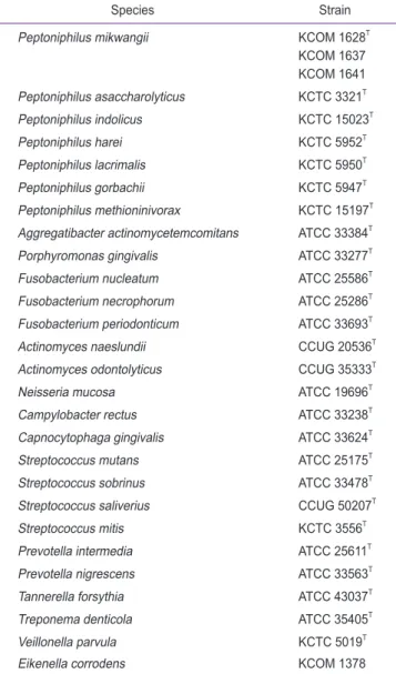
본 연구에서 사용된 P. mikwangii 및 대조군으로 이용된 세균 균주 들은 Table 1과 같다. 이 균주들은 한국구강미생물자원은행(Korean Collection for Oral Microbiology [KCOM], Gwangju, Korea), 한국 생명공학연구원 생물자원센터(Korean Collection for Type Cultures [KCTC], Biological Resource Center, Jeongeup, Korea), ATCC (American Type Culture Collection, Manassas, VA, USA) 또는 CCUG (Culture Collection, University of Göteborg, Göteborg, Sweden)에서 구입하여 사용하였다.
Streptococcus spp. 및 Neisseria mucosa 균주들은 brain heart infusion (BHI; Difco Laboratories Inc., Detroit, MI, USA) 배지를 이용하여 배양하였다. Aggregatibacter actinomycetemcomitans 균주는 tryptic soy broth (TSB; Difco Laboratories Inc.)에 0.6%
yeast extract, 5% horse serum, 75 µg/mL bacitracin 및 5 µg/
mL vancomycin (Sigma-Aldrich, St. Louis, MO, USA)이 첨가 된 배지[5]를 이용하여 배양하였다. Treponema denticola 균주는 1494 modified NOS medium (https://www.atcc.org/~/media/
C14CA5B6B5D246A6B8073D6894BFE5EB.ashx; ATCC), 그 외 균주들은 TSB에 0.5% yeast extract, 0.05% cysteine HCl-H2O, 0.5 mg/mL hemin 및 2 µg/mL vitamin K
1
이 첨가된 배지[5] 등을 이용하여 배양하였다. 이들 모든 균주들은 산소 유입이 차단되고, 혼 합가스(85% N2
, 10% CO2
, 5% H2
)가 공급되는 혐기성 세균배양기 (Bactron I; Sheldon Manufacturing Inc., Cornelius, OR, USA)를 이용하여 37℃ 조건에서 배양하여 다음의 실험에 사용하였다.2. quantitative real-time polymerase chain reaction (qPCR) 프라이머 설계 및 종-특이성 검증
P. mikwangii를 검출하기 위한 종-특이 qPCR 프라이머 쌍은 16S rDNA (GenBank accession no. KJ728812)를 표적으로 설계하 였으며, 이때 PrimerSelect 프로그램(DNASTAR Inc., Madison, WI, USA)을 이용하였다. 설계된 qPCR 프라이머 쌍은 다음과 같다;
RTB134-F4, 5′-AAG TCG AGC GAT GAA AAG G-3′; RTB134- R4, 5′-CTT CCC ATA GGA CAG AGC C-3′. 이들 프라이머 쌍 에 의해 생성이 예상되는 PCR 산물 크기는 408 bp이며, Bioneer 사 (Daejeon, Korea)에 의뢰하여 제작하였다.
본 연구에서 PCR 및 qPCR의 주형으로 사용될 P. mikwangii를 비 롯한 모든 균주들의 지놈 DNA들은 cetyltrimethylammonium bro- mide 방법에 따라 추출하였다[6].
P. mikwangii에 대한 종-특이 qPCR 프라이머의 개발을 위해 설계 된 RTB134-F4/RTB134-R4 프라이머 쌍들의 종-특이성은 P. mik- wangii 표준균주와 2개의 임상균주, 6종의 Peptoniphilus spp. 표 준균주들 및 20종의 구강 내 세균 종들의 지놈 DNA를 주형으로 일반 PCR 법으로 조사하였다. 이때 Bioneer 사의 AccuPower
®
PCR Pre- Mix 및 MyGenieTM
96 Gradient Thermal Block을 사용하여 PCR을 수행하였다. PCR 반응 용액에는 최종 1 pmole/µL씩의 RTB134-F4/RTB134-R4 프라이머들 및 4 ng의 세균 지놈 DNA를 넣어서 반응시 켰다. 종-특이 검증을 위한 PCR 조건은 다음과 같았다: 95℃에서 10 분간 전변성 처리하였고, 95℃에서 10초간 변성, 56℃에서 20초간 결 합, 72℃에서 20초간 중합 과정을 30회 시행하였으며, 72℃에서 10분 간 최종 중합과정을 시행하였다. PCR 산물은 1.5% 아가로스 젤을 매 질로 하여 전기영동 하였고, GoodView
TM
Nucleic Acid Stain (SBS Genetech Co., Ltd., Beijing, China)을 이용하여 증폭물을 염색한 후, 이를 UV transilluminator를 이용하여 관찰하였다.Table 1. The bacterial strains used in this study
Species Strain
Peptoniphilus mikwangii KCOM 1628 T
KCOM 1637 KCOM 1641 Peptoniphilus asaccharolyticus KCTC 3321 T
Peptoniphilus indolicus KCTC 15023 T
Peptoniphilus harei KCTC 5952 T
Peptoniphilus lacrimalis KCTC 5950 T
Peptoniphilus gorbachii KCTC 5947 T
Peptoniphilus methioninivorax KCTC 15197 T Aggregatibacter actinomycetemcomitans ATCC 33384 T
Porphyromonas gingivalis ATCC 33277 T
Fusobacterium nucleatum ATCC 25586 T
Fusobacterium necrophorum ATCC 25286 T Fusobacterium periodonticum ATCC 33693 T
Actinomyces naeslundii CCUG 20536 T
Actinomyces odontolyticus CCUG 35333 T
Neisseria mucosa ATCC 19696 T
Campylobacter rectus ATCC 33238 T
Capnocytophaga gingivalis ATCC 33624 T
Streptococcus mutans ATCC 25175 T
Streptococcus sobrinus ATCC 33478 T
Streptococcus saliverius CCUG 50207 T
Streptococcus mitis KCTC 3556 T
Prevotella intermedia ATCC 25611 T
Prevotella nigrescens ATCC 33563 T
Tannerella forsythia ATCC 43037 T
Treponema denticola ATCC 35405 T
Veillonella parvula KCTC 5019 T
Eikenella corrodens KCOM 1378
KCOM, Korean Collection for Oral Microbiology; KCTC, Korean Collection
for Type Cultures; ATCC, American Type Culture Collection; CCUG,
Culture Collection, University of Göteborg.
3. quantitative real-time polymerase chain reaction (qPCR)을 이용한 민감도 조사
P. mikwangii-특이 qPCR 프라이머 쌍(RTB134-F4/RTB134- R4)의 민감도(최소 검출한계)는 qPCR 법으로 측정하였다. 이때 TOP- real
TM
qPCR 2X PreMix (Enzynomics, Daejeon, Korea) 및 Exicy- clerTM
96 Real-Time Quantitative Thermal Block (Bioneer)을 이 용하여 실시하였다[7]. qPCR 반응을 위해, 60 pmoles의 RTB134- F4와 RTB134-R4 프라이머, 40 ng에서 4 fg까지 10배씩 희석된 P.mikwangii KCOM 1628
T
지놈 DNA를 각각의 qPCR 2X PreMix 가 들어 있는 PCR tube에 넣고, 최종 반응물이 50 µL가 되도록 멸균 된 이차 증류수를 첨가하였다. 이를 교반기를 이용하여 잘 혼합한 다음, 2,500 rpm에서 10초간 원심분리하였다. qPCR 조건은 다음과 같았 다: 95℃에서 10분간 전변성 처리한 후, 95℃에서 10초간 변성, 56℃에서 20초간 결합, 72℃에서 20초간 중합 과정을 40회 반복하였다. 그 후 melting curve를 얻기 위해 60–94℃에서 1℃씩 1초간 형광도를 측정하였다.
Kbp 0.5 0.3
S 1 2 3 4 5 6 7 8 9 10 11 12 13 14 15 16
17 18 19 20 21 22 23 24 25 26 27 28 29 30
S 21 22
0.5 0.3
Fig. 1. Specificity tests of the Peptoniphilus mikwangii-specific quantitative real-time polymerase chain reaction (QPCR) primers, RTB134-F4/RTB134-R4, with 4 ng of each bacterial genomic DNA. The PCR reaction products were electrophoresed on 1.5% agarose gels. Lanes: S, size marker (1 kbp ladder);
1, negative control (deionized water); 2, P. mikwangii KCOM 1628 T ; 3, P. mikwangii KCOM 1637; 4, P. mikwangii KCOM 1641; 5, Peptoniphilus asaccharo- lyticus KCTC 3321 T ; 6, Peptoniphilus indolicus KCTC 15023 T ; 7, Peptoniphilus harei KCTC 5952 T ; 8, Peptoniphilus lacrimalis KCTC 5950 T ; 9, Peptoniphilus gorbachii KCTC 5947 T ; 10, Peptoniphilus methioninivorax KCTC 15197 T ; 11, Aggregatibacter actinomycetemcomitans ATCC 33384 T ; 12, Porphyromonas gin- givalis ATCC 33277 T ; 13, Fusobacterium nucleatum ATCC 25586 T ; 14, Fusobacterium necrophorum ATCC 25286 T ; 15, Fusobacterium periodonticum ATCC 33693 T ; 16, Actinomyces naeslundii CCUG 20536 T ; 17, Actinomyces odontolyticus CCUG 35333A; 18, Neisseria mucosa ATCC 19696 T ; 19, Campylobacter rectus ATCC 33238 T ; 20, Capnocytophaga gingivalis ATCC 33624 T ; 21, Streptococcu mutans ATCC 25175 T ; 22, Streptococcu sobrinus ATCC 33478A; 23, Streptococcu saliverius CCUG 50207 T ; 24, Streptococcu mitis KCTC 3556 T ; 25, Prevotella intermedia ATCC 25611 T ; 26, P. nigrescens ATCC 33563 T ; 27, Tannerella forsythia ATCC 43037 T ; 28, Treponema denticola ATCC 35405 T ; 29, Veillonella parvula KCTC 5019 T ; 30, Eikenella corrodens KCOM 1378.
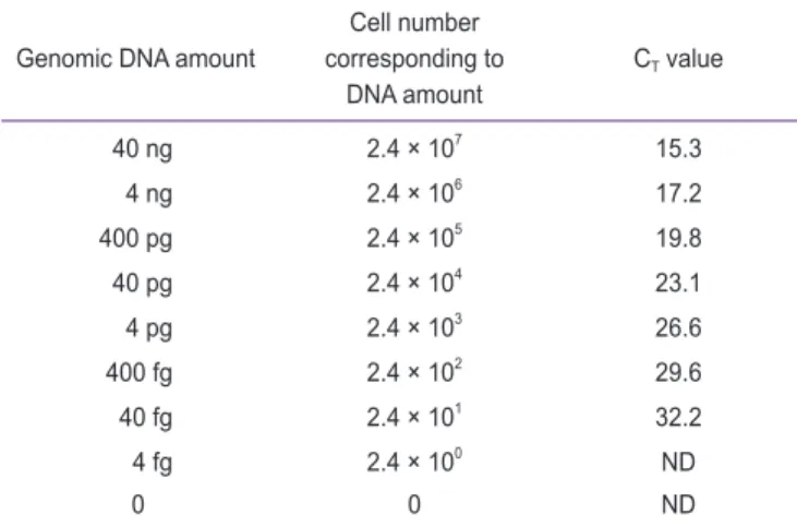
Table 2. Determination of cycle threshold (C T ) value for a dilution series of genomic DNA of Peptoniphilus mikwangii KCOM 1628 T
Genomic DNA amount Cell number corresponding to
DNA amount
C T value
40 ng 2.4 × 10 7 15.3
4 ng 2.4 × 10 6 17.2
400 pg 2.4 × 10 5 19.8
40 pg 2.4 × 10 4 23.1
4 pg 2.4 × 10 3 26.6
400 fg 2.4 × 10 2 29.6
40 fg 2.4 × 10 1 32.2
4 fg 2.4 × 10 0 ND
0 0 ND
C T values above 35 was considered “not detected” in this study.
ND, not detected.
10 20 30
40,000
30,000
20,000
10,000
Fluorescence intensity
Cycle 0
A B C
40 15 20 30
2.4 10 7
Copy number of genome
Cycle threshold (C ) T
35 60 70 80
60,000 50,000
Fluorescence intensity
0
90 25
40,000 30,000 20,000 10,000 2.4 10 6
2.4 10 5 2.4 10 4 2.4 10 3 2.4 10 2 2.4 10 1 2.4 10 0
Temperature ( )
Fig. 2. (A) Amplification plot, (B) standard curve, and (C) melting curve were obtained by quantitative real-time polymerase chain reaction using the
RTB134-F4/RTB134-R4 primers from 10-fold serial dilutions of genomic DNA of Peptoniphilus mikwangii KCOM 1628 T range from 40 ng to 4 fg. In the stan-
dard curve, the regression equation for the standard curve was: Y = –0.3395X + 11.946. The R 2 value was 0.9942.
Results
본 연구에서 P. mikwangii를 검출하기 위해 설계된 qPCR 프라이 머 쌍(RTB134-F4/RTB134-R4)의 종-특이성을 일반 PCR법으로 조 사한 결과, P. mikwangii 균주(KCOM 1628
T
, KCOM 1637, KCOM 1641)에서만 예상하였던 408 bp의 PCR 증폭물이 확인되었다(Fig.1). 또한, 본 연구에서 대조군으로 사용된 6종 Peptoniphilus spp. 표 준균주들 및 20종 구강 세균 균주들의 지놈 DNA에서는 PCR 산물이 증폭되지 않았다(Fig. 1).
P. mikwangii-특이 qPCR 프라이머 쌍(RTB134-F4/RTB134- R4)의 민감도를 측정하기 위해 P. mikwangii KCOM 1628
T
의 지 놈 DNA를 이용하여 qPCR을 수행하였다. 그 결과 RTB134-F4/RTB134-R4 프라이머 쌍은 P. mikwangii KCOM 1628
T
지놈 DNA 40 ng부터 40 fg까지 검출할 수 있었다(Table 2 and Fig. 2).RTB134-F4/RTB134-R4 프라이머 쌍에 의한 P. mikwangii KCOM 1628
T
지놈 DNA 상의 표적 유전자인 16S rDNA 이외의 영역 을 주형으로 하는 비특이적 PCR 산물이 증폭 되었는지를 알아보기 위 하여 melting curve 분석을 실시하였다. 그 결과, melting curve가 단 일한 패턴을 보였으며, 온도 변화에 따른 형광 감도의 또 다른 peak가 보이지 않았다(Fig. 2C). 이러한 결과로 RTB134-F4/RTB134-R4 프 라이머 쌍에 대한 표적 유전자인 16S rDNA를 주형으로 한 PCR 산물 만이 증폭되었음을 확인할 수 있었다.Discussion
본 연구에서 설계된 RTB134-F4/RTB134-R4 프라이머 쌍은 P.
mikwangii에 대한 종-특이성이 있음이 검증되었으며(Fig. 1), qPCR 법에 의해 P. mikwangii KCOM 1628
T
지놈 DNA를 40 fg까지 검 출할 수 있음을 알 수 있었다(Table 2 and Fig. 2). P. mikwangii KCOM 1628T
지놈 DNA 크기가 약 1.5 Mb이고, 하나의 genome 에 16S rDNA가 4 copy 존재(GenBank accession no. NZ_AUQY01000000)한다는 것[8]을 고려할 때, RTB134-F4/RTB134- R4 프라이머 쌍은 24 마리의 P. mikwangii KCOM 1628
T
및 96 copy의 16S rDNA까지 검출할 수 있음을 알 수 있었다(Table 2).PCR 또는 qPCR 프라이머 설계 프로그램을 이용할 때, 각각의 프로 그램 알고리즘에 의해 최적결합온도가 제시되지만, 실제와는 다른 경 우가 많아 gradient PCR을 수행하여 실질적인 최적결합온도를 정하는 방법이 추천되었다[7,9]. 본 연구에서 설계된 RTB134-F4/RTB134- R4 프라이머 쌍의 PrimerSelect 프로그램에서 추천된 최적결합온도는 55.1℃였다. P. mikwangii 16S rDNA 핵산염기서열과 가장 상동성 이 높은 Peptoniphilus indolicus와 Peptoniphilus asaccharolyticus 표준균주들의 지놈 DNA, 그리고 P. mikwangii KCOM 1628
T
지놈 DNA를 이용하여, 54–66℃ 사이에서 1℃ 간격으로 gradient PCR을 실시한 결과, 모든 온도에서 P. mikwangii KCOM 1628T 지놈 DNA 를 주형으로 한 경우에서만 PCR 산물이 증폭되었다. 또한, 54–62℃사이의 밴드 사이즈와 밝기가 동일하였으며, 그 보다 높은 온도에서 는 밴드 사이즈와 밝기가 작아지고 흐려졌다(data not shown). P.
mikwangii KCOM 1637와 P. mikwangii KCOM 1641 지놈 DNA 를 주형으로 56–62℃ 사이에서 2℃ 간격으로 gradient PCR을 실시한 결과, 60℃ 이상에서는 PCR 산물이 증폭되지 않았고, 56℃에서 밴드 사이즈와 밝기가 최대였다(data not shown). 그러므로, 본 연구에서 56℃를 결합온도로 최종 결정하여 사용하였다.
이상의 결과를 종합하면, 본 연구에서 개발된 RTB134-F4/
RTB134-R4 프라이머 쌍은 P. mikwangii에 대한 종-특이성 및 민감 도가 뛰어난 것으로 확인되었다. 그러므로, RTB134-F4/RTB134-R4 프라이머 쌍은 향후 P. mikwangii와 여러 구강 세균 감염성질환과의 역학 연구에 사용할 수 있을 것으로 생각된다.
Acknowledgements
이 논문은 2018학년도 조선대학교 학술연구비의 지원을 받아 연구되 었음.
Conflicts of Interest
No potential conflict of interest relevant to this article was reported.