This is an Open Access article distributed under the terms of the Creative Commons Attribution Non-Commercial License (http: //creativecommons.org/licenses/by- nc/4.0/) which permits unrestricted non-commercial use, distribution, and reproduction in any medium, provided the original work is properly cited.
© 2018 THE KOREAN SOCIETY OF MYCOLOGY.
https://doi.org/10.4489/KJM.20180034
RESEARCH ARTICLE
구상나무와 주목의 침엽에서 분리된 국내 미기록 내생균
박혁, 김동여, 김새롬, 엄안흠*
한국교원대학교 생물교육과
New Records of Endophytic Fungi Isolated from Leaves of Abies koreana and Taxus cuspidata in Korea
Hyeok Park, Dong-Yeo Kim, Sae-Rom Kim, Ahn-Heum Eom*
Department of Biology Education, Korea National University of Education, Cheongju 28173, Korea
*Corresponding author: [email protected]
ABSTRACT
We isolated endophytic fungi from the coniferous leaves of Abies koreana and Taxus cuspidata inhabiting Mt. Hallasan, Korea. The isolated fungal strains were identified based on a phylogenetic analysis using nucleotide sequences of the internal transcribed spacer region, large subunit region of ribosomal DNA, and translation elongation factor region.
Results confirmed four prevously unreported endophytic fungi in Korea: Lachnellula hyalina, Ochrocladosporium elatum, Phacidium lacerum, and Phyllosticta cussoniae. In this report, we describe the morphological characteristics of these fungi and the results of the phylogenetic analysis.
Keywords: Lachnellula hyalina, Ochrocladosporium elatum, Phacidium lacerum, Phyllosticta cussoniae
서론
내생균(endophyticfungi)은 식물에 공생하는 균류이다. 내생균은 식물의 모든 조직 내에서 병증을 일으키지 않고 서식하고 있으며[1], 포식자인 초식동물 혹은 식물에 병을 일으키는 병원성 미생물 에 대한 화학적 방어 등의 이점을 제공한다[2, 3]. 특히 주목의 침엽에 서식하는 내생균이 분비하는 taxol과 같이[4], 침엽수에 서식하는 내생균은 항생 혹은 항암 작용을 할 수 있는 물질을 분비하기도 하며[5], 이러한 점에서 연구의 가치가 있다고 생각된다. 본 연구에서는 제주도 한라산에 서식하는 침엽수 잎에서 내생균을 분리하던 중에 구상나무(Abieskoreana)와 주목(Taxuscuspidata)의 침엽에서 분리된 국내 미기록종 4종의 형태적 특성 및 계통적 분석에 대해 서술하고자 한다.
Accepted: August 25, 2018 Revised: August 23, 2018 Received: August 14, 2018
Kor. J. Mycol. 2018 September, 46(3): 241-248 OPEN ACCESS
pISSN : 0253-651X eISSN : 2383-5249
재료 및 방법
한라산의 해발 1,300~1,400m 부근에 서식하는 구상나무와 주목의 침엽 중에서, 외관상 병증이 나 타나지 않는 건강한 잎을 채취하여 실험실로 운반하였다. 시료를 1%의 NaClO 용액과 70% EtOH 로 표면살균한 뒤 potatodextroseagar (PDA) 배지에 4조각씩 치상하였다. 25℃의 암소에서 3일 이상 배양하면서 균사가 뻗어 나오면 새로운 PDA 배지에 계대하여 순수 분리하였고, 확보된 균주를 다 시 PDA 배지와 maltextractagar (MEA) 배지에 3점 계대하여 7일간 동일한 조건으로 배양한 뒤 형태 적 특징을 관찰하였다(Tables1, 2, Fig. 1). 염기서열 분석을 위하여 DNeasyPlantminikit (Qiagen, Germantown, MD, USA)의 protocol에 따라 균사에서 genomicDNA를 추출한 뒤 균 특이적인 프라이 머인 ITS1F와 ITS4를 이용하여 internaltranscribedspacer (ITS) 영역을 증폭하였고[6], 프라이머 LR0R 과 LR16을 이용하여 28SrDNA를 포함하는 largesubunit (LSU) 영역을 증폭하였으며[7], 추가적인 염기서열 분석이 필요한 균주들은 primer526F와 1567R을 이용하여[8] translationelongationfactor (TEF) α-1 영역을 증폭하였다. Annealing 온도는 ITS 영역은 50°C, LSU 영역은 44°C, TEF 영역은 57°C로 설정하여 수행하였다. PCR 산물은 1.5% agarosegel에서 22분간 전기영동을 실시하였고, 전 기영동 결과 증폭된 DNA 단편의 크기를 확인한 후 염기서열 분석을 의뢰하였다(SolGent, Daejeon, Korea). 분석된 염기서열은 NCBI 상에서 BLAST를 이용하여 유사도를 확인한 후 각 종들 간의 계
Table 1. Morphological characteristics of fungal strains isolated from Abies koreana
Strain 16H321 Lachnellula hyalina [10] 16H339 Phyllosticta cussoniae [14, 15]
Colony MEA, 25°C, 7 days MEA, 24°C PDA, 25°C, 7 days PDA, 27°C, 7 days
Color Beige White Olive to dark green, reverse grey Iron-grey
Size 11~14 mm in diam. Unrecoded 30~34 mm in diam. Covering the dish in 1 month
Shape Flat, margins irregular Cottony mycelium Umbonate, margin irregular
undulate Erumpent, spreading, with sparse aerial mycelium, margin feathery
Conidia Hyaline, globose to ellipse, 1-septate, (3.4~5.5) × (1.5~2.0) μm in diam.
Hyaline, ellipsoidal oblong,
3.0 × 1.5 μm in diam. Hyaline, aseptate, ellipsoid to obovoid, (8.5~10.8) × (5.0~6.0) μm in diam.
Thin and smooth walled, hyaline, aseptate, ellipsoid to obovoid, (10~)12~15(~17) × (6~)7(~8) μm in diam.
MEA, malt extract agar; PDA, potato dextrose agar; diam., diameter.
Table 2. Morphological characteristics of fungal strains isolated from Taxus cuspidata.
Strain 17C006 Ochrocladosporium elatum [11] 16H339 Phyllosticta cussoniae [14, 15]
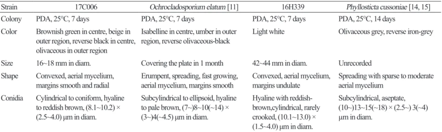
Colony PDA, 25°C, 7 days PDA, 25°C, 7 days PDA, 25°C, 7 days PDA, 25°C, 14 days
Color Brownish green in centre, beige in outer region, reverse black in centre, olivaceous in outer region
Isabelline in centre, umber in outer
region, reverse olivaceous-black Light white Olivaceous grey, reverse iron-grey Size 16~18 mm in diam. Covering the plate in 1 month 42~44 mm in diam. Unrecorded
Shape Convexed, aerial mycelium,
margins smooth and radial Erumpent, spreading, fast growing,
aerial mycelium, margins smooth Convexed, aerial mycelium,
margins undulate Spreading with sparse to moderate aerial mycelium
Conidia Cylindrical to coniform, hyaline to reddish brown, (8.1~10.2) × (2.5~4.0) μm in diam.
Subcylindrical to ellipsoid, hyaline to pale brown, (7~)8~10(~14) × (3~)4(~4.5) μm in diam.
Hyaline with reddish- brown,cylindrical, rarely crooked, (10.1~13.0) × (1.5~4.0) μm in diam.
Subcylindrical, aseptate, (10~)13~15(~18) × (2.5~) 3(~4) µm in diam.
PDA, potato dextrose agar; diam., diameter.
통 분석을 통한 유연 관계를 확인하기 위해 MEGA7를 이용하여[9] concatenatedalignment를 만든 후, neighbor-joinning 방법으로 계통수를 작성하였다. 분리된 균주는 국립생물자원관(NIBR)에 기탁하 였으며, DNA 염기서열은 미국 국립생물정보센터(NCBI)에 제출하였다.
결과 및 고찰
Lachnellula hyalina Dharne, Phytopathologische Zeitschrift 53: 119 (1964)
구상나무의 침엽에서 분리된 균주이다. PDA 배지에서 7일간 배양된 균총의 크기는 10~12mm 정 도로 매우 느리게 자라고, 균총의 색은 앞면은 전체적으로 옅은 베이지색을 띠고 뒷면은 연한 갈색 을 띤다. 균총의 가장자리는 매우 불규칙하며, 균총의 고도는 배지에 납작하게 붙어 있다(Fig. 1A).
MEA 배지에서 7일간 배양된 균총의 크기는 11~14mm 정도로 PDA 배지에서와 마찬가지로 매우 느린 속도로 자란다. 균총의 색은 앞·뒷면 모두 베이지색에 가깝고, 균총의 고도는 배지에 납작하 게 붙어 있다. 균총의 가장자리는 매우 불규칙하다(Fig. 1E). 균사 생장 방향의 측면에서 구형 혹은 불규칙한 타원형의 분생자(conidia)가 형성된다. 분생자는 투명한 유리질이며, 격막(septate)으로 분 리된다. 분생자의 크기는 (3.4~5.5) × (1.5~2.0) μm 정도이다(Fig. 1I).
Specimenexamined: Mt. Hallasan, Jeju-do, Korea, 33°22'03.5"N, 126°32'37.1"E, August23, 2016, isolated fromleavesofAbieskoreana, strain16H321, NIBRFG0000502333, GenBankno. MH734785.
Notes: L. hyalina는 1964년 Dharne에 의해 보고된 종이다. 분생자의 투명한(hyaline) 형태적 특성으로 부터 종명이 유래했으며, 스위스에 분포하는 소나무과의 침엽수인 무고소나무(Pinusmugo)의 가지 로부터 분리되었다[10]. 본 연구에서 확인된 유리질로 된 분생자의 형태와 크기는 원 기재문의 설 Fig. 1. Colonies of strain 16H321 (Lachnellula hyalina) grown for 7 days on potato dextrose agar (PDA) (A) and malt extract agar (MEA) (E), conidia (I). Colonies of strain 17C006 (Ochrocladosporium elatum) grown for 7 days on PDA (B) and MEA (F), conidiophore (J). Colonies of strain 17C009 (Phacidium lacerum) grown for 7 days on PDA (C) and MEA (G), conidia (K). Colonies of strain 16H339 (Phyllosticta cussoniae) grown for 7 days on PDA (D) and MEA (H), conidia (L) (scale bars = 10 μm).
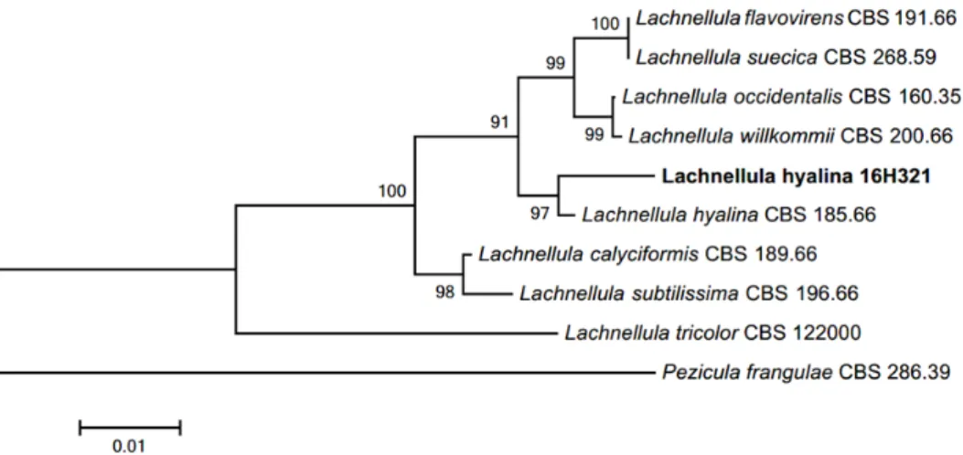
명과 일치하였으며, ITS 영역과 LSU 영역의 염기서열의 분석 결과 ITS 영역의 DNA 염기서열은 L. hyalinaKC464638.1과 98%의 일치도를 보였고, LSU 영역의 염기서열은 L. hyalinaKC492976.1과 99%의 일치도를 보였으며, 모두 같은 계통을 형성했다(Fig. 2).
Ochrocladosporium elatum (Harz) Crous & U. Braun, Studies in Mycology 58: 46 (2007)
주목의 침엽에서 분리된 균주이다. PDA 배지에서 7일간 배양된 균총의 직경은 16~18mm 정도이 며, 균총의 색은 앞면은 중앙부에서는 짙은 녹갈색을 띠고, 가장자리에는 옅은 베이지색의 띠가 형 성된다. 뒷면은 중앙부에서 검은색을 띠고, 바깥쪽은 올리브색이며 가장자리에 앞면과 마찬가지 로 베이지색의 띠가 형성된다. 고도는 배지에서 살짝 융기되어 있고, 가장자리는 촘촘한 균사가 방 사형으로 뻗어 있다(Fig. 1B). MEA 배지에서 7일간 배양된 균총의 직경은 15~16mm 정도이며, 균 총의 색은 앞면은 전체적으로 밝은 흰색을 띠고 뒷면은 중앙부에서 황갈색을, 가장자리에서 베이 지색을 띤다. 균총의 고도는 중앙부에서 볼록 융기되어 있으며, 가장자리의 형태는 균사들이 불규 칙하게 뻗어 나가는 형태이다(Fig. 1F). 균사 생장 방향의 측면에서 불규칙한 타원형으로 비대해진 분생자경(conidiophore)이 형성되며, 분생자경으로부터 원추형 혹은 원통형의 투명한 유리질 분생 자가 형성된다. 분생자의 색은 적갈색을 띠거나 혹은 무색이고, 분생자의 크기는 (8.1~10.2) × (2.5~4.0) μm 정도이다(Fig. 1J).
Specimenexamined: Mt. Hallasan, Jeju-do, Korea, 33°23'08.6"N, 126°32'14.0"E, August23, 2016, isolated fromleavesofTaxuscuspidata, strain17C006, NIBRFG0000502331, GenBankno. MH734786.
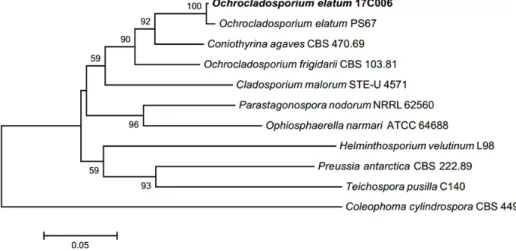
Notes: O. elatum은 2007년 Crous & Braun에 의해 새로 명명되었다. 본래 Cladosporium속에 속해 있었 으며, 연한 갈색의 분생자로부터 기인하여, 속명에 황갈색을 뜻하는 그리스어 접두어 Ochro-가 붙 어 속명이 새로 정립되었다[11]. 분생자의 형태적 특징은 대체로 원 기재문과 일치하였으며, ITS 영 역과 LSU 영역의 염기서열의 분석 결과 ITS 영역의 DNA 염기서열은 O. elatumGU248334.1과 99% 의 일치도를 보였고, LSU 영역의 염기서열은 O. elatumEU040233.1과 98%의 일치도를 보였으며, 모두 같은 계통을 형성했다(Fig. 3).
Fig. 2. Neighbor-joining phylogenetic tree based on a combined alignment of both internal transcribed spacer and large subunit sequences. Pezicula frangulae was used as an outgroup. Numbers on branches indicate bootstrap values (1,000 replicates). Fungal strain isolated in this study is in bold.
Phacidium lacerum Fr., Observationes mycologicae 2: 313 (1818)
주목의 침엽에서 분리된 균주이다. PDA 배지에서 7일간 배양된 균총의 크기는 42~44mm 정도이 고, 균총의 색은 앞·뒷면 모두 밝은 흰색을 띤다. 균총의 고도는 배지에서 살짝 융기되어 있으며, 가 장자리는 공중 균사가 발달하여 물결 모양으로 퍼져 나간다(Fig. 1C). MEA 배지에서 7일간 배양된 균총의 크기는 40~43mm 정도이고, 균총의 색은 앞면은 흰색을 띠고 뒷면은 베이지색에 가깝다.
균총의 고도는 배지에서 살짝 융기되어 있고, 가장자리는 불규칙하다. 균총의 뒷면에는 중앙을 기 준으로 불규칙한 방사형의 주름들이 형성된다(Fig. 1G). 분생자경은 균사 생장 방향을 따라 형성되 거나 혹은 분지되며, 전체적으로 적갈색 빛이 감도는 투명한 유리질로 이루어져 있다. 분생자경에 서 형성되는 분생자는 투명한 유리질의 길쭉한 원통형이며 초승달처럼 끝이 휘어지기도 한다. 분 생자의 크기는 (10.1~13.0) × (1.5~4.0) μm 정도이다(Fig. 1K).
Specimenexamined: Mt. Hallasan, Jeju-do, Korea, 33°23'08.6"N, 126°32'14.0"E, August23, 2016, isolated fromleavesofTaxuscuspidata, strain17C009, NIBRFG0000502330, GenBankno. MH734787.
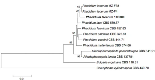
Notes: P. lacerum은 1818~1823년경 Fries에 의해 보고된 것으로 알려져 있으며, 유럽에 널리 분포하 는 구주소나무(Pinussylvestris)에서 최초 분리된 것으로 보고되어 있다[12, 13]. Phacidium속에 속하 는 종들은 길쭉한 유리질의 분생자를 형성하는 것이 특징이며, 본 연구에서 확인된 길쭉한 원통형 의 분생자는 Crous 등[12]의 연구와 일치한다. ITS 영역과 LSU 영역의 염기서열의 분석 결과 ITS 영 역의 DNA 염기서열은 P. lacerumKU942438.1과 99%의 일치도를 보였고, LSU 영역의 염기서열은 P. lacerumMG720334.1과 99%의 일치도를 보였으며, 모두 같은 계통을 형성했다(Fig. 4).
Phyllosticta cussoniae Cejp, Bothalia 10: 341 (1971)
구상나무의 침엽에서 분리된 균주이다. PDA 배지에서 7일간 배양된 균총의 크기는 30~34mm 정 도이고, 균총의 앞면은 연한 올리브색과 어두운 녹색이 섞여 있으며, 뒷면은 전체적으로 회색을 띤 다. 균총의 고도는 배지에서 살짝 융기되어 있으며, 가장자리는 불규칙한 물결 형태이다(Fig. 1D).
MEA 배지에서 7일간 배양된 균총의 크기는 42~45mm 정도로 PDA 배지에서보다 빠른 속도로 자 란다. 앞면은 전체적으로 흰색을 띠며 뒷면은 중앙부는 검은색을 띠고 가장자리는 흰색을 띤다. 균
Fig. 3. Neighbor-joining phylogenetic tree based on a combined alignment of both internal transcribed spacer and large subunit sequences. Coleophoma cylindrospora was used as an outgroup. Numbers on branches indicate bootstrap values (1,000 replicates). Fungal strain isolated in this study is in bold.
총의 고도는 중앙부에서는 납작하고 가장자리에서 공중 균사가 발생하여 융기되는 형태이다(Fig. 1H). 균사의 끝부분에서 둥근 형태의 분생자경이 발생하며, 분생자경에서 격막이 없는 투명한 유 리질의 계란형 혹은 짧은 타원형 분생자를 형성한다. 분생자의 크기는 (8.5~10.8) × (5.0~6.0) μm 정 도이다(Fig. 1L).
Specimenexamined: Mt. Hallasan, Jeju-do, Korea, 33°22'03.5"N, 126°32'37.1"E, August23, 2016, isolated fromleavesofAbieskoreana, strain16H339, NIBRFG0000502334, GenBankno. MH734930.
Notes: P. cussoniae는 1971년 Cejp[14]에 의해 보고된 종이다. 남아프리카 공화국의 Cussonia속 목본 식물의 잎에서 분리된 것에서 기원하였다. 분생자의 형태 비교는 원 기재문 외에 Wikee 등[15]의 연 구를 참고하였고, 본 연구에서 확인된 분생자의 형태가 참고문헌과 대체로 일치하는 것을 확인하 였다. ITS 영역과 LSU 영역의 염기서열의 분석 결과 ITS 영역의 DNA 염기서열은 P. cussoniae KF170311.1과 98%의 일치도를 보였고, LSU 영역의 염기서열은 P. cussoniaeKF206278.1과 99%의 일치도를 보였으며, TEF 영역의 염기서열은 P. cussoniaeKF289223.1과 98%의 일치도를 보였고 모 두 같은 계통을 형성했다(Fig. 5).
적요
제주도 한라산의 구상나무와 주목의 침엽에서 내생균을 분리하였다. 분리된 균주들은 형태적 특 성 및 internaltranscribedspacer 영역, largesubunitrDNA 영역 및 translationelongationfactor 영역 염기서 열의 계통분석을 통해 종을 동정하였다. 그 결과 4종의 국내 미기록 내생균을 확인하였고, 확인된 종은 각각 Lachnellulahyalina, Ochrocladosporiumelatum, Phacidiumlacerum, Phyllostictacussoniae이다.
확인된 4종의 미기록 내생균 균주의 형태적 특성 및 염기서열 계통분석의 결과에 대해 기술하였 다.
Fig. 4. Neighbor-joining phylogenetic tree based on a combined alignment of both internal transcribed spacer and large subunit sequences. Coleophoma cylindrospora was used as an outgroup. Numbers on branches indicate bootstrap values (1,000 replicates). Fungal strain isolated in this study is in bold.
ACKNOWLEDGEMENTS
This study was supported by the Project on Survey and Discovery of Indigenous Fungal Species of Korea funded by NIBR of the Ministry of Environment and by the Project on Conservation and Adaptation of Forest Species Vulnerable to Climate Changes funded by National Arboretum of Korea Forest Service.
REFERENCES
1. Carroll G. Fungal endophytes in stems and leaves: from latent pathogen to mutualistic symbiont. Ecology 1988;69:2-9.
2. Schulz B, Boyle C, Draeger S, Römmert AK, Krohn K. Endophytic fungi: a source of novel biologically active secondary metabolites. Mycol Res 2002;106:996-1004.
3. Christensen MJ. Antifungal activity in grasses infected with Acremonium and Epichloë endophytes. Australas Plant Pathol 1996;25:186-91.
4. Strobel GA, Hess WM, Ford E, Sidhu RS, Yang X. Taxol from fungal endophytes and the issue of biodiversity. J Ind Microbiol Biotechnol 1996;17:417-23.
5. Findlay JA, Buthelezi S, Lavoie R, Peña-Rodriguez L, Miller JD. Bioactive isocoumarins and related metabolites from conifer endophytes. J Nat Prod 1995;58:1759-66.
6. White TJ, Bruns T, Lee S, Taylor J. Amplification and direct sequencing of fungal ribosomal RNA genes for phylogenetics. In: Innis MA, Gelfand DH, Sninsky JJ, White TJ, editors. PCR protocols: a guide to methods and applications. San Diego: Academic Press; 1990. p. 315-22.
7. Moncalvo JM, Lutzoni FM, Rehner SA, Johnson J, Vilgalys R. Phylogenetic relationships of agaric fungi based on nuclear large subunit ribosomal DNA sequences. Syst Biol 2000;49:278- Fig. 5. Neighbor-joining phylogenetic tree based on a combined alignment of both internal transcribed spacer, large subunit and translation elongation factor sequences. Dothidea ribesia was used as an outgroup. Numbers on branches indicate bootstrap values (1,000 replicates). Fungal strain isolated in this study is in bold.
305.
8. Rehner SA, Buckley E. A Beauveria phylogeny inferred from nuclear ITS and EF1-α sequences: evidence for cryptic diversification and links to Cordyceps teleomorphs. Mycologia 2005;97:84-98.
9. Kumar S, Stecher G, Tamura K. MEGA7: Molecular Evolutionary Genetics Analysis Version 7.0 for Bigger Datasets. Mol Biol Evol 2016;33:1870-4.
10. Dharne CG. Taxonomic investigations on the discomycetous genus Lachnellula Karst. J Phytopathol 1965;53:101-44.
11. Crous PW, Braun U, Schubert K, Groenewald JZ. Delimiting Cladosporium from morphologically similar genera. Stud Mycol 2007;58:33-56.
12. Crous PW, Quaedvlieg W, Hansen K, Hawksworth DL, Groenewald JZ. Phacidium and Ceuthospora (Phacidiaceae) are congeneric: taxonomic and nomenclatural implications. IMA
Fungus 2014;5:173-93.
13. Fries E. Observationes mycologicae praecipue ad illustrandam Floram Suecicam. Copenhagen:
Gerhardi Bonnier; 1818.
14. Cejp K. Some members of the Sphaeropsidales from South Africa. Bothalia 1971;10:341-5.
15. Wikee S, Lombard L, Nakashima C, Motohashi K, Chukeatirote E, Cheewangkoon R, McKenzie EH, Hyde KD, Crous PW. A phylogenetic re-evaluation of Phyllosticta (Botryosphaeriales). Stud Mycol 2013;76:1-29.