http://dx.doi.org/10.11620/IJOB.2015.40.4.223
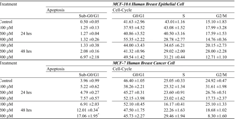
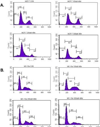
Apoptotic Effects of 6-Gingerol in Human Breast Cancer Cells
Hyun-Woo Kim
1, Deuk-Hee Oh
1, Jeong-Tae Koh
2, and Young-Chai Lim
1*1
Department of Pharmacology, Chonnam National University Medical School, Gwangju, Korea
2