Abstract. Previously, we reported that 20-O-(β -D-gluco-pyranosyl)-20(S)-protopanaxadiol (Compound K, a meta bolite of ginseng saponin) induces mitochondria-dependent and caspase-dependent apoptosis in HT-29 human colon cancer cells via the generation of reactive oxygen species. The aim of the present study was to elucidate the mechanism underlying apop-tosis induced by Compound K with respect to endoplasmic reticulum (ER) stress in HT-29 cells. In the present study, Compound K induced apoptotic cell death as confirmed by DNA fragmentation and apoptotic sub-G1 cell population.
Compound K also induced ER stress as indicated by staining with ER tracker, cytosolic and mitochondrial Ca2+
over-loading, phosphorylation of protein-kinase-like endoplasmic reticulum kinase (PERK), phosphorylation of eukaryotic initiation factor-2α (eIF-2α), phosphorylation of IRE-1, splicing of ER stress-specific X-box transcription factor-1 (XBP-1), cleavage of activating transcription factor-6 (ATF-6), upregulation of glucose-regulated protein-78 (GRP-78/BiP) and CCAAT/enhancer-binding protein-homologous protein (CHOP), and cleavage of caspase-12. Furthermore, downregu-lation of CHOP expression using siCHOP RNA attenuated Compound K-induced apoptosis. Taken together, these results support the important role of ER stress response in mediating Compound K-induced apoptosis in human colon cancer cells.
Introduction
The endoplasmic reticulum (ER) is a secretory organelle, which plays a critical role in lipid synthesis and protein folding and modification before export to the Golgi body. Protein folding is impaired under unbalanced redox state, failure of protein synthesis, misfolding, transport or degradation and Ca2+
over-loading, which can result in ER stress (1-3). ER stress leads to the accumulation of unfolded proteins in the ER lumen where they form highly toxic aggregates. If these toxic aggregates reach critical levels, ER stress response and apoptotic signaling are initiated (4).
The ER stress response is primarily regulated by three ER transmembrane proteins; protein kinase-like endoplasmic reticulum kinase (PERK), activating transcription factor-6 (ATF-6), and inositol requiring kinase-1 (IRE-1) (5). In normal condition, these proteins remain inactive and bound to ER-resident chaperone glucose-regulated protein-78 (GRP-78/BiP). During ER stress, GRP-78 dissociates from these proteins (5). Dissociated RERK and IRE-1 are then free to form homo-dimers, which lead to their autophosphorylation and activation (5). Activated PERK then phosphorylates and inactivates eukaryotic initiation factor-2α (eIF-2α), leading to an overall suppression in translation (6,7), whereas acti-vated IRE-1 converts X-box transcription factor-1 (XBP-1) pre-mRNA into mature mRNA by unconventional splicing. The protein translated from this spliced XBP-1 mRNA medi-ates transcriptional activation of ER-associated degradation component genes (8). Dissociation of ATF-6 from GRP-78 permits its translocation to the Golgi apparatus where it is sequentially cleaved (activated) by proteases (9). The activated cytoplasmic fragment of ATF-6 then binds to the ER stress response element in the nucleus to activate transcription of ER chaperone genes such as GRP-78 and transcription factor genes such as XBP-1, and expression of CCAAT/enhancer-binding protein-homologous protein (CHOP/GADD153) (9,10). Severe or prolonged ER stress stimulates RERK, ATF-6 and IRE-1
20-O-(
β
-D-glucopyranosyl)-20(S)-protopanaxadiol
induces apoptosis via induction of endoplasmic
reticulum stress in human colon cancer cells
RUI ZHANG1, YOUNG CHUNG2, HEE SUN KIM3, DONG HYUN KIM4, HYE SUN KIM5, WEON YOUNG CHANG1 and JIN WON HYUN1
1School of Medicine and Applied Radiological Science Research Institute, Jeju National University,
Jeju 690-756, Republic of Korea; 2Pharmaceutical Chemistry, University of California, Davis, CA 95616, USA; 3Department of Neuroscience, College of Medicine, Ewha Womans University, Seoul 110-783;
4Department of Microbial Chemistry, College of Pharmacy, Kyung Hee University, Seoul 130-701; 5Cancer Research Institute, Seoul National University College of Medicine, Seoul 110-799, Republic of Korea
Received July 4, 2012; Accepted September 14, 2012 DOI: 10.3892/or.2013.2270
Correspondence to: Professor Jin Won Hyun or Professor Weon Young Chang, School of Medicine, Jeju National University, 66 Jejudaehakno, Jeju 690-756, Republic of Korea
E-mail: [email protected] E-mail: [email protected]
Key words: Compound K, apoptosis, endoplasmic reticulum stress, CCAAT/enhancer-binding protein-homologous protein
apoptotic signaling and increases CHOP expression. CHOP is the major component of the ER stress pathway, and CHOP knockdown cells are resistant to ER stress-induced apoptosis (11). Caspase-12 has also been implicated in ER stress-mediated cell death, and caspase-12 knockdown mice are resistant to ER stress-induced apoptosis (12).
Accumulating evidence suggests that anticancer agents are toxic to cancer cells because they increase oxidative stress, pushing the cancer cells beyond their limits (13-15). Cytotoxic reactive oxygen species (ROS) appears to be triggered by activation of the mitochondrial-dependent cell death pathway via bcl-2 family proteins, with consequent mitochondrial membrane permeabilization and apoptosis (16).
20-O-(βDglucopyranosyl)20(S)protopanaxadiol (Com -pound K, Fig. 1A) is the main metabolite of protopanaxadiol- type ginsenoside formed in the intestine after oral administra-tion (17-20). Ginseng saponin was reported to show beneficial roles on abnormal coronary contraction (21) and possess therapeutic effect on skin wound healing (22). Our previous studies show that Compound K enhances gamma ray-induced apoptosis via generation of ROS and disruption of the mito-chondrial membrane in human lung cancer cells (23), and induces mitochondria- and caspase-dependent apoptosis via the generation of ROS in human colon cancer cells (24). Furthermore, Compound K exhibits anti-proliferative effects against cancer cells, which are mediated via apoptosis (23-29). Despite evidence for the anti-proliferative effects of Compound K, the underlying cytotoxic mechanisms with respect to ER stress-mediated apoptosis have not been reported. Therefore, the aim of the present study was to deter-mine the role of ER stress in mediating Compound K-induced apoptosis in HT-29 human colon cancer cells.
Materials and methods
Preparation of Compound K. Compound K was provided by Professor Dong Hyun Kim (Kyung Hee University, Seoul, Republic of Korea). Compound K was prepared by the incuba-tion of protopanaxadiol type ginsenosides with Bacteroides JY-6, a human intestinal bacterium, subcultured in a general anaerobic medium for 24 h at 37˚C. The incubated medium was extracted with n-butanol. The supernatant was concentrated in vacuo and processed using silica gel column chromatog-raphy with chloroform/methanol/H2O (65:35:10). The isolated
Compound K was characterized by mass spectroscopy and 1H
and 13C nuclear magnetic resonance (NMR) spectrometry.
Reagents. Propidium iodide (PI) was purchased from Sigma Chemical Company (St. Louis, MO, USA). CHOP, caspase-12, ATF-6 and ß-actin antibodies were purchased from Cell Signaling Technology (Beverly, MA, USA). Phospho PERK, phospho eIF-2α, phospho IRE-1, XBP-1 and GRP-78 antibodies were purchased from Santa Cruz Biotechnology (Santa Cruz, CA, USA). All other chemicals and reagents were of analytical grade.
Cell culture. HT-29 human colon cancer cells were obtained from the American Type Culture Collection (Rockville, MD, USA) and maintained at 37˚C in an incubator with a humidified atmosphere of 5% CO2 in air. Cells were cultured in RPMI-1640
containing 10% heat-inactivated fetal calf serum, streptomycin (100 µg/ml) and penicillin (100 U/ml).
DNA fragmentation. Cells were seeded at a concentration of 5x10⁴ cells/ml, and 16 h after plating, were treated with
Compound K at 20 µg/ml and incubated for 48 h. Cellular DNA fragmentation was assessed using the cytoplasmic histone-associated DNA fragmentation kit from Roche Diagnostics (Mannheim, Germany) according to the manufacturer's instructions.
Detection of sub-G1 hypodiploid cells. The amount of apoptotic
sub-G1 hypodiploid cells was determined by flow cytometry
(30). Cells were seeded at a six-well plate at a concentration of 1x10⁵ cells/ml, and 16 h after plating, were treated with
Compound K at 20 µg/ml for 48 h. Harvested cells were washed twice with phosphate buffered saline (PBS) and fixed in 70% ethanol for 30 min at 4˚C. Cells were incubated for 30 min in the dark at 37˚C in 1 ml PBS containing 100 µg PI and 100 µg RNase A. Flow cytometric analysis was performed using a FACS Calibur flow cytometer (Becton Dickinson, Mountain View, CA, USA). The proportion of sub-G1 hypo-diploid cells
was assessed by the histograms generated using the computer program Cell Quest and Mod-Fit (Becton Dickinson).
Cytosolic Ca2+ measurement. Cytosolic Ca2+ was detected
with the fluorescent probe Fluo4 AM. Cells were seeded at a concentration of 1x10⁵ cells/ml, and 16 h after plating, were
treated with Compound K at 20 µg/ml and incubated for 24 h. Cells were then harvested and loaded with 10 µM of Fluo4 AM for 30 min at 37˚C and the supernatant was removed by suction and after trypsin treatment, cells were washed with PBS. The fluorescence of Fluo4 AM-loaded cells was measured using a flow cytometer.
Measurement of mitochondrial Ca2+. A Rhod2 AM probe
(Molecular Probes, Eugene, OR, USA) was used to determine mitochondrial Ca2+ level (31). Rhod2 AM has a net positive
charge, which facilitates its sequestration into mitochondria via membrane potential-driven uptake. The use of Rhod2 AM enhances the selectivity for mitochondrial loading because the dye exhibits Ca2+-dependent fluorescence only after it is
oxidized, and this occurs preferentially within mitochondria. Cells were seeded at a concentration of 1x10⁵ cells/ml, and 16 h
after plating, were treated with Compound K at 20 µg/ml and incubated for 24 h. Cells were harvested, washed, and resus-pended in PBS containing Rhod2 AM (1 µM). After 15 min of incubation at 37˚C, cells were washed, suspended in PBS and analyzed by flow cytometry. For image analysis, cells were loaded with Rhod2 AM and incubated for 30 min at 37˚C. Cells were then washed, and the stained cells mounted onto micro-scope slides with mounting medium (Dako, Carpinteria, CA, USA). Microscopic images were examined using a confocal laser scanning microscope and the 5 PASCAL program (Carl Zeiss, Jena, Germany).
Fluorescent microscopy and ER staining. Cells were seeded in Lab-Tek chamber slides (Nalge Nunc International, Naperville, IL, USA) at a density of 1x10⁵ cells/ml, and 16 h after plating,
24 h. For ER staining, the ER-Tracker Blue-White DPX probe (Molecular Probes) was added to the cells and incubated for 30 min under the same growth conditions. The loading solu-tion was removed and cells were then washed with PBS before adding fresh medium without the stain. Microscopic images were collected using the laser scanning microscope 5 Pascal program (Carl Zeiss) as described above.
Western blot analysis. Cells were seeded at a concentration of 1.5x10⁵ cells/ml, and 16 h after plating, were treated with
Compound K at 20 µg/ml, After 3, 6, 12, 24 or 48 h, cells were harvested, washed twice with PBS, lysed on ice for 30 min in 100 µl of lysis buffer [120 mM NaCl, 40 mM Tris (pH 8.0), 0.1% NP40] and then centrifuged at 13,000 x g for 15 min. The supernatants were collected from the lysates and the protein concentrations were determined. Aliquots of the
lysates (40 µg of protein) were boiled for 5 min and electro-phoresed in 10% SDS-PAGE gel. The proteins in the gels were transferred onto nitrocellulose membranes and incubated with the primary antibodies. The membranes were subsequently incubated with the secondary immunoglobulin-G-horseradish peroxidase conjugates (Pierce, Rockford, IL, USA). Protein bands were detected using an enhanced chemiluminescence western blotting detection kit (Amersham, Little Chalfont, Buckinghamshire, UK), and then exposed to X-ray film. Transient transfection of small RNA interference (siRNA). Cells were seeded in 24-well plate at a density of 1.5x10⁵ cells/ml
and allowed to reach ~50% confluence on the day of trans-fection. The siRNA construct used were: mismatched siControl RNA (Santa Cruz Biotechnology) and siCHOP RNA (Bioneer Corporation, Bioneer, South Korea). Cells were transfected with 10-50 nM siRNA using Lipofectamine™ 2000 (Invitrogen, Carlsbad, CA, USA) according to the manufacturer's instruc-tions. After 24 h, cells were treated with Compound K for 48 h and examined by western blot analysis, DNA fragmentation assay and PI staining.
Statistical analysis. All the measurements were made in tripli-cate and all values are represented as the mean ± standard error of the mean (SEM). Differences in results were analyzed using analysis of variance (ANOVA) and the Tukey test. p<0.05 was considered significant.
Results
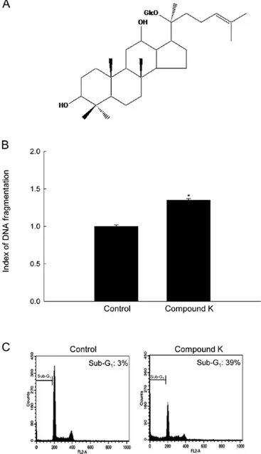
Induction of apoptosis in human colon cancer cells by Compound K treatment. A recent study showed that Compound K induces mitochondria- and caspase-dependent apoptosis in HT-29 cells via the generation of ROS (24). The results of the present study showed that 20 µg/ml of Compound K, which is a concentration of 50% growth inhibi-tion, increased DNA fragmentation and sub-G1 phase of cell
population, which are hallmarks of apoptosis (Fig. 1B and C). Compound K induces cytosolic and mitochondrial Ca2+
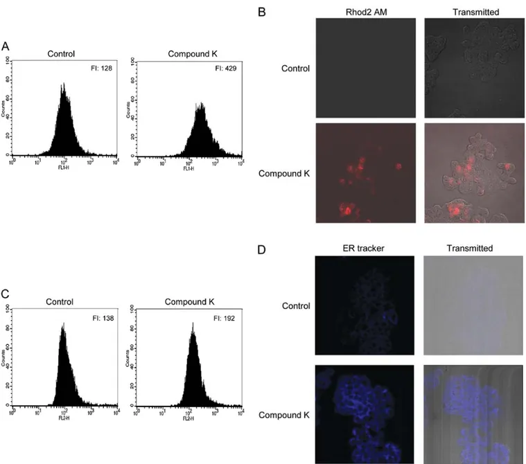
over-loading and ER stress. Depletion of ER calcium stores induces ER stress, which leads to an increase in cytosolic and mitochon-drial Ca2+ levels (32,33). Therefore, the effect of Compound K
on the mobilization of Ca2+ was examined. Compound K
resulted in significant increases in cytosolic (Fig. 2A) and mito-chondrial Ca2+ levels (Fig. 2B and C) at 24 h after Compound K
treatment. ER stress increases the fluorescence intensity of the ER-Tracker Blue-White DPX dye (34). As shown in Fig. 2D, Compound K significantly increased the staining intensity of this dye, suggesting the induction of ER stress.
Compound K increases the level of ER stress-related proteins. During ER stress, the activated phospho form of PERK phos-phorylates eIF-2α, leading to the attenuation of translational initiation and protein synthesis (35,36). As shown in Fig. 3, Compound K induced the expression of both phosphorylated PERK and phosphorylated eIF-2α in a time-dependent manner. Also, the activated phospho form of IRE-1 splices XBP-1, leading to increase in membrane phospholipids and expansion of the surface area and volume of the rough ER (37). As shown in
Figure 1. Chemical structure of Compound K and induction of apoptosis by Compound K. (A) The chemical name for Compound K is 20-O-D-gluco-pyranosyl-20(S)-protopanaxadiol. (B) DNA fragmentation was quantified using an ELISA kit and (C) the apoptotic sub-G1 DNA content was detected
by flow cytometry after PI staining. Significantly different from control (*p<0.05).
Fig. 3, Compound K induced the expression of phosphorylated IRE-1 and spliced XBP-1 protein in a time-dependent manner. Other hallmarks of the ER stress responses include activation of ATF-6, the subsequent induction of GRP-78 and CHOP, and the activation of caspase-12 (32,38,39). Compound K also enhanced ATF-6 activation, induced the expression of GRP-78 and CHOP, and activated caspase-12 (Fig. 3).
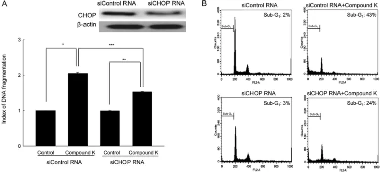
Suppression of CHOP expression attenuates Compound K- induced apoptosis. CHOP plays a proapoptotic role during ER stress (38,40). Suppression of siRNA-mediated CHOP expres-sion attenuated the apoptotic cell death induced by Compound K, which was confirmed by the DNA fragmentation pattern and
the sub-G1 cell population (Fig. 4A and B). These results
suggest that upregulation of CHOP may, in part, be involved in Compound K-induced apoptosis.
Discussion
Although Compound K induces apoptosis in many cancer cell lines (23-29), the underlying mechanisms are not well understood. The central novel finding of this study provides important evidence to support the involvement of ER stress in the induction of apoptosis by Compound K in HT-29 cells.
The ER is the primary site of protein synthesis, folding and trafficking. Under various stressful conditions, the accumulation
Figure 2. Compound K enhanced cytosolic and mitochondrial Ca2+ overloading and ER staining. Cells were treated with 20 µg/ml of Compound K for 24 h,
and then harvested and treated with the fluorescent probes Fluo4 AM and Rhod2 AM, respectively. (A) Cytosolic Ca2+ levels were measured by flow cytometry
and mitochondrial Ca2+ levels were measured by (B) confocal microscopy and (C) flow cytometry. FI indicates the fluorescence intensity of Fluo4 AM and
Rhod2 AM, respectively. The representative confocal microscopic images illustrate the increase in red fluorescence intensity of Rhod2 AM produced by mitochondrial Ca2+ overloading in Compound K-treated cells compared to the control. (D) Cells were treated with Compound K for 24 h, followed by the ER
Tracker Blue-White DPX dye and fluorescence intensity measured by confocal microscopy. The representative confocal microscopic images illustrate the increase in blue fluorescence intensity of the ER Tracker in Compound K-treated cells compared to the control.
of unfolded or misfolded proteins in the ER results in ER stress (1,2). Under conditions of ER stress, the elevation of cytosolic or mitochondrial Ca2+ levels, or the depletion of ER Ca2+ stores
are typical ER stress responses of cells. The present study shows that Compound K induced the elevation of cytosolic and mitochondrial Ca2+ levels and ER staining. Compound K also
induced apoptosis as assessed by increased DNA fragmentation and sub-G1 phase of cell population. The experimental evidence
presented here shows that the induction of ER stress-related proteins may be involved in Compound K-induced apoptosis as: i) Compound K induces phosphorylation of PERK and eIF-2α;
ii) Compound K induces phosphorylation of IRE-1 and the spliced XBP transcription factor; iii) Compound K induces cleavage of ATF-6, and subsequent GRP-78 and CHOP expres-sion; and iv) Compound K induces caspase-12 cleavage. Among ER-associated apoptotic molecules, CHOP and caspase-12 are major proapoptotic factors that are closely associated with ER stress (11). Taken together, these observations demonstrate that Compound K induces ER-mediated apoptosis. ER stress response pathways are normally activated as a protective mechanism to ensure cell survival (41). However, during severe ER stress, activation of these pathways leads to increased CHOP expression, which is a crucial element that switches ER stress signaling from pro-survival to proapoptosis (42). The CHOP protein is a member of the CCAAT/enhancer-binding proteins and functions as a dominant-negative inhibitor of gene transcription (38). Expression of CHOP is mainly regulated at the transcriptional level through the PERK/eIF-2α/ATF-6 pathway (38). CHOP knockout mice show reduced apoptosis in response to ER stress (43). Therefore, CHOP is one of the components of the ER stress-mediated apoptosis pathway. In the present study, suppression of CHOP using CHOP siRNA attenuated Compound K-induced apoptosis.
In summary, Compound K induces the apoptosis of HT-29 colon cancer cells which is mediated by ER stress signaling pathway and this is the first report to reveal that the association between the capacity of Compound K to induce ER stress and apoptosis in colon cancer cells.
Acknowledgements
This work was supported by the research grant from Jeju National University Hospital. And this study was supported by a grant from the National R&D Program for Cancer Control, Ministry for Health and Welfare, Republic of Korea (1120340).
Figure 3. Compound K induced ER stress-related proteins. Cell lysates were subjected to electrophoresis, and phosphorylated PERK, phosphorylated eIF-2α, phosphorylated IRE-1, spliced XBP-1, cleaved ATF-6, GRP-78, CHOP and cleaved caspase-12 were detected using their respective specific antibodies.
Figure 4. Downregulation of CHOP attenuated Compound K-induced apoptosis. Cells were transfected with siCHOP RNA or siControl RNA after treatment with Compound K for 48 h, (A) DNA fragmentation was quantified using an ELISA kit and (B) the apoptotic sub-G1
DNA content was detected by flow cyto-metry after PI staining. Significantly different from siControl RNA-treated cells (*p<0.05), significantly different from siCHOP RNA-treated cells (**p<0.05),
References
1. Ilieva EV, Kichev A, Naudi A, Ferrer I, Pamplona R and Portero-Otin M: Mitochondrial dysfunction and oxidative and endoplasmic reticulum stress in argyrophilic grain disease. J Neuropathol Exp Neurol 70: 253-263, 2011.
2. Dolai S, Pal S, Yadav RK and Adak S: Endoplasmic reticulum stress-induced apoptosis in Leishmania through Ca2+-dependent and caspase-independent mechanism. J Biol Chem 286: 13638-13646, 2011.
3. Song HS, Kim HM, Jung SK and Lee DH: Characterization of tunicamycin as anti-obesity agent. Biomol Ther 17: 162-167, 2009.
4. Yoshida H: ER stress and diseases. FEBS J 274: 630-658, 2007. 5. Szegezdi E, Logue SE, Gorman AM and Samali A: Mediators of
endoplasmic reticulum stress-induced apoptosis. EMBO Rep 7: 880-885, 2006.
6. Vattem KM and Wek RC: Reinitiation involving upstream ORFs regulates ATF4 mRNA translation in mammalian cells. Proc Natl Acad Sci USA 101: 11269-11274, 2004.
7. Ma K, Vattem KM and Wek RC: Dimerization and release of molecular chaperone inhibition facilitate activation of eukaryotic initiation factor-2 kinase in response to endoplasmic reticulum stress. J Biol Chem 277: 18728-18735, 2002.
8. Calfon M, Zeng H, Urano F, et al: IRE1 couples endoplasmic reticulum load to secretory capacity by processing the XBP-1 mRNA. Nature 415: 92-96, 2002.
9. Kim R, Emi M, Tanabe K and Murakami S: Role of the unfolded protein response in cell death. Apoptosis 11: 5-13, 2006.
10. Okada T, Yoshida H, Akazawa R, Negishi M and Mori K: Distinct roles of activating transcription factor 6 (ATF6) and double-stranded RNA-activated protein kinase-like endoplasmic reticulum kinase (PERK) in transcription during the mammalian unfolded protein response. Biochem J 366: 585-594, 2002. 11. Wali VB, Bachawal SV and Sylvester PW: Endoplasmic reticulum
stress mediates gamma-tocotrienol-induced apoptosis in mam mary tumor cells. Apoptosis 14: 1366-1377, 2009.
12. Nakagawa T, Zhu H, Morishima N, et al: Caspase-12 mediates endoplasmic-reticulum-specific apoptosis and cytotoxicity by amyloid-beta. Nature 403: 98-103, 2000.
13. Cordero MD, Sanchez-Alcazar JA, Bautista-Ferrufino MR, et al: Acute oxidant damage promoted on cancer cells by amitriptyline in comparison with some common chemotherapeutic drugs. Anticancer Drugs 21: 932-944, 2010.
14. Cvorovic J, Tramer F, Granzotto M, Candussio L, Decorti G and Passamonti S: Oxidative stress-based cytotoxicity of delphinidin and cyanidin in colon cancer cells. Arch Biochem Biophys 501: 151-157, 2010.
15. Zhang R, Niu Y and Zhou Y: Increase the cisplatin cytotoxicity and cisplatin-induced DNA damage in HepG2 cells by XRCC1 abrogation related mechanisms. Toxicol Lett 192: 108-114, 2010. 16. Roos WP and Kaina B: DNA damage-induced cell death by
apoptosis. Trends Mol Med 12: 440-450, 2006.
17. Akao T, Kida H, Kanaoka M, Hattori M and Kobashi K: Intestinal bacterial hydrolysis is required for the appearance of compound K in rat plasma after oral administration of ginsenoside Rb1 from Panax ginseng. J Pharm Pharmacol 50: 1155-1160, 1998. 18. Hasegawa H, Sung JH and Huh JH: Ginseng intestinal bacterial
metabolite IH901 as a new anti-metastatic agent. Arch Pharm Res 20: 539-544, 1997.
19. Hasegawa H, Sung JH and Benno Y: Role of human intestinal Prevotella oris in hydrolyzing ginseng saponins. Planta Med 63: 436-440, 1997.
20. Akao T, Kanaoka M and Kobashi K: Appearance of compound K, a major metabolite of ginsenoside Rb1 by intestinal bacteria, in rat plasma after oral administration-measurement of compound K by enzyme immunoassay. Biol Pharm Bull 21: 245-249, 1998. 21. Kim HB, Kang CW, Kim BS, et al: Beneficial role of ginseng
saponin on hemodynamic functions of porcine blood vessel. J Ginseng Res 34: 314-320, 2010.
22. Kim YS, Cho IH, Jeong MJ, et al: Therapeutic effect of total ginseng saponin on skin wound healing. J Ginseng Res 35: 360-367, 2011.
23. Chae S, Kang KA, Chang WY, et al: Effect of compound K, a metabolite of ginseng saponin, combined with gamma-ray radiation in human lung cancer cells in vitro and in vivo. J Agric Food Chem 57: 5777-5782, 2009.
24. Lee IK, Kang KA, Lim CM, et al: Compound K, a metabolite of ginseng saponin, induces mitochondria-dependent and caspase-dependent apoptosis via the generation of reactive oxygen species in human colon cancer cells. Int J Mol Sci 11: 4916-4931, 2010. 25. Kang KA, Lim HK, Kim SU, et al: Induction of apoptosis by
ginseng saponin metabolite in U937 human monocytic leukemia cells. J Food Biochem 29: 27-40, 2005.
26. Kim AD, Kang KA, Zhang R, et al: Ginseng saponin metabolite induces apoptosis in MCF-7 breast cancer cells through the modulation of AMP-activated protein kinase. Environ Toxicol Pharmacol 30: 134-140, 2010.
27. Choo MK, Sakurai H, Kim DH and Saiki I: A ginseng saponin metabolite suppresses tumor necrosis factor-α-promoted metas-tasis by suppressing nuclear factor-κB signaling in murine colon cancer cells. Oncol Rep 19: 595-600, 2008.
28. Kim do Y, Yuan HD, Chung IK and Chung SH: Compound K, intestinal metabolite of ginsenoside, attenuates hepatic lipid accumulation via AMPK activation in human hepatoma cells. J Agric Food Chem 57: 1532-1537, 2009.
29. Kim do Y, Park MW, Yuan HD, Lee HJ, Kim SH and Chung SH: Compound K induces apoptosis via CAMK-IV/AMPK pathways in HT-29 colon cancer cells. J Agric Food Chem 57: 10573-10578, 2009.
30. Nicoletti I, Migliorati G, Pagliacci MC, Grignani F and Riccardi C: A rapid and simple method for measuring thymocyte apoptosis by propidium iodide staining and flow cytometry. J Immunol Methods 139: 271-279, 1991.
31. Hajnóczky G, Robb-Gaspers LD, Seitz MB and Thomas AP: Decoding of cytosolic calcium oscillations in the mitochondria. Cell 82: 415-424, 1995.
32. Cullinan SB and Diehl JA: PERK-dependent activation of Nrf2 contributes to redox homeostasis and cell survival following endoplasmic reticulum stress. J Biol Chem 279: 20108-20117, 2004.
33. Cullinan SB and Diehl JA: Coordination of ER and oxidative stress signaling: the PERK/Nrf2 signaling pathway. Int J Biochem Cell Biol 38: 317-332, 2006.
34. Abdelrahim M, Newman K, Vanderlaag K, Samudio I and Safe S: 3,3'-diindolylmethane (DIM) and its derivatives induce apoptosis in pancreatic cancer cells through endoplasmic reticulum stress-dependent upregulation of DR5. Carcinogenesis 27: 717-728, 2006.
35. Jiang HY and Wek RC: Phosphorylation of the alpha-subunit of the eukaryotic initiation factor-2 (eIF2alpha) reduces protein synthesis and enhances apoptosis in response to proteasome inhibition. J Biol Chem 280: 14189-14202, 2005.
36. Moenner M, Pluquet O, Bouchecareilh M and Chevet E: Integrated endoplasmic reticulum stress responses in cancer. Cancer Res 67: 10631-10634, 2007.
37. Sriburi R, Jackowski S, Mori K and Brewer JW: XBP1: a link between the unfolded protein response, lipid biosynthesis, and biogenesis of the endoplasmic reticulum. J Cell Biol 167: 35-41, 2004.
38. Oyadomari S and Mori M: Roles of CHOP/GADD153 in endo-plasmic reticulum stress. Cell Death Differ 11: 381-389, 2004. 39. Rao R, Nalluri S, Kolhe R, et al: Treatment with panobinostat
induces glucose-regulated protein 78 acetylation and endo-plasmic reticulum stress in breast cancer cells. Mol Cancer Ther 9: 942-952, 2010.
40. Ariyama Y, Tanaka Y, Shimizu H, et al: The role of CHOP messenger RNA expression in the link between oxidative stress and apoptosis. Metabolism 57: 1625-1635, 2008.
41. Boyce M and Yuan J: Cellular response to endoplasmic reticulum stress: a matter of life or death. Cell Death Differ 13: 363-373, 2006.
42. Xu C, Bailly-Maitre B and Reed JC: Endoplasmic reticulum stress: cell life and death decisions. J Clin Invest 115: 2656-2664, 2005. 43. Oyadomari S, Koizumi A, Takeda K, et al: Targeted disruption
of the Chop gene delays endoplasmic reticulum stress-mediated diabetes. J Clin Invest 109: 525-532, 2002.