Relationship between Reactive Oxygen Species and Adenosine
Monophosphate-activated Protein Kinase Signaling in Apoptosis Induction of Human Breast Adenocarcinoma MDA-MB-231 Cells by Ethanol Extract of Citrus unshiu Peel
Min Yeong Kim
1,2, Hyun HwangBo
1,2, Seon Yeong Ji
1,2, Su-Hyun Hong
1,2, Sung Hyun Choi
3, Sung Ok Kim
4, Cheol Park
5and Yung Hyun Choi
1,2*
1Department of Biochemistry, Dong-eui University College of Korean Medicine, Busan 47227, Korea
2Anti-Aging Research Center, Dong-eui University, Busan 47340, Korea
3Department of System Management, Korea Lift College, Geochang 50141, Korea
4Department of Food Science and Biotechnology, College of Engineering, Kyungsung University, Busan 48434, Korea
5Department of Molecular Biology, College of Natural Sciences, Dong-eui University, Busan 47340, Korea Received January 9, 2019 /Revised February 9, 2019 /Accepted February 11, 2019
Citrus unshiu peel extracts possess a variety of beneficial effects, and studies on their anticancer activ-
ity have been reported. However, the exact mechanisms underlying this activity remain unclear. In the current study, the apoptotic effect of ethanol extract of C. unshiu peel (EECU) on human breast ad- enocarcinoma MDA-MB-231 cells and related mechanisms were investigated. The results showed that the survival rate of MDA-MB-231 cells treated with EECU was significantly inhibited in a concentration- dependent manner, which was associated with the induction of apoptosis. EECU-induced apoptosis was associated with the activation of caspase-8 and caspase-9, which initiate extrinsic and intrinsic apoptosis pathways, respectively, and caspase-3, a representative effect caspase. EECU suppressed the expression of the inhibitor of apoptosis family of proteins, leading to an increased Bax/Bcl-2 ratio and proteolytic degradation of poly (ADP-ribose) polymerase. EECU also enhanced the loss of the mi- tochondrial membrane potential and cytochrome c release from the mitochondria to the cytosol, along with truncation of Bid. In addition, EECU activated AMP-activated protein kinase (AMPK), and com- pound C, an AMPK inhibitor, significantly weakened EECU-induced apoptosis and cell viability reduction. Furthermore, EECU promoted the generation of reactive oxygen species (ROS), which acted as upstream signals for AMPK activation as pretreatment of cells, with the antioxidant N-acetyl cys- teine reversing both EECU-induced AMPK activation and apoptosis. Collectively, these findings sug- gest that EECU inhibits MDA-MB-231 adenocarcinoma cell proliferation by activating intrinsic and ex- trinsic apoptotic pathways, which was mediated through ROS/AMPK-dependent pathways.
Key words : AMPK, apoptosis, breast cancer cells, Citrus unshiu peel, ROS
*Corresponding author
*Tel : +82-51-850-7413, Fax : +82-51-853-4036
*E-mail : [email protected]
This is an Open-Access article distributed under the terms of the Creative Commons Attribution Non-Commercial License (http://creativecommons.org/licenses/by-nc/3.0) which permits unrestricted non-commercial use, distribution, and reproduction in any medium, provided the original work is properly cited.
Journal of Life Science 2019 Vol. 29. No. 4. 410~420 DOI : https://doi.org/10.5352/JLS.2019.29.4.410
Introduction
Breast cancer is known to be one of the most common types of malignancies in women worldwide, and is a leading cause for cancer-related deaths [31]. Although advances in recent therapies have increased the survival rates for women with breast cancer, the incidence and mortality rates for breast cancer in both developed and developing countries
have also rapidly increased [18, 24]. It is therefore imperative to understand the basic mechanisms of breast cancer pro- gression, and to find new biological targets and effective treatment strategies for breast cancer prevention and treatment.
Recently, several types of programmed cell death asso-
ciated with inhibition of the proliferation of cancer cells have
been described [28, 32]. Among them, apoptosis, the most
typical cell death mechanism, is characterized by the activa-
tion of common caspases [5, 23]. The two major effector cas-
cades involved in apoptosis are roughly divided into death
receptor (DR)-initiated extrinsic and mitochondria-mediated
intrinsic pathways [11, 14]. The extrinsic pathway triggers
apoptosis through the binding of death ligand to the DRs,
which activates the caspase cascade from the upstream ini-
tiator caspase-8 to the downstream effector caspases such as caspase-3 and -7 [14, 35]. The intrinsic pathway, also known as the mitochondrial pathway, is regulated by changes in the expression of Bcl-2 family members com- posed of proteins capable of promoting or inhibiting apopto- sis, and the activation of caspase-9 [5, 10]. Such apoptosis induction is regulated by the complicated activation and in- activation of various signal pathways in the cell.
One of the major signaling systems for cell fate control, which plays a key role in the energy homeostasis of cells, is 5‘-AMP-activated protein kinase (AMPK) [16, 30]. AMPK is activated under conditions where the AMP:ATP ratio in- creases with various environmental changes in the cell.
When phosphorylation occurs at the Thr 172 residue of α subunit among three subunits, it has enzymatic activity to increase cell energy level [2, 3, 27]. AMPK activation may play a decisive role in the regulation of apoptosis in cancer cells, and excessive reactive oxygen species (ROS) have been reported to stimulate the activation of this kinase [12, 19].
Therefore, the importance of the AMPK signaling pathway has emerged as a potential therapeutic target for inducing apoptosis associated with the inhibition of proliferation of cancer cells.
Over the years, plants that have been used worldwide in traditional medicine have been constantly reviewed as a resource for the development of new drugs for the control of a variety of diseases, including cancer [21, 36]. Citrus un-
shiu Markovich, which belongs to the Rutaceae family, is aseedless and easily peeled citrus fruit that is cultivated in East Asia, including Korea [26, 34]. For thousands of years, citrus and dried peels have been used as traditional medi- cines to treat common colds, indigestion, and bronchial dis- comfort [20, 25, 29, 33]. Recently, it was reported that extracts of C. unshiu peel reduce tumor growth, which is associated with increased production of cytokines in a tumor-bearing mouse model [17]. In addition, C. unshiu peel has been re- ported to inhibit inflammatory responses in tumor-bearing mice, and reduce the production of pro-cachectic factors in tumors, which was associated with the prevention of skeletal muscle weight loss and atrophy [15]. Nevertheless, the evi- dence for the anti-cancer effect of C. unshiu peel in human cancer cells and the underlying mechanism remain unclear.
Therefore, as part of the search for traditional medicinal products with anti-cancer activity, we investigated the an- ti-cancer activity of ethanol extract of C. unshiu peel (EECU) against MDA-MB-231 human breast adenocarcinoma cancer
cells. In this study, we found for the first time that AMPK activation by EECU triggers apoptotic cell death in MDA- MB-231 cells, and suggest that ROS production is involved in AMPK activation by EECU.
Materials and Methods
Preparation of EECU
For the preparation of EECU, the dried peels of C. unshiu were provided by Dong-eui Korean Medical Center (Busan, Republic of Korea), and pulverized into a fine powder. The powder (100 g) was extracted in 1 l of 70% ethanol by soni- cation for 24 hr at room temperature. After filtering, the fil- trate was concentrated with a vacuum rotary evaporator (BUCHI, Switzerland), and the residue was freeze-dried in a freezing-dryer, and then stored at -80℃. The powder was dissolved in dimethylsulfoxide (DMSO, Sigma-Aldrich Chemical Co., St. Louis, MO, USA) to a final concentration of 100 mg/ml (extract stock solution), and was stored at 4°C.
The stock solution was diluted to the desired concentration in the medium before use.
Cell culture
MDA-MB-231 breast adenocarcinoma cancer cell line was obtained from the American Type Culture Collection (Man- assas, VA, USA). The cells were cultured in RPMI 1640 me- dium (WelGENE Inc., Daegu, Republic of Korea), supple- mented with 10% fetal bovine serum (FBS, WelGENE Inc.), 2 mM L-glutamine, 100 U/ml penicillin, and 100 mg/ml streptomycin (WelGENE Inc.) at 37℃ in a humidified atmos- phere with 5% CO
2.
Cell viability assay
To assess the effects of EECU on MDA-MB-231 cell via-
bility, the cells were treated with various concentrations of
EECU for 24 hr and then the medium was replaced with
solutions containing different concentrations of EECU for 24
hr. The cells were treated with 0.1 mg/ml of 3-(4,5-dime-
thylthiazol)-2,5-diphenyltetrazolium bromide (MTT, Sigma-
Aldrich Chemical Co.) for 2 hr at 37℃, and the media were
carefully removed. The formazan crystals were then dis-
solved in DMSO. The plate was shaken, and the optical den-
sity (OD) of each culture well was measured using an en-
zyme-linked immunosorbent assay (ELISA) reader (Molecular
Devices, Silicon Valley, CA, USA) at 540 nm. The relative
percentage of viable cells was calculated by dividing the ab-
sorbance resulting from the treated cells by that of the con- trol included in each experiment.
Apoptosis analysis
The Annexin V-fluorescein isothiocyanate (FITC) Apopto- sis Detection Kit (BD Pharmingen San Diego, CA, USA) was used to determine the magnitude of the apoptosis by EECU.
Briefly, after collecting cells treated with EECU, the cells were washed with phosphate-buffered saline (PBS) and binding buffer, and then stained with FITC-conjugated an- nexin V and propidium Iodide (PI) for 20 min in the dark.
The mixture was then analyzed using a flow cytometer (Becton Dickinson, San Jose, CA, USA), according to the manufacturer’s protocol. The Annexin V-FITC
−/PI
−cell population was considered as normal, while the Annexin V-FITC
+/PI
−and Annexin V-FITC
+/PI
+cell populations were considered as apoptotic cells.
Western blot analysis
The cells were lysed in a protein extraction buffer (20 mM sucrose, 1 mM ethylenediaminetetraacetic acid, 20 μM Tris- Cl, pH 7.2, 1 mM dithiothreitol, 10 mM KCl, 1.5 mM MgCl
2, 5 μg/ml pepstatin A, 10 μg/ml leupeptin, and 2 μg/ml apro- tinin), and centrifuged at 15,000 rpm for 30 min at 4℃. In a parallel experiment, the mitochondrial and cytosolic pro- teins were isolated using a mitochondrial fractionation kit (Active Motif, Carlsbad, CA, USA), according to the manu- facturer’s instructions. Equal amounts of proteins were elec- trophoresed on sodium dodecyl sulfate-polyacrylamide gel, and were transferred onto polyvinylidene fluoride (PVDF) membrane (Schleicher & Schuell, Keene, NH, USA), using an electrophoretic transfer system. After blocking with TBS- T buffer [20 mM Tris (pH 7.4), 150 mM NaCl, 0.1% Tween 20] containing 5% skim milk, the membranes were probed with specific primary antibodies at 4℃ overnight, and then incubated with the appropriate horseradish peroxidase (HRP)-conjugated secondary antibodies (Amersham Life Science, Arlington Heights, IL, USA). The protein bands were visualized by an enhanced chemiluminescence (ECL) kit (Amersham Life Science), following the manufacturer’s protocol instructions.
Analysis of caspase activity
The activities of the caspases (caspase-3, -8 and -9) were detected using colorimetric assay kits (R&D Systems, Min- neapolis, MN, USA), which utilize synthetic tetrapeptides
[Asp-Glu-Val-Asp (DEAD) for caspase-3; Ile-Glu-Thr-Asp (IETD) for caspase-8; and Leu-Glu-His-Asp (LEHD) for cas- pase-9] labeled with p-nitroaniline (pNA) that is linked to the end of the caspase-specific substrate, according to the manufacturer’s instructions. Briefly, the cells were lysed in the supplied lysis buffer. The equal amounts of proteins were incubated with the supplied reaction buffer containing dithiothreitol and DEAD-pNA, IETD-pNA, or LEHD-pNA as substrates at 37℃ for 2 hr in the dark. The degree of enzymatic activity was compared with the changes in ab- sorbance at 405 nm using an ELISA reader.
Analysis of mitochondrial membrane potential (MMP) The values of MMP were determined with dual-emission potential-sensitive probe, 5,5',6,6'-tetrachloro-1,1',3,3'-tetraethyl- imidacarbocyanine iodide (JC-1; Sigma-Aldrich Chemical Co). Briefly, the cells treated with EECU were collected, and washed with cold PBS. One hundred ml of 10 μM JC-1 sol- ution was loaded for 30 min at 37℃ in the dark. After, the cells were washed with PBS to remove unbound dye, and the amount of JC-1 retained by 10,000 cells per sample was measured using a flow cytometer (at 488 and 575), by follow- ing the manufacturer’s protocol.
Analysis of ROS generation
2',7'-dichlorofluorescin diacetate (DCF-DA, Molecular Probes, Leiden, Netherlands) dye was used to detect intra- cellular ROS production, according to the manufacturer’s instructions. In brief, after collecting the cells treated with EECU for a certain period of time, the cells were harvested, rinsed with PBS, and then stained with 10 μM DCF-DA for 20 min at 37℃ in a dark room. The cells were immediately washed, resuspended in PBS, and analyzed for fluorescence intensity using a flow cytometer. The values were expressed as a percentage of fluorescence intensity relative to blank control cells. To confirm whether intracellular ROS levels play any role in the cytotoxicity of EECU, cells were pre- treated with N-acetyl-L-cysteine (NAC, Sigma-Aldrich Chemicals Co.), a well-established antioxidant, for 1 hr prior to treatment with EECU.
Data analysis
The experimental results were presented as mean ± stand-
ard deviation (S.D.) of experiments repeated at least three
times. In each treatment group, the statistical significance
was compared and verified using a one-way ANOVA or
A
B
C
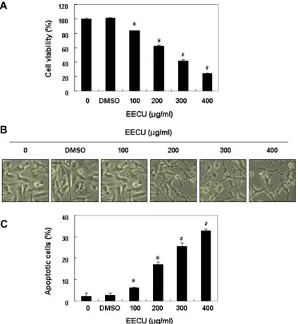
Fig. 1. Inhibition of cell viability and induction of apoptosis by EECU in MDA-MB-231 cells.
MDA-MB-231 was treated with various con- centrations of EECU for 24 hr. (A) The cell viability was measured by MTT assay. (B) The morphological changes of MDA-MB-231 cells treated with EECU in various concen- trations were observed under an inverted mi- croscope (magnification, x200). (C) The per- centages of Annexin V-FITC positive cells cultured under the same conditions were in- dicated. The data were expressed as the mean
± SD of three independent experiments (*p<0.05, #p<0.01 vs. untreated control).
Student t-test method (p<0.05 or p<0.01).
Results
Inhibition of cell viability and induction of apopto- sis by EECU in MDA-MB-231 cells
The effect of EECU on MDA-MB-231 cell viability was analyzed by MTT assay. Fig. 1A shows that EECU signifi- cantly decreased the MDA-MB-231 cell survival rate in a concentration-dependent manner, and was accompanied by various morphological changes, including membrane bleb- bing, diminished cell density, and an increased number of floating cells (Fig. 1B). To determine whether EECU treat- ment led to growth inhibition due to apoptosis induction, the apoptosis rate was measured by a flow cytometer. The results showed that compared with the untreated control group, EECU markedly enhanced the percentage of apop- totic cells (Fig. 1C), indicating that EECU suppressed cell viability by inducing apoptosis in MDA-MB-231 cells.
Activation of caspases and inhibition of expression of IAP family proteins by EECU in MDA-MB-231 cells
Because the activation of caspase cascades plays a key role
in two representative apoptosis-inducing pathways [5, 23], we next investigated whether caspase activation is involved in the induction of apoptosis by EECU in MDA-MB-231 cells.
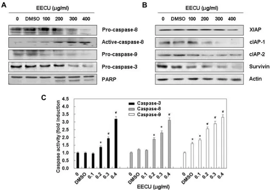
Our immunoblotting results showed that expression of pro-caspase-8, an initiator caspase of the extrinsic apoptosis pathway, was apparently decreased with increasing EECU concentration, while expression of active-caspase-8 was in- creased. Although no expression was observed of the active- caspase-9 or active-caspase-3, an initiator caspase of the ex- trinsic apoptosis pathway and a typical effector caspase, re- spectively, the expression of their pro-forms was suppressed, depending on the EECU treatment concentration (Fig. 2A).
The expression of inhibitor of apoptosis proteins (IAP) fam-
ily members, which play a role in inhibiting caspase activity
[6, 22], was also decreased by treatment with EECU in a
concentration-dependent manner (Fig. 2B). A subsequent in-
crease was also observed in the degradation of poly(ADP-ri-
bose) polymerase (PARP), which is a representative sub-
strate protein of activated caspase-3. Consistent with Western
blot analysis results, the in vitro activity of the three inves-
tigated caspases was significantly enhanced by EECU treat-
ment (Fig. 2C), indicating that both pathways are activated
during the induction of apoptosis by EECU.
A B
C
Fig. 2. Activation of caspases and inhibition of IAP family proteins expression by EECU in MDA-MB-231 cells. MDA-MB-231 were treated with the indicated concentrations of EECU for 24 hr. (A and B) The cell lysates were prepared, and equal amounts of cellular proteins were separated on SDS-polyacrylamide gels, and transferred to PVDF membranes. The membranes were probed with the indicated antibodies, and the proteins were visualized using an ECL detection system. Actin was used as an internal control. (C) The activities of caspases were evaluated using caspases colorimetric assay kits. The data were expressed as the mean ± SD of three independent experiments (*p<0.05, #p<0.01 vs. untreated control).
Modulation of DR-related and Bcl-2 family proteins expression by EECU in MDA-MB-231 cells
The results of Fig. 2 indicated that the possibility that two apoptotic pathways were activated in the induction of apop- tosis by EECU, so we next investigated the effect of EECU on the expression of DR-related and Bcl-2 family proteins.
The immunoblotting data indicated that the expressions of Fas, Fas-associated protein with death domain (FADD), DR4, DR5, and TNF-related apoptosis-inducing ligand (TRAIL) were concentration-dependently increased by EECU treat- ment, even though the expression of Fas ligand (FasL) was not changed (Fig. 3), suggesting that EECU might regulate the extrinsic pathway. Among the Bcl-2 family proteins, an- ti-apoptotic Bcl-2 expression was remarkably reduced by EECU treatment, but the expression of pro-apoptotic Bax was increased to some extent. In addition, total Bid ex- pression was decreased by EECU treatment, but truncated Bid (tBid) expression was progressively increased depending on EECU treatment concentration, presumably resulting from truncation by activated caspase-8.
Fig. 3. Effects of EECU on the levels of DR-related and Bcl-2 family proteins in MDA-MB-231 cells. After 24 hr in- cubation with the indicated concentrations of EECU, the cells were lysed, and cellular proteins were separated by SDS-polyacrylamide gel electrophoresis, and trans- ferred to membranes. The membranes were probed with the indicated antibodies. Proteins were visualized using an ECL detection system. Equal protein loading was confirmed by the analysis of actin in the protein extracts.
A
B C
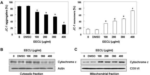
Fig. 4. Effects of EECU on the levels of MMP values and cytochrome c expression in MDA-MB-231 cells. (A) After 24 hr incubation with the indicated concentrations of EECU, the cells were stained with JC-1 dye, and were then analyzed on a flow cytometer, in order to evaluate the changes in MMP. The data were expressed as the mean ± SD of three independent experiments (*p<0.05, #p<0.01 vs. untreated control). (B and C) Cells cultured under the same conditions were lysed, and cytosolic and mitochondrial proteins were separated by SDS polyacrylamide gel electrophoresis, and transferred to membranes. The mem- branes were probed with anti-cytochrome c antibody. Proteins were visualized using an ECL detection system. Equal protein loading was confirmed by the analysis of actin and cytochrome oxidase subunit VI (COX VI) in each protein extract.
Loss of MMP and release of cytochrome c to cytosol by EECU in MDA-MB-231 cells
Recent accumulation studies have shown that the loss of MMP associated with the cytosolic release of cytochrome c is a hallmark for the activation of intrinsic apoptosis path- way [10, 35], we therefore investigated whether these phe- nomena were involved in the EECU-induced apoptosis in MDA-MB-231 cells. Our flow cytometry analysis revealed that EECU markedly destroys the integrity of the mitochon- dria measured by concentration-dependent loss of MMP (Fig. 4A). Subsequently, the release of cytochrome c from the mitochondria into the cytosol was markedly enhanced in a concentration-dependent manner (Fig. 4B, Fig. 4C), in- dicating that mitochondrial dysfunction may also contribute to EECU-induced apoptosis in MDA-MB-231 cells.
The role of AMPK in EECU-induced apoptosis in MDA-MB-231 cells
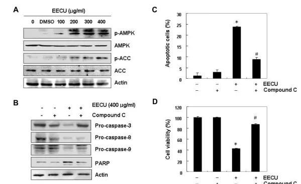
Because recent accumulation studies have shown that AMPK activation under stress conditions promotes apopto- sis and the growth inhibition of cancer cells [12, 30], we next examined whether EECU activates AMPK, which is reflected by increased phosphorylation of AMPKα and its down- stream target kinase acetyl-CoA carboxylase (ACC), using phosphorylation-specific antibodies. Our immunoblotting
results demonstrated that EECU remarkably enhanced the phosphorylation of AMPKα (Thr 172), as well as ACC (Ser 79), with increasing EECU concentration (Fig. 5A), indicating that they were converted to the activated state. To address whether AMPK activation is a key pathway for EECU-in- duced MDA-MB-231 cell apoptosis, the effects of EECU on the expression of caspases and PARP after pretreatment of an inhibitor of AMPK, compound C, were investigated. As shown in Fig. 6B, the EECU-induced down-regulation of cas- pase-3, -8 and -9, and degradation of PARP were partially prevented in the presence of compound C (Fig. 5B), implying a linkage between caspase and AMPK activation. In addi- tion, the increased apoptotic cell death was attenuated in the presence of compound C (Fig. 5C), and the suppression of cell viability was also significantly abrogated in EECU- treated cells (Fig. 5D). These data indicate that activation of AMPK is important for EECU-induced cytotoxicity in MDA-MB-231 cells.
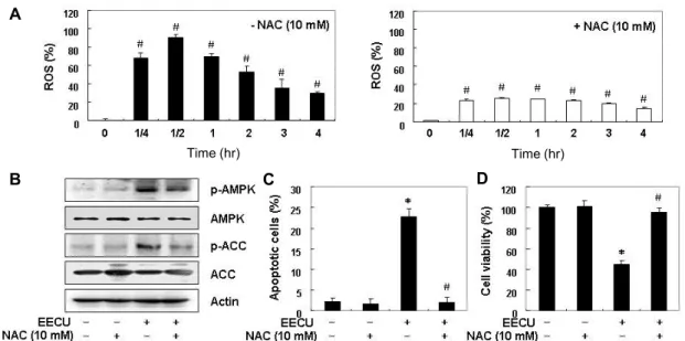
Induction of apoptosis by EECU through activation of ROS-dependent AMPK in MDA-MB-231 cells
Accumulated evidence has demonstrated that ROS, which
are mainly produced by the mitochondria during the ex-
ecution phase of apoptosis, are a known activator of AMPK
[12, 19]. Therefore, we further wanted to determine whether
A
B
C
D
Fig. 5. Involvement of AMPK activation in EECU-induced apoptosis in MDA-MB-231 cells. MDA-MB-231 were (A) treated with different concentrations of EECU for 24 hr, or (B)–(D) pre-treated with 10 μM compound C, an AMPK inhibitor, for 1 hr, and then treated with 400 μg/m EECU for 24 hr. (A and B) Equal amounts of cell lysate were resolved by SDS-poly- acrylamide gels, transferred to membranes, and probed with the indicated antibodies. The proteins were visualized using an ECL detection system. Actin was used as an internal control. (C) The percentages of apoptotic cells were measured using flow cytometric analysis. (D) The cell viability was measured by MTT assay. The data are expressed as the mean ± SD of three independent experiments (*p<0.05 vs. untreated control; #p<0.05 vs. EECU-treated cells).
the induction of apoptosis by EECU associated with AMPK activation is a ROS-dependent signaling pathway. To de- termine whether ROS accumulates in cells by EECU, the in- tracellular ROS levels were measured using DCF-DA probe.
As shown in Fig. 6A, the results of flow cytometry indicated that ROS accumulation levels increased within 25 min after EECU treatment, and the levels of ROS generation were pro- gressively reduced relative to untreated cells. However, when the generation of ROS was blocked by NAC, a ROS scavenger, the increase of ROS contents by EECU was great- ly reduced.
Next, we attempted to determine whether the activation of AMPK is associated with excessive ROS generation, and ROS generation plays a critical role for the induction of apoptosis by EECU. Fig. 6B demonstrates that EECU-in- duced phosphorylation of ACC as well as AMPK was great- ly attenuated in the presence of NAC. Furthermore, the EECU-induced apoptotic cell death and reduction in cell via- bility were suppressed when ROS production was artificially blocked (Fig. 6C, Fig. 6C). Taken together, these findings suggest that EECU increases ROS generation, which is re- quired for AMPK activation, and then induces apoptosis in
MDA- MB-231 cells.
Discussion
Recently, much attention has focused on the search for anti-cancer active substances that have been used for a long time to treat various diseases. In the present study, we inves- tigated the anti-cancer activity of EECU, ethanol extract of
C. unshiu peel, and showed that EECU could inhibit the pro-liferation of MDA-MB-231 adenocarcinoma cells, and induce apoptosis. The principal finding of this study is that both extrinsic and intrinsic apoptosis pathways can be activated to induce apoptosis of MDA-MB-231 cells by EECU (Fig. 7).
It also demonstrated that EECU activates AMPK in a ROS-dependent manner, and contributes to EECU-mediated suppression of MDA-MB-231 cell viability.
Of the two typical apoptotic pathways, the DR-mediated
extrinsic pathway is activated by binding of the cell-surface
DRs of the death ligands to the activation of caspase-8 after
the recruitment of the adapter molecules containing FADD,
which in turn leads to the activation of caspase-3 and cleav-
age of death substrates [11, 14]. On the other hand, the in-
A
B C D
Time (hr) Time (hr)
Fig. 6. ROS-dependent activation of AMPK by EECU in MDA-MB-231 cells. (A) MDA-MB-231 cells were either treated with 400 μg/m EECU for the indicated times, or pre-treated with NAC (10 mM) for 1 hr before EECU treatment, and then collected.
The medium was discarded, and the cells were incubated at 37℃ in the dark for 20 min with new culture medium containing 10 μM DCF-DA. ROS generation was measured by a flow cytometer. The data are the means of the two different experiments.
(B) The cells were pre-treated with 10 mM NAC for 1 hr, before 400 μg/m EECU treatment. After 24 hr incubation, the cellular proteins were separated by SDS-polyacrylamide gel electrophoresis, and transferred to membranes. The membranes were probed with the indicated antibodies, and the proteins were visualized using an ECL detection system. Actin was used as an internal control. (C and D) MDA-MB-231 cells were pre-treated with 10 mM NAC for 1 hr, before 400 μg/m EECU treatment for 24 hr. (C) The percentage of apoptotic cells was analyzed by flow cytometer. (D) Cell viability was determined by MTT assay. Each point represents the mean ± SD of three independent experiments (*p<0.05 vs. untreated control; #p<0.05 vs. EECU-treated cells).
Fig. 7. A suggested schematic model for EECU-induced apopto- sis in MDA-MB-231 cells.
ternal pathway is initiated by the activation of caspase-9 with the release of pro-apoptotic factors such as cytochrome
c from the mitochondria to the cytoplasm following loss ofmitochondrial membrane integrity [5, 10]. The insertion of mitochondrial membrane and the oligomerization of Bax, a pro-apoptotic protein belonging to the Bcl-2 family, are re-
quired for the release of cytochrome c. Therefore, increased
ex-
pression of Bax plays a key role in the activation of the in-
trinsic pathway, and Bcl-2 is a typical anti-apoptotic protein
that inhibits this process [10, 35]. In addition, the truncation
of Bid, a pro-apoptotic BH3-interacting domain death ago-
nist, by activated caspase-8 results in activation of caspase
cascade by caspase-9 and caspase-3, following initiation of
cytosolic release of cytochrome c, which means that Bid acts
as a linker molecule that connects the DR and the mitochon-
dria-dependent pathways [1, 13]. In the present study, the
activation of caspases (-8, -9 and -3) by EECU clearly demon-
strated its apoptotic effects, which was associated with the
degradation of PARP, a marker for apoptotic cells and sub-
strate of activated caspase that triggers cellular disassembly
and viability reduction [4, 9]. EECU also down- regulated
the IAP family proteins, which selectively bind to caspases
and block apoptosis, due to their ability to act directly as
inhibitors [6, 22]. With the activation of caspases, EECU also
increased the expression of FADD, as well as the expression
of DR-related proteins, except FasL, suggesting that extrinsic
pathway may be involved in apoptosis by EECU in
MDA-MB-231 cells. Current data also clearly revealed that EECU treatment significantly promotes the ratio of Bax/Bcl-2 expression, followed by a loss of MMP associated with increased release of cytochrome c and cleavage of Bid.
These results suggest that the intrinsic pathway is also in- volved in the induction of EECU-induced apoptosis in MDA-MB-231 cells, and that the extrinsic pathway even- tually amplifies the intrinsic pathway through caspase-8- mediated truncation of Bid.
Recent studies have shown that the activation of AMPK is directly related to the apoptosis induced by various stim- uli, including chemotherapeutic drugs [7, 8]. Activation of AMPK requires phosphorylation by upstream AMPK kinase, and regulates the activity of various downstream targets that regulate cell fate [2, 3]. Therefore, the relation of AMPK path- way to EECU-induced apoptosis in MDA-MB-231 cells was investigated, and it was found that EECU promotes phos- phorylation of AMPK and its downstream target ACC.
However, blockade of AMPK activation by compound C, an AMPK inhibitor, significantly prevented EECU-induced apoptosis and decreased cell viability, which was accom- panied by the reduction of caspase activation and PARP degradation. These data indicate that AMPK activation is involved in EECU-mediated apoptosis in MDA-MB-231 cells, and that AMPK is likely to act upstream of caspase activa- tion in the signaling pathways involved in apoptosis by EECU.
Since mitochondrial electron transport chains are a major source of ATP production, mitochondrial dysfunction pro- motes the reduction of ATP synthesis, and leads to activation of AMPK, so that the activity of AMPK is dependent on mitochondrial function [12, 30]. In addition, the loss of MMP in the apoptosis induction process of cancer cells by various substances having anti-cancer activity is directly related to the increase of ROS generation, and, as is well known in many previous studies, ROS acts as a powerful AMPK acti- vator [12, 19]. We therefore investigated the possible rele- vance of ROS production in EECU-induced AMPK activa- tion. As noted in the results, the accumulation of ROS by treatment with EECU disappeared in cultured MDA-MB-231 cells in medium containing anti-oxidant NAC, and markedly attenuated EUCC-induced phosphorylation of AMPK and ACC. Moreover, EECU-induced apoptosis was effectively prevented by blocking ROS production, and the reduced cell viability was also significantly restored to the control level.
These results indicate that the activation of ROS-dependent
AMPK in MDA-MB-231 cells is a major mediator of apopto- sis, and that ATP depletion due to mitochondrial damage caused by EECU probably induced AMPK activity.
The extracts of C. unshiu Peel are composed of various phytochemicals such as polyphenols, terpenoids and fla- vonoids, and their anticancer potentials are well known [15, 17]. However, since these compounds are not present only in C. unshiu Peel, the apoptotic effect of WECU in MDA- MB-231 cells seems to be due to the overall effect of various substances contained in WECU. Therefore, further inves- tigation of the relevance of other intracellular signaling path- ways and identification of key active compounds in the WECU should be undertaken in the future.
In conclusion, the present results demonstrate that EECU induces apoptosis in MDA-MB-231 adenocarcinoma cells through intrinsic and extrinsic pathways by increasing DR- related regulators, MMP loss, the cytosolic release of cyto- chrome c, and ROS generation, combined with an increase in the Bax/Bcl-2 ratio and Bid truncation. EECU also pro- moted AMPK activation, and the inhibition of AMPK activ- ity blocked EECU-induced activation of caspase, and pre- vented the induction of apoptosis. Consequently, when ROS production was blocked, EECU-induced activation of AMPK was inhibited, and the survival rate of MDA-MB-231 ad- enocarcinoma cells was improved, indicating ROS is a poten- tial upstream molecule for EECU-induced AMPK activation and its cytotoxic effects.
Acknowledgement
This research was supported by Basic Science Research Program through the National Research Foundation of Korea (NRF) grant funded by the Korea government (2018R1A2B 2005705).
References
1. Billen, L. P., Shamas-Din, A. and Andrews, D. W. 2008. Bid:
a Bax-like BH3 protein. Oncogene 27, S93-104.
2. Carling, D., Sanders, M. J. and Woods, A. 2008. The regu- lation of AMP-activated protein kinase by upstream kinases.
Int. J. Obes. (Lond) 32, S55-S59.
3. Crute, B. E., Seefeld, K., Gamble, J., Kemp, B. E. and Witters, L. A. 1998. Functional domains of the alpha1 catalytic sub- unit of the AMP-activated protein kinase. J. Biol. Chem. 273, 35347-35354.
4. Decker, P. and Muller, S. 2002. Modulating poly (ADP-ri- bose) polymerase activity: potential for the prevention and
therapy of pathogenic situations involving DNA damage and oxidative stress. Curr. Pharm. Biotechnol. 3, 275-283.
5. Fulda, S. and Debatin, K. M. 2006. Extrinsic versus intrinsic apoptosis pathways in anticancer chemotherapy. Oncogene 25, 4798-4811.
6. Fulda S. and Vucic, D. 2012. Targeting IAP proteins for ther- apeutic intervention in cancer. Nat. Rev. Drug Discov. 11, 109-124.
7. Garcia-Gil, M., Pesi, R., Perna, S., Allegrini, S., Giannecchini, M., Camici, M. and Tozzi, M. G. 2003. 5'-aminoimidazole- 4-carboxamide riboside induces apoptosis in human neuro- blastoma cells. Neuroscience 117, 811-820.
8. Hadad, S. M., Baker, L., Quinlan, P. R., Robertson, K. E., Bray, S. E., Thomson, G., Kellock, D., Jordan, L. B., Purdie, C. A., Hardie, D. G., Fleming, S. and Thompson, A. M. 2009.
Histological evaluation of AMPK signalling in primary breast cancer. BMC Cancer 9, 307.
9. Hajra, K. M. and Liu, J. R. 2004. Apoptosome dysfunction in human cancer. Apoptosis 9, 691-704.
10. Hata, A. N., Engelman, J. A. and Faber, A. C. 2015. The BCL2 family: Key mediators of the apoptotic response to targeted anticancer therapeutics. Cancer Discov. 5, 475-487.
11. Hengartner, M. O. 2000. The biochemistry of apoptosis.
Nature 407, 770-776.
12. Kaminskyy, V. O. and Zhivotovsky, B. 2014. Free radicals in cross talk between autophagy and apoptosis. Antioxid.
Redox. Signal. 21, 86-102.
13. Kantari, C. and Walczak, H. 2011. Caspase-8 and bid: caught in the act between death receptors and mitochondria.
Biochim. Biophys. Acta. 1813, 558-563.
14. Kaufmann, T., Strasser, A. and Jost, P. J. 2012. Fas death receptor signalling: roles of Bid and XIAP. Cell Death Differ.
19, 42-50.
15. Kim, A., Im, M., Gu, M. J. and Ma, J. Y. 2016. Citrus unshiu peel extract alleviates cancer-induced weight loss in mice bearing CT-26 adenocarcinoma. Sci. Rep. 6, 24214.
16. Kim, J., Yang, G., Kim, Y., Kim, J. and Ha, J. 2016. AMPK activators: mechanisms of action and physiological activities.
Exp. Mol. Med. 48, e224.
17. Lee, S., Ra, J., Song, J. Y., Gwak, C., Kwon, H. J., Yim, S.
V., Hong, S. P., Kim, J., Lee, K. H., Cho, J. J., Park, Y. S., Park, C. S. and Ahn, H. J. 2011. Extracts from Citrus unshiu promote immune-mediated inhibition of tumor growth in a murine renal cell carcinoma model. J. Ethnopharmacol. 133, 973-979.
18. Li, C., Yang, L., Zhang, D. and Jiang, W. 2016. Systematic review and meta-analysis suggest that dietary cholesterol intake increases risk of breast cancer. Nutr. Res. 36, 627-635.
19. Liangpunsakul, S., Wou, S. E., Zeng, Y., Ross, R. A., Jayaram, H. N. and Crabb, D. W. 2008. Effect of ethanol on hydrogen peroxide-induced AMPK phosphorylation. Am. J. Physiol.
Gastrointest. Liver Physiol. 295, G1173-1181.
20. Min, K. Y., Kim, H. J., Lee, K. A., Kim, K. T. and Paik, H.
D. 2014. Antimicrobial activity of acid-hydrolyzed Citrus un- shiu peel extract in milk. J. Dairy Sci. 97, 1955-1960.
21. Ming-Hua, C., Bao-Hua, Z. and Lei, Y. 2016. Mechanisms of anorexia cancer cachexia syndrome and potential benefits of traditional medicine and natural herbs. Curr. Pharm.
Biotechnol. 17, 1147-1152.
22. Nachmias, B., Ashhab, Y. and Ben-Yehuda, D. 2004. The in- hibitor of apoptosis protein family (IAPs): an emerging ther- apeutic target in cancer. Semin. Cancer Biol. 14, 231-243.
23. Nakajima, Y. I. and Kuranaga, E. 2017. Caspase-dependent non-apoptotic processes in development. Cell Death Differ.
24, 1422-1430.
24. Niraula, S. and Ocana, A. 2016. Mechanism of drug resist- ance in relation to site of metastasis: Meta-analyses of randomized controlled trials in advanced breast cancer ac- cording to anticancer strategy. Cancer Treat. Rev. 50, 168-174.
25. Oh, Y. C., Cho, W. K., Jeong, Y. H., Im, G. Y., Yang, M.
C., Hwang, Y. H. and Ma, J. Y. 2012. Anti-inflammatory effect of Citrus unshiu peel in LPS-stimulated RAW 264.7 macrophage cells. Am. J. Chin. Med. 40, 611-629.
26. Omura, M. and Shimada, T. 2016. Citrus breeding, genetics and genomics in Japan. Breed Sci. 66, 3-17.
27. Ota, S., Horigome, K., Ishii, T., Nakai, M., Hayashi, K., Kawamura, T., Kishino, A., Taiji, M. and Kimura, T. 2009.
Metformin suppresses glucose-6-phosphatase expression by a complex I inhibition and AMPK activation-independent mechanism. Biochem. Biophys. Res. Commun. 388, 311-316.
28. Ouyang, L., Shi, Z., Zhao, S., Wang, F. T., Zhou, T. T., Liu, B. and Bao, J. K. 2012. Programmed cell death pathways in cancer: a review of apoptosis, autophagy and pro- grammed necrosis. Cell Prolif. 45, 487-498.
29. Park, H. J., Jung, U. J., Cho, S. J., Jung, H. K., Shim, S. and Choi, M. S. 2013. Citrus unshiu peel extract ameliorates hy- perglycemia and hepatic steatosis by altering inflammation and hepatic glucose- and lipid-regulating enzymes in db/db mice. J. Nutr. Biochem. 24, 4194-4127.
30. Shackelford, D. B. and Shaw, R. J. 2009. The LKB1-AMPK pathway: metabolism and growth control in tumour sup- pression. Nat. Rev. Cancer 9, 563-575.
31. Siegel, R. L., Miller, K. D. and Jemal, A. 2016. Cancer statistics. 2016. CA Cancer J. Clin. 66, 7-30.
32. Su, Z., Yang, Z., Xu, Y., Chen, Y. and Yu, Q. 2015. Apoptosis, autophagy, necroptosis, and cancer metastasis. Mol. Cancer 14, 48.
33. Suzuki, M., Sasaki, K., Yoshizaki, F., Oguchi, K., Fujisawa, M. and Cyong, J. C. 2005. Anti-hepatitis C virus effect of Citrus unshiu peel and its active ingredient nobiletin. Am.
J. Chin. Med. 33, 87-94.
34. Tanaka, T., Yasui, Y., Ishigamori-Suzuki, R. and Oyama, T.
2008. Citrus compounds inhibit inflammation- and obesity- related colon carcinogenesis in mice. Nutr. Cancer 60, S70-80 35. Tummers, B. and Green, D. R. 2017. Caspase-8: regulating
life and death. Immunol. Rev. 277, 76-89.
36. Wang, C. Y., Bai, X. Y. and Wang, C. H. 2014. Traditional Chinese medicine: a treasured natural resource of anticancer drug research and development. Am. J. Chin. Med. 42, 543- 559.
초록:진피 추출물에 의한 인간유방암 MDA-MB-231 세포의 apoptosis 유도에서 ROS 및 AMPK의 역할
김민영
1,2․황보현
1,2․지선영
1,2․홍수현
1,2․최성현
3․김성옥
4․박철
5․최영현
1,2*
(1동의대학교 한의과대학 생화학교실, 2동의대학교 항노화연구소, 3한국승강기대학교 승강기시스템관리과, 4경성
대학교 공과대학 식품응용공학부, 5동의대학교 자연과학대학 분자생물학과)