1. Introduction
Ionizing radiation is one of the most commonly used treatments for a wide variety of tumors. Exposure of cells to ionizing radiation results in the simultaneous activation or down regulation of multiple signaling pathways, which play critical role in controlling cell death and cell survival after irradiation in a cell type specific manner. The molecular mechanism by which apoptotic cell death occurs in response to ionizing radiation has been widely explored but not precisely deciphered. Therefore an improved understanding of the mechanisms involved in radiation-induced apoptosis may ultimately provide novel strategies of intervention in specific signal transduction pathways to favorably alter the therapeutic ratio in the treatment of human malignancies.
The aim of our investigation was to elucidate molecular mechanisms of the mitochondrial dysfunction-mediated apoptotic cell death triggered by ionizing radiation in human cervical cancer cells. We demonstrated that ionizing radiation utilizes PI3K-JNK signaling pathway for amplifying mitochondrial dysfunction and susequent apoptotic cell death: We showed that PI3K-dependent JNK activation leads to transcriptional upregulation of Fas and the phosphorylation/inactivation of Bcl-2, resulting in mitochondrial dysfunction-mediated apoptotic cell death in response to ionizing radiation.
2. Methods
Radiation-induced apoptotic cell death was determined by flow cytometric analysis. Involvement of the mitochondrial pathway in radiation-induced cell death was examined by monitoring of the mitochondria membrane potential, cytochrome c release, Bax translocation, and Bcl-2 phosphorylation. Subcellular redistributions of apoptosis inducing factor (AIF) were detected using Western blot analysis after subcellular fractionation and confocal microscopic analysis. Phosphorylation of Bcl-2 by JNK after irradiation was determined by immune complex kinase assay.
3. Results and Conclusion
Ionizing radiation caused loss of mitochondrial membrane potential, release of cytochrome c and AIF from mitochondria, and apoptotic cell death in human
cervical cancer cells. Fas (APO1/CD95) expression, caspase-8 activation, Bid cleavage, and Bax and Bak activations were also observed in response to radiation (Figure 1).
Figure 1. Ionizing radiation induces mitochondrial dysfunction-mediated apoptotic cell death through apoptotic conformational changes of Bax and Bak in human cervical cancer cells.
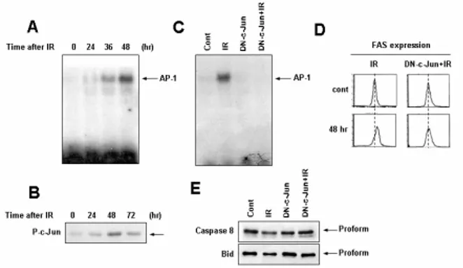
Constitutively expressed endogenous c-Jun was found to be phosphorylated after irradiation, resulting in increased activator protein 1 (AP-1) DNA binding activity. Overxpression of dominant negative forms of c-Jun inhibited radiation-induced Fas expression, caspase-8 activation, Bid cleavage, and mitochondrial membrane potential loss (Figure 2).
Figure 2. Radiation-induced transcriptional upregulation of Fas is dependent on AP-1 activation.
Furthermore, radiation also caused phosphorylation of
Ionizing radiation induces PI3K-dependent JNK activation for amplifying mitochondrial
dysfunction in human cervical cancer cells*
Min-Jung Kim1, Soon-Young Choi1, Sangwoo Bae2, Chang-Mo Kang3, Yun-Sil Lee2, and Su-Jae Lee1 1Laboratory of Radiation Experimental Therapeutics, 2Laboratory of Radiation Effect, Laboratory of Radiation
Cytogenetics & Epidemiology, Korea Institute of Radiological & Medical Sciences, Seoul 139-706, Korea
Transactions of the Korean Nuclear Society Autumn Meeting Busan, Korea, October 27-28, 2005
serine 70 of Bcl-2. Overexpression of serine 70 specific-mutant forms of Bcl-2 (S70A) significantly inhibited radiation-induced mitochondrial membrane potential loss and apoptotic cell death (Figure 3).
Figure 3. Ionizing radiation induces phosphorylation of Bcl-2 on serine 70.
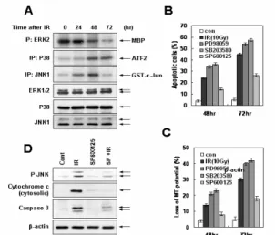
On the other hand, exposure of cells to radiation markedly induced JNK and p38MAPK activation (Figure 4). Inhibition of JNK effectively blocked radiation-induced mitochondrial membrane potential loss, cytochrome c release and apoptotic cell death. However, inhibition of p38MAPK did not attenuate radiation-induced mitochondrial dysfunction-mediated apoptotic cell death.
Figure 4. JNK activation is required for mitochondrial dysfunction-mediated apoptotic cell death in response to ionizing radiation exposure.
We also found that inhibition of JNK attenuated radiation-induced Bcl-2 phosphorylation as well as Fas expression (Figure 5). Moreover, inhibition of JNK blocked radiation-induced FADD-caspase-8 interaction and Bid activation. In addition, JNK directly phosphorylated serine 70 residue of Bcl-2 in response to
radiation.
Figure 5. JNK plays a critical role in Fas expression-mediated Bax and Bak activations and Bcl-2 phosphorylation in response to radiation treatment.
Phosphatidyl inositol-3 kinase (PI3K) was found to be activated in response to radiation. Interestingly, inhibition of PI3K attenuated radiation-induced JNK activation, Fas expression, Bcl-2 phosphorylation, and mitochondrial dysfunction-mediated apoptotic cell death (Figure 6).
Figure 6. The PI3K activation is critical for the activation of JNK and subsequent cell death machinery.
Taken together, we demonstrated in the present study that ionizing radiation can utilize PI3K-JNK signaling pathway for amplifying mitochondrial dysfunction-mediated apoptotic cell death in human cervical cancer cells. We showed that mitochondrial dysfunction in response to radiation is induced by activations of Bax and Bak initiated by Fas upregulation, and by phosphorylation/inactivation of Bcl-2, in a JNK dependent manner. An improved understanding of the mechanisms involved in radiation-induced apoptosis may ultimately provide novel strategies of intervention in
specific signal transduction pathways to favorably alter the therapeutic ratio in the treatment of human malignancies.
4. Acknowledgements
*This work was supported by Korea Science and Engineering Foundation (KOSEF) and Ministry of Science and Technology (MOST), Korean government, through its National Nuclear Technology Program.
5. References
1. Willers H, Dahm-Daphi J, Powell SN. Repair of radiation damage to DNA. Br J Cancer. 2004;90: 297-1301.
2. Wheeler JA, Stephens LC, Tornos C, et al. ASTRO Research Fellowship: apoptosis as a predictor of tumor response to radiation in stage IB cervical carcinoma. American Society for Therapeutic Radiology and Oncology. Int J Radiat Oncol Biol Phys. 1995;32:1487–1493.
3. Coleman CN, Turrisi AT. Radiation and chemotherapy sensitizers and protectors. Crit Rev Oncol Hematol. 1990;10:225-252.
4. Green DR, Reed JC. Mitochondria and apoptosis. Science. 1998;281:1309–1312.
5. Crompton M. The mitochondrial permeability transition pore and its role in cell death. Biochem J. 1999;341:233-249.
6. Gottlieb RA. Mitochondria: execution central. FEBS Lett. 2000;482:6-12.
7. Li P, Nijhawan D, Budihardjo I, Srinivasula SM, et al. Cytochrome c and dATP-dependent formation of Apaf-1/caspase-9 complex initiates an apoptotic protease cascade. Cell. 1997;91:479-489.
8. Susin SA, Lorenzo HK, Zamzami N, et al. Molecular characterization of mitochondrial apoptosis-inducing factor. Nature. 1999; 397:441-446.
9. Susin SA, Lorenzo HK, Zamzami N, et al. Mitochondrial release of caspase-2 and -9 during the apoptotic process. J Exp Med. 1999;189:381-394. 10. Sakahira H, Enari M, Nagata S. Cleavage of CAD
inhibitor in CAD activation and DNA degradation during apoptosis. Nature. 1998;391:96-99.
11. Ferri KF, Jacotot E, Blanco J, et al. Apoptosis control in syncytia induced by the HIV type 1-envelope glycoprotein complex: role of mitochondria and caspases. J Exp Med. 2000;192:1081-1092.
12. Schulz JB, Bremen D, Reed JC, et al. Cooperative interception of neuronal apoptosis by BCL-2 and BAG-1 expression: prevention of caspase activation and reduced production of reactive oxygen species. J Neurochem. 1997;69:2075-2086.