Effect of Increase in Occlusal Vertical Dimension on Appendage Muscle Strength
Su-Jin Ahna, DMD, MSD, PhD, Richard Sung-bok Leeb, DMD, MSD, PhD, Suk-Won Leec, DMD, MSD, PhD
aClinical Assistant Professor, Department of Biomaterials & Prosthodontics, East West Neo Medical Center, School of Dentistry Kyung Hee University, Seoul, Korea
bProfessor & Chairman, Department of Biomaterials & Prosthodontics, East West Neo Medical Center, School of Dentistry Kyung Hee University, Seoul, Korea
cAssistant Professor, Department of Biomaterials & Prosthodontics, East West Neo Medical Center, School of Dentistry Kyung Hee University, Seoul, Korea
Objective. This study was conducted to observe the effect on appendage muscle strength according to increase in occlusal vertical dimension.
Materials and methods. Ten males with a mean age of 21 were selected. The tested occlusal splints were made at the position of increased occlusal vertical dimension of 2mm,3.5mm and 5mm from the intercuspal position. Before and after wearing occlusal splints, the appendage muscle strength were tested by Cybex II dynamometer (Lumex Inc., Ronkonkoma, NY, USA).
Results. Statistical analysis using the paired t-test revealed significant differences for flexion and extension of the hip, pronation of the forearm, internal rotation of the shoulder, external and internal rotation of the knee, and dorsiflexion and plantarflexion of the ankle (p<0.05).
Conclusions. As the result of this study, we conclude that when occlusal vertical dimension was increased, most of mean muscular strength values were increased. Especially at the position of 3.5mm increased vertical dimension displayed the highest mean muscular strength value than other positions.
Key words: appendage muscle strength, occlusal splint, occlusal vertical dimension, sports dentistry, temporomandibular joint (Journal of Dental Rehabilitation and Applied Science 2010:26(2):169~178)
Correspondence to : Dr. Richard Sung-bok, Lee
Richard Leesungbok, Professor & Chairman, Department of Biomaterials & Prosthodontics, East West Neo Medical Center, School of Dentistry Kyung Hee University, Sangil-Dong 149, Kangdong-Gu, Seoul134-727, Korea.
Tel : 82-2-440-7500, 82-10-3237-2869, Fax : 82-2-440-7549, E-mail: [email protected] Received: March 05, 2010, Last Revision: May 20, 2010, Accepted: June 25, 2010
INTRODUCTION
Sport is an important part of life not only for professional athletes, but also for ordinary people.
Sports dentistry is not a well-known field in Korea and many dentists are not yet paying enough attention to the area. In the United States, however, members of the American Dental Association (ADA) have actively participated in enhancing the oral health of athletes since the 1950s. Later, when mouth protectors and face guards were widely used to protect the maxillofacial areas from trauma during exercise, sports dentistry began to boom.1
Sports dentistry in developed countries has been making progress in two areas. One area involves protective appliances, in terms of whether mouthpieces and face guards are effective in protecting the maxillofacial and cranial areas where athletes are often injured during sports.2-9The other area concerns the relationship between oral function and overall athletic performance, as in a recent study to investigating the changes made in oral condition, such as changes in mandibular position and occlusion, affect performance capability.10,11
An occlusal splint is one of the most common appliances used to change the position of the mandible. Repositioning of the temporomandibular joint (TMJ) using an occlusal splint to adapt the location of the mandible is the most universal form of sports dentistry aimed at releasing stress, correcting body posture, and enhancing various physiological and functional exercises. Dentists who recommend the use of occlusal splints argue that there are complicated correlations between the TMJ and muscles of the maxillofacial area and overall body, stating that an ideal physiological condition can be acquired by correcting improper relationships between the maxilla and mandible.12-14
According to a study of the vertical dimension,
most participants can tolerate splints of <6 mm in thickness.15 Therefore, in this study the horizontal relation was chosen as the most stable centric relation, and distances of 2, 3.5 and 5 mm were chosen for the vertical relation of the mandible.
After splints were constructed accordingly and inserted into the subjects, the muscle strength for seven appendage parts was measured using a Cybex II Dynamometer. The aim of this study was to determine the amount of increase in occlusal vertical dimension that has the greatest effect on appendage muscle strength.
MARERIALS AND METHODS 1. Subjects
Ten healthy adult males aged between 20 and 22 years were selected from the Division of Dentistry of Kyung Hee University (Seoul, Korea). All participants gave informed consent after receiving a full explanation of the goal and structure of the study, which was approved by the Ethics Committee of Kyung Hee University. None of the participants had previously been treated for disorders of the TMJ, occlusion, or masticatory system. They had no missing teeth except for the third molar.
2. Measurement Equipment
A Cybex II dynamometer was used to record the appendage muscle strength of the subjects (Fig. 1).
This instrument has been used in many studies since Smith first used it in 1978 to measure muscle strength.10 The Cybex II dynamometer was manufactured for the treatment and evaluation of muscle strength around the shoulder, elbow, forearm, wrist, ankle, knee, and hip. It is an isokinetic muscle-loading device that determines torque as a
Fig. 1. Cybex II dynamometer (Lumex Inc., Ronkonkoma, NY, USA).
measure of the muscle capability of a subject against fixed speed and varying resistance. The speed can range from 5deg/s to 500deg/s and the manufacturer's recommendation of 60deg/s was used in the present study. Torque was measured in ft.lb or Nm.
3. Construction of Occlusal Splints Maxilla cover-type splints were constructed for this study. Muscle strength was measured with teeth together in an intercuspal position and with the increases of 2, 3.5 and 5 mm in the occlusal vertical dimension.
Alginate impressions of both the maxillary and mandibular dentitions of the subjects were taken to obtain stone casts. The maxillary casts were then mounted on a semi-adjustable articulator using a face bow transfer. Aluwax (Aluwax Dental Products co, Michigan, USA), a bite registration material, was used to record the centric relation occlusion in accordance with Dr. P.E. Dawson's bilateral manipulation method.16 Then the mandibular cast was mounted on the articulator.
Marks were made on attached gingivae around the
right canines and first premolars of the mounted maxillary and mandibular casts. From these marks, the distance between the maxilla and mandible in the centric occlusion position was measured, and then the incisal pin of the articulator was opened by the distance of 2, 3.5 and 5 mm.
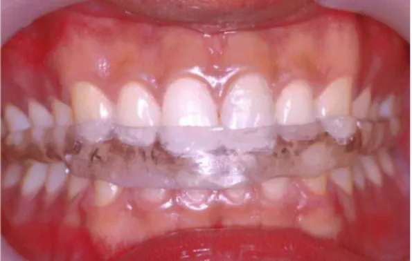
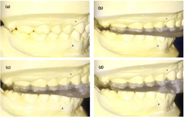
All teeth were in even contact in the centric occlusion position and the canine guidance for lateral movements and the anterior guidance for protrusive movements. In general, stabilization splints that are widely used for the treatment of temporomandibular disorders are not provided with the fossae formed by the occlusal surfaces of the opposing teeth. However, several fossae were reported to be necessarily made in the centric relation splint so that all cusp tips of opposing teeth can be evenly aligned on the splint.16,17 Flasked and cured in the usual manner, three maxilla cover-type splints yielding increases of 2, 3.5 and 5 mm in the occlusal vertical dimension were constructed for each subject. As for the stabilization splints, heat-curing clear acrylic resin was used. To maintain the occlusal vertical dimension, the splints were repositioned on the cast after the heat curing (Fig. 3). They were then adjusted on the articulator before being adapted and occlusally adjusted in the subject's mouth (Fig. 2).
Fig. 2. Splint placed in the upper dentition of the subject.
(a) (b)
(c) (d)
Fig. 3. Maxillary clear acrylic resin splint.
(a) Intercuspal position (b) 2mm increase of vertical dimension (c) 3.5mm increase of vertical dimension (d) 5mm increase of vertical dimension
4. Measuring Muscle Strength
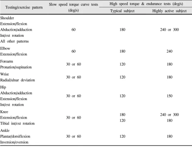
The muscle strength of four parts of the upper appendage (elbow, wrist, forearm, shoulder) and three parts of the lower appendage (hip, knee, ankle) was measured. Before measurement, the subject's weight in pounds and the target speed were input to the computer terminal attached to the Cybex II dynamometer (Table I). At high speed, the peak torque and work rate are low and the mean muscle strength is high. In contrast, the peak torque and work rate are high and the mean muscle strength is low at low speed. Thus, athletes with high muscle strength should be tested at high speed and non-athlete subjects should be tested at low speed so that the peak torque can be easily compared.
Preliminary tests revealed that a speed of 60deg/s was most appropriate for the present study.
On the first day, muscle strength was measured in the intercuspal position. For each body part, a maximum of five movement repetitions were performed, with a 3-min rest period after each. Since only one subject was tested on each day, all subjects had a 10-day rest after making these 14 movements.
The next step was to measure muscle strength after insertion of the splint yielding a 2-mm increase in occlusal vertical dimension. In this case, muscle strength was immediately measured, since warm-up tests revealed that muscle strength did not depend on the length of time for which the splint was worn.
The same procedure was repeated for the splints yielding increases of 3.5 and 5 mm in occlusal
Testing/exercise pattern Slow speed torque curve tests (deg/s)
High speed torque & endurance tests (deg/s) Typical subject Highly active subject Shoulder
Extension/flexion Abduction/adduction Int/ext rotation All other patterns
60 180 240 or 300
Elbow
Extension/flexion 60 180 240
Forearm
Pronation/supination 30 or 60 120 180
Wrist
Radial/ulnar deviation 30 or 60 120 180
Hip
Abduction/adduction Extension/flexion Int/ext rotation
30 or 60 120 150
Knee
Extension/flexion Tibial int/ext rotation
30 or 60 180
120
240 or 300 180 Ankle
Plantar/dorsiflexion Inversion/eversion
30 or 60 120 180
deg/s : degree/second
Table I. Suggested Cybex test speeds
vertical dimension. To prevent an increase in muscle strength due to repeated exercise, the intercuspal position was first measured as the control condition, and measurements for increases of 2, 3.5 and 5 mm in occlusal vertical dimension were then taken randomly.
5. Stastistical analysis
Data were statistically analyzed using the SPSS statistical softwere for windows (SPSS Japan Inc., Tokyo, Japan). Significance was determined with a
paired t-test and a value of p<0.05 was considered statistically significant.
RESULTS
Muscle strength for movements of the hip, elbow, wrist, forearm, shoulder, knee, and ankle was measured and compared with and without the insertion of splints to increase the occlusal vertical dimension by 2, 3.5 and 5 mm.
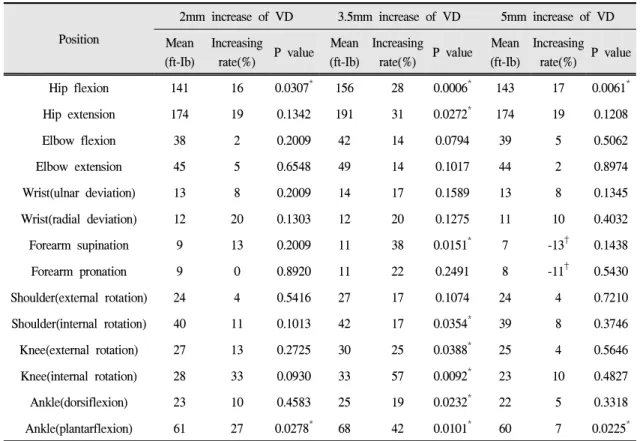
Table Ⅱ shows the muscle strength measured for each increase in occlusal vertical dimension. The
Position
2mm increase of VD 3.5mm increase of VD 5mm increase of VD Mean
(ft-Ib)
Increasing
rate(%) P value Mean (ft-Ib)
Increasing
rate(%) P value Mean (ft-Ib)
Increasing rate(%) P value
Hip flexion 141 16 0.0307* 156 28 0.0006* 143 17 0.0061*
Hip extension 174 19 0.1342 191 31 0.0272* 174 19 0.1208
Elbow flexion 38 2 0.2009 42 14 0.0794 39 5 0.5062
Elbow extension 45 5 0.6548 49 14 0.1017 44 2 0.8974
Wrist(ulnar deviation) 13 8 0.2009 14 17 0.1589 13 8 0.1345
Wrist(radial deviation) 12 20 0.1303 12 20 0.1275 11 10 0.4032
Forearm supination 9 13 0.2009 11 38 0.0151* 7 -13† 0.1438
Forearm pronation 9 0 0.8920 11 22 0.2491 8 -11† 0.5430
Shoulder(external rotation) 24 4 0.5416 27 17 0.1074 24 4 0.7210
Shoulder(internal rotation) 40 11 0.1013 42 17 0.0354* 39 8 0.3746
Knee(external rotation) 27 13 0.2725 30 25 0.0388* 25 4 0.5646
Knee(internal rotation) 28 33 0.0930 33 57 0.0092* 23 10 0.4827
Ankle(dorsiflexion) 23 10 0.4583 25 19 0.0232* 22 5 0.3318
Ankle(plantarflexion) 61 27 0.0278* 68 42 0.0101* 60 7 0.0225*
VD : Vertical Dimension ft-Ib : foot-pound
†decreased rate of muscle strength
*p<0.05(paired t-test)
Table Ⅱ. Mean values, increasing rate and p value of muscle strength at each VD
mean muscle strength increased for most movements when the occlusal vertical dimension was increased.
In particular, the mean muscle strength and amount of the increase were greatest for a 3.5-mm increase in vertical dimension, whereas the values for forearm supination and pronation movements were decreased with the 5 mm increase in vertical dimension.
For the hip, the 3.5-mm increase in the occlusal vertical dimension was clearly effective for flexion movement. However, quite similar results were observed for all three splints for extension
movement. In the case of the elbow, flexion and extension had similar muscle strength for all splints and showed little effect. Ulnar and radial deviation of the wrist with the 3.5-mm increase in occlusal vertical dimension exhibited the greatest increase in mean muscle strength.
Results for the forearm differed. Significantly greater muscle strength was observed for supination on splint insertion, whereas pronation showed similar or lower strength on splint insertion. External rotation of the shoulder exhibited significantly higher
muscle strength with the 3.5mm increase in the occlusal vertical dimension, whereas the splint was not effective for internal rotation. Internal rotation of the knee exhibited an increase of 57%, which was the greatest strength of all 14 movements evaluated for a 3.5-mm increase in the occlusal vertical dimension.
Dorsiflexion of the ankle showed a similar mean increase in strength for a 2-mm increase in vertical dimension, whereas plantarflexion showed the highest mean increase for a 3.5-mm increase in the occlusal vertical dimension.
Statistical analysis using the paired t-test revealed significant differences for flexion and extension of the hip, pronation of the forearm, internal rotation of the shoulder, external and internal rotation of the knee, and dorsiflexion and plantarflexion of the ankle (p<0.05). Hip flexion and ankle plantarflexion movements showed that splints yielding increases of 2, 3.5 and 5 mm in occlusal vertical dimension were indeed effective (p<0.05). Apart from the latter two, movements that exhibited significant differences were those measured for the splint that yielded a 3.5-mm increase in the occlusal vertical dimension.
DISCUSSION
Muscle activity was investigated for 14 movements of four parts of the upper appendage (elbow, wrist, forearm, shoulder) and three parts of the lower appendage (hip, knee, and ankle) after insertion of the splints yielding 2,3.5 and 5mm increases in occlusal vertical dimension. Muscle strength was evaluated using a Cybex II dynamometer.
Occlusal splints were effective in all movements, except for forearm supination and shoulder internal rotation, where they were associated with the upper appendage. Therefore, it is suggested that the splints
had a greater effect on muscle strength for the legs than for the arms. This result is in contrast to the study by Williams et al.10on the effect of mandibular position on appendage muscle strength, for which the authors observed significant differences for the arms.
Various factors may account for the contradictory results of Williams et al to ours, one of which is that different movements were tested for the same parts of the body.
The mechanisms by which splints affect the whole body have been controversial. Stenger et al.12 reported that mouth guards alter not only the placement of the cranium over the cervix, but also the angle of the body within the cervix itself, and stated that these changes affect shoulder function when maximum torque is demonstrated at certain moments. According to Burfoot 18, a change in mandibular position influences the cranial/sacral mechanism, so that splint insertion affects the auditory apparatus, vestibular mechanism, vagus nerve, and respiratory mechanism, all of which are related to cranial decussation. The author also stated that gait patterns can have a dramatic impact on certain types of physical performance. Meanwhile, Eversaul suggested that a change in mandibular position affects afferent fibers of the muscle spindles for mastication and increases the action potential, increasing the gamma efferent discharge and the sensitivity of the overall muscle group.19
The results of this study differs from others in that normal people instead of athletes were chosen as subjects. In a study with athletes by Schubert et al.20 it was difficult to distinguish the effect of a mandibular orthopedic repositioning appliance from that of everyday exercise. In fact, the mean increase in appendage muscle strength in the present study was greater than that in other studies with athletes as subjects.
One of the limitations on this study was that we
did not check the direction and angle of changes in TMJ position after the splint insertion. Methods used to check condyle position include transcranial projection, tomography, computed tomography, anthrography, and MRI(Magnetic resonance imaging). Although many studies have used transcranial projection and tomography to quantitatively analyze the joint space, MRI, which does not require any injections and can reveal changes in soft tissues, is the most appropriate method for investigating TMJ changes.21-24 Thus, MRI of the TMJ before and after the insertion of splints of varies occlusal vertical dimensions is required. Furthermore, research concerning whether splints can protect teeth and bones from trauma is also necessary.
CONCLUSIONS
Based on the results obtained in the present study, the following conclusions can be made:
1. When the occlusal vertical dimension was increased, the mean muscular strength of most movements increased, with the exception of forearm supination and pronation for an occlusal vertical increase of 5 mm.
2. Statistical analysis demonstrated that an increase in occlusal vertical dimension was significantly better than the intercuspal position at improving muscle strength for flexion and extension of the hip, pronation of the forearm, internal rotation of the shoulder, external and internal rotation of the knee, and plantarflexion and dorsiflexion of the ankle (p<0.05).
3. A 3.5-mm increase in occlusal vertical dimension yielded the highest mean muscle strength of all positions.
4. The differences in movement on splints insertion, except for forearm supination and internal
rotation of the shoulder, were more significant for the lower appendage, indicating that splints had a greater effect on the lower than on the upper appendage.
5. As demonstrated by the mean increase in muscle strength for the knee (57%), ankle (47%) and wrist (42%), body parts using small muscle groups exhibited a greater increase in strength than that for the hip (31%), elbow (14%) and shoulder (17%).
REFERENCES
1. Godwin WC. The role of the sports team dentist. Dent Clin North Am 1991; 35: 701-705.
2. Holmes C. Mouth protection in sport in Scotland a review. Br Dent J 2000; 13: 473-474.
3. Labella CR, Smith BW, Sigurdsson A. Effect of mouthguards on dental injuries and concussion in college basketball. Med Sci Sports Exerc 2002; 34:
41-44.
4. Newsome PR, Tran DC, Cooke MS. The role of the mouthguard in the prevention of sports-related dental injuries: a review. Int J Paediatr Dent 2001; 11:
396-404.
5. Winters J. Role of properly fitted mouthguards in prevention of sport-related concussion. J Athl Train 2001; 36: 339-341.
6. Elliot MA. Professional responsibility in sports dentistry. Dent Clin North Am 1991; 35: 831-840.
7. Pollack BR. Legal considerations in sports dentistry.
Dent Clin North Am 1991; 35: 809-829.
8. Adair SM, Durr DP. Practical clinic applications of sports dentistry in private practice. Dent Clin North Am 1991; 35: 757-770.
9. Pinknam JR, Kohn DW. Epidemiology and prediction of sports-related traumatic injuries. Dent Clin North Am 1991; 35: 609-626.
10. Williams MO, Chaconas SJ, Bader P. The effect of mandibular position on appendage muscle strength. J Prosthet Dent 1983; 49: 560-567.
11. Greenberg MS, Cohen SG, Springer P, Kotwick JE,
Vegso JJ. Mandibular position and upper body strength: a controlled clinical trial. J Am Dent Assoc 1981;103 : 576-579.
12. Stenger JM, Lawson, EA Wright JM, Ricketts J.
Mouthguards: protection against shock to head, neck and teeth. J Am Dent Assoc 1964; 69: 273-281.
13. Chu SA, Suvinen TI, Clement JG, Reade PC. The effect of interocclusal appliances on temporomandi- bular joints as assessed by 3D reconstruction of MRI scans. Aust Dent J 2008; 46: 18-23.
14. McArdle WD. Temporomandibular joint repositioning and exercise performance: a double-blind study. Med Sci Sports Exerc 1983; 16: 228-233.
15. Olthoff LW, Van Der Glas HW, Van Der Bilt A.
Influence of occlusal vertical dimension on the masticatory performance during chewing with maxillary splints. J Oral Rehabil 2007; 34: 560-565.
16. Dawson PE. Functional Occlusion : From TMJ to Smile Design. St. Louis: C.V. Mosby: 2007; p.
92-101.
17. Dawson PE. Optimum TMJ condyle position in clinical practice. Int J Periodont Restor Dent 1985; 5:
1131.
18. Burfoot A. A miracle device that can improve your running. Runner's World 1981; 16: 50-54.
19. Eversaul GA. Dental Kinesiology, 2nd ed. Private publication: 1980.
20. Schubert MM, Guttu RL, Hunter LH, Hall R, Thomas R. Changes in shoulder and leg strength in athletes wearing mandibular orthopedic repositioning appliances. J Am Dent Assoc 1984; 108: 334-337.
21. Tognini F, Manfredini D, Melchiorre D, Bosco M.
Comparison of ultrasonography and magnetic resonance imaging in the evaluation of temporoman- dibular joint disc displacement. J Oral Rehabil 2005;
32: 248-253.
22. Westesson PL. Reliability and validity of imaging diagnosis of temporomandibular joint disorder. Adv Dent Res 1993; 7: 137-151.
23. Katzberg WR, Schenck J, Roberts D, Tallents RH, Manzione JV, Hart HR. Magnetic resonance imaging of the temporomandibular joint meniscus. Oral Surg Oral Med Oral Pathol 1985; 59: 332-335.
24. Tasaki MM, Westesson PL. Temporomandibular joint:
diagnostic accuracy with saggital and coronal MR imaging. Radiology 1993; 186: 723-729.
수직적 교합고경의 증가가 사지 근력에 미치는 영향에 관한 연구
경희대학교 치의학전문대학원 부속 동서신의학병원 치과병원 생체재료보철과 안수진․이성복․이석원
연구목적 : 이 연구는 수직적 교합고경의 증가에 따른 사지 근력에 대한 영향을 관찰하고자 하였다.
연구재료 및 방법 : 평균 연령 21세의 10명의 남자를 선발하였다. 상악피개형의 교합장치를 제작하여 장치를 착용 하지 않은 교두감합 (Intercuspal Position, ICP)시와 2mm, 3.5mm 그리고 5mm 수직적 교합고경을 증가시킨 위치에서 각각의 근력을 Cybex II dynamometer (Lumex Inc., Ronkonkoma, NY, USA)를 이용하여 측정하였다.
결과 : 측정한 14가지 동작 중 hip의 굴곡운동과 신전운동, forearm의 회외운동, shoulder의 내전운동, knee의 외전 운동과 내전운동, ankle의 배측굴곡과 족측굴곡에서 교합장치의 장착시 근력의 유의한 증가를 보였다 (p<0.05).
결론 : 이 연구의 결과로 볼 때 수직적 교합고경을 증가시켰을 때 대부분의 동작에서 평균근력이 증가된다고 결 론지을 수 있었다. 특히 3.5mm 수직적 교합고경을 증가시켰을 때 가장 높은 평균근력 증가율을 보였다.
주요어 : 사지근력, 교합장치, 수직적 교합고경, 스포츠치의학, 측두하악관절
교신저자:이성복
경희대학교 치의학전문대학원 보철과
E-mail : [email protected], Tel : 02-440-7500, Fax : 02-440-7549
원고접수일: 2010년 03월 05일, 원고수정일: 2010년 05월 20일, 원고채택일: 2010년 06월 25일