INTRODUCTION
Despite of the substantial developments for the diagno- sis and treatment of head and neck cancer, there has been no clear improvement in its prognosis. There have been many studies carried out on the various tumor
markers for head and neck cancer, including angiogenic factors, for establishing effective treatment strategies.
Angiogenesis is the formation of new blood vessels from existing vascular networks, and there are a variety of angiogenic factors involved in tumor angiogenesis. The key factor released from the malignant tumor cells is vas- cular endothelial growth factor (VEGF). It has been reported that VEGF is a specific mitogen for vascular endothelial cells. It primarily binds to two types of recep- tor, the vascular endothelial growth factor receptor-2 (VEGFR-2 or Flk-1/KDR) and the vascular endothelial growth factor receptor-1 (VEGFR-1 or Flt-1). VEGF
* Corresponding author June-Ho Byun
Dept. of OMFS, College of Medicine, Gyeongsang National Univ.
90 Chilam-dong, Jinju, 660-702, South Korea Tel: +82-55-751-8264 Fax: +82-55-761-7024 E-mail: [email protected]
Expression of vascular endothelial growth factor receptors in tumor and stromal cells of tongue squamous cell carcinoma
Bong-Wook Park, June-Ho Byun, Young-Sool Hah*, Deok-Ryong Kim*, In-Kyo Chung**, Jong-Ryoul Kim**, Uk-Kyu Kim**, Bong-Soo Park***, Gyoo-Cheon Kim***
Department of Oral and Maxillofacial Surgery, College of Medicine and Institute of Health Science, Research Institute of Life Science, Gyeongsang National University, Jinju, Korea
*Department of Biochemistry and RINS, College of Medicine and Institute of Health Science, Gyeongsang National Universityy, Jinju, Korea
**Department of Oral and Maxillofacial Surgery, College of Dentistry, Pusan National University, Pusan, Korea
***Department of Oral Anatomy, College of Dentistry, Pusan National University, Pusan, Korea
This study was to evaluate the expression of vascular endothelial growth factor receptors (VEGFRs) in tumor and stromal cells of tougue squamous cell carcinoma (SCC). We also wanted to characterize the differences, from the angiogenic aspect, between cancer-associated stromal cells and non-malignant stromal cells. Paraffin-embedded tumor specimens from eleven patients with tongue SCCs were studied. Immunohistochemical staining for VEGFR-1,-2, and -3 was performed on the tumor cells, stromal fibroblasts and tumor-associated macrophages of the specimens. The expression of all 3 receptors was detected in the tumor cells themselves of the biopsy specimens. All 3 receptors were also expressed on stromal cells, except that VEGFR-2 was not expressed in stromal fibroblasts. In radical excision specimens, the staining intensity for VEGFR-1, -2 in the tumor cells and VEGFR-1,-3 in the tumor-associated macrophages was significantly lower than that in the biopsy specimens (P < 0.05).
By using the general marker of fibroblast and macrophage, 5B5 and CD68, respectively, we performed double immunofluores- cence staining for 5B5 and each VEGFR in the stromal fibroblasts and for CD68 and each VEGFR in the tumor-associated macrophages of the radical excision specimens. We used 4 cases of fibroma and 4 cases of chronic inflammation tissue as the controls. It was found that only each marker was expressed in the control group, however, 5B5/VEGFR-1 and 5B5/VEGFR-3 in the stromal fibroblasts, and CD68/VEGFR-1 and CD68/VEGFR-3 in the tumor-associated macrophages were double stained in the radical excision specimens.
Although our study used small number of specimens, the results of our study showed that in tongue SCC, in association with the angiogenesis, the stromal cells showed the activated phenotype and this was different from the nonmalignant stromal cells.
Key words
Vascular endothelial growth factor receptor (VEGFR), Stromal fibroblast, Tumor-associated macrophage Abstract
induces an increase of the microvessel density through the multiple steps such as the growth, differentiation and maturation of new vessels. It has also been reported that VEGF binds to another receptor, the vascular endothelial growth factor receptor-3 (VEGFR-3 or Flt-4), and this plays an important role in the growth of lymphatic ves- sels1,2).
The expression of VEGF has been reported in many studies to be associated with tumors, however, studies on the expression of VEGFRs are not abundant. In tumors, VEGFRs are present on the vascular endothelial cells and the lymphatic endothelial cells, and this implies that VEGF mediates its function via a paracrine growth mechanism. However, the expression of such receptors within the tumor cells themselves has been recently reported in some tumor types3). This may represent that VEGF functions as an autocrine growth mechanism and it directly mediates the activation and growth of tumor cells. Thus, it may be considered that VEGFRs also play an important role in the biological aspects of tumor.
Stromal cells outnumber malignat cells in some tumors, and they play an important role in angiogenesis throught tumor-stroma interaction. In the tumor environment, stroma is basically different from its counterpart in non- malignancy tissue, and these differences are charac- terised by the modified composition of extracellular matrix, the increased microvessel density and the stro- mal cells that show an activated phenotype4,5). The expression of VEGF in tumor cells and stromal cells has been reported to some degree, however, the expression of VEGFRs in tumor cells and stromal cells has rarely been shown.
Therefore, we examined VEGFR-1,-2, and -3 expression in tumor cells, stromal fibroblasts and tumor-associated macrophages of paraffin-embedded biopsy specimens and paraffin-embedded radical excision specimens from 11 patients with tongue squamous cell carcinomas (SCCs). All of these patients had received neoadjuvant chemotherapy prior to radical excision. In addition, by using the general marker of fibroblast and macrophage, 5B5 and CD68, respectively, we performed double stain- ing for 5B5 and each VEGFR in the stromal fibroblasts and for CD68 and each VEGFR in the tumor-associated macrophages of the radical excision specimens. The aim of this study was to investgate the VEGFR-related angio- genic phenotye of the stromal cells in tongue SCC.
MATERIALS AND METHODS
Paraffin-embedded biopsy specimens and paraffin- embedded radical excision specimens were obtained from the 11 patients who were diagnosed with tongue SCC from November 1998 to September 2003. Before rad- ical excision, all of the patients had received neoadjuvant chemotherapy using cisplatin and 5-fluorouracil (1-4 courses). Cisplatin 70 mg/m2was infused over 120 min- utes and 5-fluorouracil 1000 mg/m2per day was given as a 24-hour infusion for 5 days. This treatment was repeat- ed every 3 weeks for further courses.
1. Immunohistochemical analysis
After initial review of all avialable hematoxylin-eosin- stained slides of the specimens, one representative paraf- fin block was selected from each case. 5-μm-thick sec- tions of the paraffin-embedded specimens were obtained and immunostained for VEGFR-1, VEGFR-2, and VEG- FR-3. Briefly, the sections were incubated for 10 minutes in phosphate-buffered saline (PBS) containing 0.3% H2O2 to block the endogenous peroxidase activity. After wash- ing in PBS, the sections were incubated with a 5% normal horse serum. The excess solution was shaken off and the sections were incubated overnight at 4℃ with the prima- ry antibodies, rabbit polyclonal antihuman VEGFR-1 (Neomarkers, CA, USA) diluted at 1:200, rabbit polyclon- al antihuman VEGFR-2 (Neomarkers, CA, USA) diluted at 1:200, and rabbit polyclonal antihuman VEGFR-3 (Santa Cruz, CA, USA) diluted at 1:200. After washing in PBS three times, biotinylated goat anti-rabbit IgG (Vector Lab, CA, USA) at 1: 200 was added. The sections were then rinsed in PBS and incubated with avidin-biotin complex (ABC) reagent (Vector Lab, CA, USA) for 60 minutes at room temperature. Following washing in PBS, the sections were developed with a 0.05% 3,3’- diaminobenzine-H2O2-medium under micoscopic control at room temperature. The sections were subsequently mounted in a xylene-based mounting medium. The expression of VEGFRs in the tumoral cells, stromal fibroblasts, and tumor-associated macrophages was assessed by the staining intensity. The intensity was graded on a scale of 0 to 3, with 0 representing no detectable stain and 3 representing the strongest stain.
Immunohistochemical staining was evaluated indepen- dently by two pathologists who were blind to the clinical outcomes of the patients. Discrepant immunohistochemi-
cal scores were resolved at the 2-headed microscope and a consensus was achieved in these cases.
2. Double immunofluorescence
By using the specific marker of fibroblast and macrophage, we performed the double immunofluores- cence staining for fibroblasts and each VEGFR, and for macrophages and each VEGFR in the radical excision specimens. From 4 cases of fibroma and 4 cases of chron- ic inflammation tissue, paraffin-embedded specimens were used as controls. After blocking unspecific antibody bindng with 1% normal goat serum, mouse monoclonal antihuman fibroblast (5B5, diluted 1:200, Dako- Cytomation, Glostrup, Denmark) or mouse monoclonal antihuman macrophage (CD68, diluted 1:200, BioGenex, CA, USA) and rabbit polyclonal antihuman VEGFR-1 (diluted 1:200, Neomarkers, CA, USA), rabbit polyclonal antihuman VEGFR-2 (diluted 1:200, Neomarkers, CA, USA), or rabbit polyclonal antihuman VEGFR-3 (diluted 1:200, Santa Cruz, CA, USA) were mixed at equal ratios, and applied to reveal the presence of fibroblast and macrophage with each VEGFR. Then the sections were incubated overnight at room temperature in the primary antibodies at the dilutions above. The mouse monoclonal antibodies against fibroblasts and macrophages were visualized with rhodamine-conjugate anti-mouse IgG (diluted 1 : 200, KOMA Biotech, Seoul, Korea), and the rabbit polyclonal antibodies against each VEGFR were visualized with fluorescein isothiocyanate (FITC)-conju- gate anti-rabbit IgG (diluted 1 : 200, KOMA Biotech, Seoul, Korea), respectively. The tissues completed stain- ing were sealed by adding a drop of 50% glycerine and analyzed under fluorescent microscope (AxioSkop, Carl Zeiss, Germany) equipped with a AxioCam MRc camera (Carl Zeiss, Germany). Images were captured on a com- puter using AxioVision release 4.3 software (Carl Zeiss, Germany). Images were treated and assembled using Adobe Photoshop software version 7.0.
3. Statistical analysis
We compared the staining intensity for each VEGFR in the tumor cells, stromal fibroblasts and tumor-associated macrophages of the radical excision specimens against that in the tumor cells, stromal fibroblasts and tumor- associated macrophages of the biopsy specimens. These comparions were analyzed by Wilcoxon’s signed rank
RESULTS
1. The expression of VEGFRs in the biopsy specimens and in the radical excision specimens
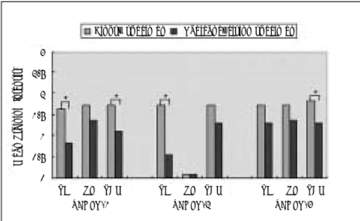
We examined the expression of VEGFRs on the tumor cells, stromal fibroblasts and tumor-associated macrophages in the biopsy and radical excision speci- mens. In the biopsy specimens, all 3 receptors were expressed in the tumor cells, stromal fibroblasts and tumor-associated macrophages, except that VEGFR-2 was not expressed in the stromal fibroblasts. In the radical excision specimens, the staining intensity for VEGFR-1 and VEGFR-2 in the tumor cells was significantly lower than that in the biopsy specimens (P < 0.05). The staining intensity for VEGFR-1 and -3 in the tumor-associated macrophages was lower than that in the biopsy specimens (P < 0.05). In stromal fibroblasts, the specific change of expression of VEGFRs was not detected (Fig. 1).
(1) VEGFR-1 expression
The expression of VEGFR-1 was detected on the tumoral cells, stromal fibroblasts and tumor-associated macrophages in all the biopsy specimens, and the mean staining intensity in each cell type was almost similar.
However, a difference of expression according to the cell type was detected in the radical excision specimens. The
Fig. 1.Mean staining intensity for the vascular endothelial growth factor receptors (VEGFRs) in tumoral cells, stromal fibroblasts, and tumor-associated macrophages of biopsy and radical excision specimens. An asterisk indicates statistically significant difference (P < 0.05) when the radical exicion speci- mens are compared to the biopsy specimens.
VEGFR: vascular endothelial growth factor receptor, TC:
tumor cell, SF: stromal fibroblast, TAM: tumor-associated macrophage.
Biopsy specimen Radical excision specimen
TC SF TAM VEGFR-1 3
2.5 2 1.5 1 0.5 0
TC SF TAM VEGFR-2
TC SF TAM VEGFR-3
Mean Staining Intensit)
staining intensity for VEGFR-1 in the tumor cells was significantly lower than that in the tumor cells of the biopsy specimens (P < 0.05). The expression of VEGFR-1 in the tumor cells of 4 specimens was not deteced. For the stromal fibroblasts, similar to the biopsy specimens, the expression of VEGFR-1 was detected in all the speci- mens, and the mean staining intensity was not signifi- cantly different. For the tumor-associated macrophages, 2 specimens did not express VEGFR-1, and in addition, the staining intensity showed a significant difference (P <
0.05, Fig. 2).
(2) VEGFR-2 expression
The expression of VEGFR-2 in the tumor cells and the tumor-associated macrophages was detected in all biop- sy specimens and the mean staining intensity of the tumor cells and the tumor-associated macrophages was almost similar. However, the expression of VEGFR-2 was not shown in the stromal fibroblasts of all specimens except 1 specimen, which was also similar in the radical excision specimens. In the radical excision specimens, the staining intensity for VEGFR-2 in the tumor cells was
significantly lower than that in the tumor cells of the biopsy specimens (P < 0.05). The expression of VEGFR-2 in the tumor cells was not detected in 5 specimens. VEG- FR-2 was not expressed in the tumor-associated macrophages of 2 specimens, however the mean staining intensity in the tumor-associated macrophages was not statistically different (Fig. 3).
(3) VEGFR-3 expression
The expression of VEGFR-3 was detected in the tumor cells, stromal fibroblasts and tumor-associated macrophages of all biopsy specimens, and the mean staining intensity in each cell type was almost similar. 2 radical excision specimens did not express VEGFR-3 in the tumor cells, however, the mean staining intensity of VEGFR-3 in the tumor cells was not significantly differ- ent. For the stromal fibroblasts, the mean staining inten- sity of VEGFR-3 also did not show a significant differ- ence. VEGFR-3 was not expressed in the tumor-associat- ed macrophages of 2 radical excision specimens, and a statistical difference of the expression was detected (P <
0.05, Fig. 4).
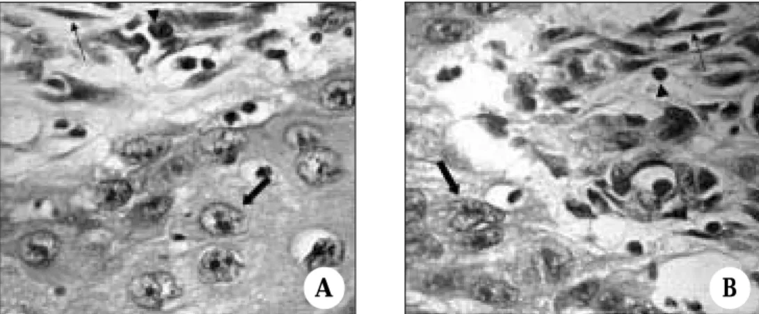
Fig. 2.Representative photomicrographs of staining for vascular endothelial growth factor receptor-1 (VEGFR-1) in biopsy (A) and radical excision specimen (B).
A, VEGFR-1 immunoreactivity was detected in tumor cells (thick arrow), stromal fibroblasts (thin arrow), and tumor-associated macrophages (arrowhead) of biopsy specimen (immunohis- tochemical stain, original magnification × 400).
B, VEGFR-1 immunoreactivity was seen in stromal fibroblasts (thin arrow), however, it demon- strated lack of staining in tumor cells (thick arrow) and tumor-associated macrophages (arrow- head) of radical excision specimen (immunohistochemical stain, original magnification × 400).
A B
2. The double immunoflourescence staining for 5B5 and each VEGFR in the stromal fibroblasts and for CD68 and each VEGFR in the tumor-associated macrophages of the radical excision specimens All the control specimens were negative for the expres-
sion of 3 VEGFRs, and only each marker, 5B5 and CD68, was expressed in the fibromas and the chronic inflamma- tion tisssues, respectively (data not shown). On the other hand, double staining was observed in the radical exci- sion specimens (Table 1). The double staining for 5B5/VEGFR-1 and 5B5/VEGFR-3 in the stromal fibrob- Fig. 3.Representative photomicrographs of staining for vascular endothelial growth factor
receptor-2 (VEGFR-2) in biopsy (A) and radical excision specimen (B).
A, VEGFR-2 immunoreactivity was detected in tumor cells (thick arrow) and tumor-associated macrophages (arrowhead), whereas no expression was detected in any stromal fibroblast (thin arrow, immunohistochemical stain, original magnification × 400).
B, VEGFR-2 immunoreactivity was seen in tumor-associated macrophages (arrowhead), howev- er, it demonstrated lack of staining in tumor cells (thick arrow) of radical excision specimen (immunohistochemical stain, original magnification × 400).
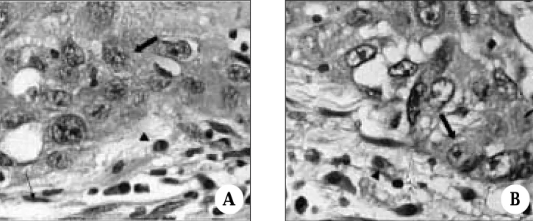
Fig. 4.Representative photomicrographs of staining for vascular endothelial growth factor receptor-3 (VEGFR-3) in biopsy (A) and radical excision specimen (B).
A, VEGFR-3 immunoreactivity was detected in tumor cells (thick arrow), stromal fibroblasts (thin arrow), and tumor-associated macrophages (arrowhead, immunohistochemical stain, orig- inal magnification × 400).
B, VEGFR-3 immunoreactivity was detected in tumor cells (thick arrow) and stromal fibroblasts (thin arrow), however, it demonstrated lack of staining in tumor-associated macrophages (arrowhead) of radical excision specimen (immunohistochemical stain, original magnification
× 400).
A B
A B
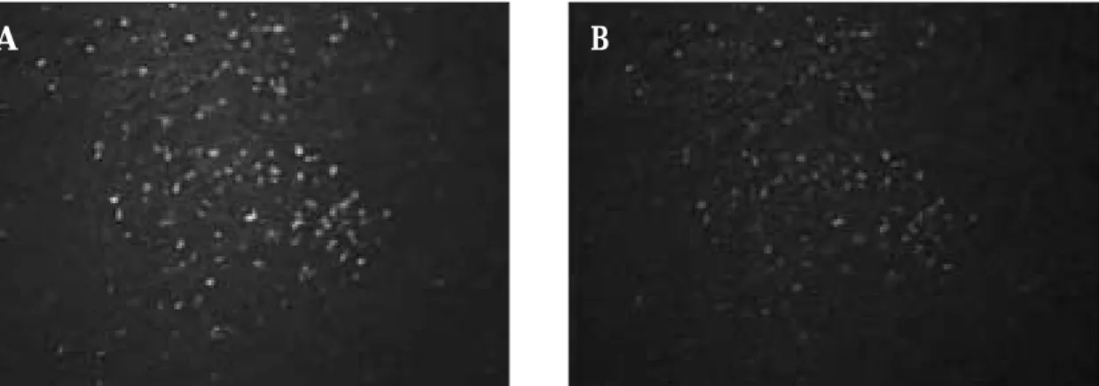
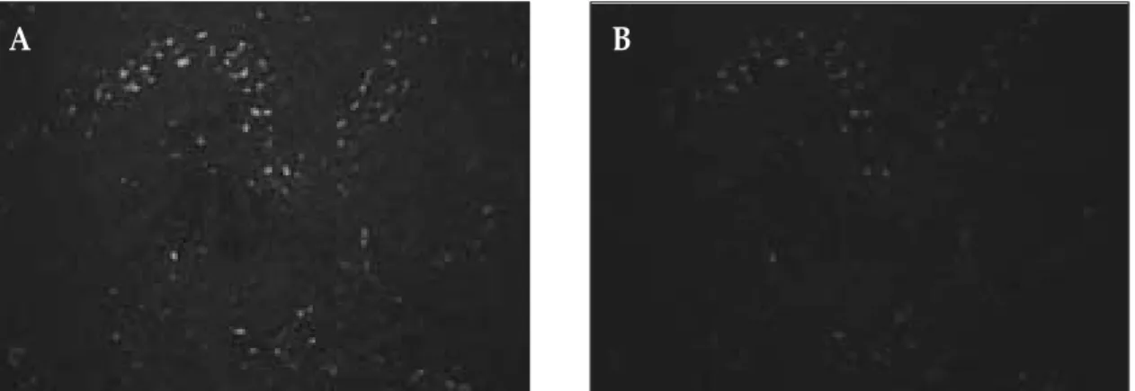
Fig. 5. Double immunofluorescence staining for vascular endothelial growth factor receptor-1 (VEGFR-1, green) and CD68 (red) in the tumor-associated macrophages.
A, VEGFR-1 immunoreactivity was detected in the tumor-associated macrophages (original magnification × 200).
B, CD68 immunoreactivity was detected in the tumor-associated macrophages (original magnification × 200).
The staining pattern corresponds to the areas of VEGFR-1 staining.
Table 1. Evaluation of double immunoflourescence staining for 5B5 and each VEGFR in the stromal fibrob- lasts and for CD68 and each VEGFR in the tumor-associated macrophages of radical excision specimens.
5B5 5B5 5B5 CD68 CD68 CD68
stromal cell + + + + + +
VEGFR1 VEGFR2 VEGFR3 VEGFR1 VEGFR2 VEGFR3
stromal fibroblast + - +
tumor-associated macrophage + - +
Data are given as positive (+) or negative (-).
VEGFR: vascular endothelial growth factor receptor.
A B
Fig. 6.Double immunofluorescence staining for vascular endothelial growth factor receptor-3 (VEGFR-3, green) and CD68 (red) in the tumor-associated macrophages.
A, VEGFR-3 immunoreactivity was detected in the tumor-associated macrophages (original magnification × 200).
B, CD68 immunoreactivity was detected in the tumor-associated macrophages (original magnification × 200).
The staining pattern corresponds to the areas of VEGFR-3 staining.
A B
lasts was detected in 8 specimens and 9 specimens, respectively. In addition, the double staining for CD68/VEGFR-1 and CD68/VEGFR-3 in the tumor-asso- ciated macrophages was detected in 9 specimens and 9 specimens, respectively (Figs. 5-8).
DISCUSSION
VEGF is one of the most important factors in tumor angiogenesis, and it primarily binds to two types of receptor that are present on vascular endothelial cells, VEGFR-2 and VEGFR-1, and VEGF mediates the migra- tion and proliferation of blood vessels. It also reacts with
another receptor that is present on lymphatic endothelial cells and it plays an important function in the growth of lymphatic vessels2,5). The importance of VEGFRs has been already reported in many studies. Zhu et al.6)who report- ed on the importance of the VEGFR-2 in tumor angio- genesis, showed that intervening between the interac- tions of VEGF/VEGFR with using anti-KDR antibody blocked the VEGF-stimulated phosphorylation of VEG- FR-2 and the mitogenesis of endothelials cells. Hence, they have suggested the potential clinical application of anti-KDR antibody. In regard to VEGFR-1, its function has not yet been elucidated. Clauss et al.7)suggested that VEGFR-1, but not the VEGFR-2, act as a functional recep- Fig. 7. Double immunofluorescence staining for vascular endothelial growth factor receptor-1 (VEGFR-3,
green) and 5B5 (red) in the stromal fibroblasts.
A, VEGFR-1 immunoreactivity was detected in the stromal fibroblasts (original magnification × 200).
B, 5B5 immunoreactivity was detected in the stromal fibroblasts (original magnification × 200). The staining pattern corresponds to the areas of VEGFR-1 staining.
A B
Fig. 8. Double immunofluorescence staining for vascular endothelial growth factor receptor-3 (VEGFR-3, green) and 5B5 (red) in the stromal fibroblasts.
A, VEGFR-3 immunoreactivity was detected in the stromal fibroblasts (original magnification × 200).
B, 5B5 immunoreactivity was detected in the stromal fibroblasts (original magnification × 200). The staining pattern corresponds to the areas of VEGFR-3 staining.
A B
tor for VEGF in endothelial cells and monocyte/
macrophage lineage cells. However, Hiratsuka et al.8) reported the negative effect of VEGFR-1 on the prolifera- tion of endothelial cells. In head and neck cancer, instead of the direct invasion to the adjacent tissues, metastasis is developed through the local lymph node in most cases, and so the concern with VEGFR-3 is recently on the increase. VEGFR-3 is expressed on the blood vessels and lymphatic vessels, yet its expression in the lymphatic vessels is generally greater that in the blood vessels2,9). Okamoto et al.10)examined the factors associated with delayed neck metastasis in the early stage of tongue SCC, and they reported that tumor thickness over 4 mm and the expression of VEGFR-3 are the risk factors for devel- oping delayed neck metastasis.
Malignant cells generally secrete various factors such as VEGF that binds to VEGF receptors on endothelial cells to induce angiogenesis for their growth and metas- tasis. This indicates that VEGF is an important paracrine mediator for angiogenesis. However, in some tumors, the expression of VEGF and VEGFRs on the tumor cell itself has been reported. Masood et al.3)have reported the expression of VEGF and VEGFRs on melanoma, ovarian carcinoma, pancreatic carcinoma, and Kaposi’s sarcoma in experiments using various tumor cell lines. This means VEGF is an autocrine growth factor for tumor cell lines that express VEGFRs. The secretion of VEGF from tumor cells and stromal cells is already common known, so examining whether tumor cells and stromal cells also express VEGFRs may be important11-14). It has been recent- ly reported that VEGFR-1 is expressed on monocyte/
macrophage lineage cells to a certain degree, neverthe- less, the expression of VEGFRs on stromal cells has been rarely reported15,16). In the present study, all 3 receptors were expressed in tumor cells, stromal fibroblasts and tumor-associated macrophages in the biopsy specimens of tongue SCC, except that VEGFR-2 was not expressed in the stromal fibroblasts. According to our results, in tongue SCC, the VEGF-driven autocrine growth mecha- nism may be present not only in the tumor cells, but also in stromal cells. Particularly, we found that VEGFRs were expressed on stromal cells which are non-endothe- lial cells. It is likely that the phenotypic change associat- ed with angiogenesis in stromal cells occurs under tumor environment.
In tumor, both inflammation and fibrosis response occur after chemotherapy that induces tumor cell apop- tosis and necrosis. This results in an increase in the
macrophage and fibroblast population17). Generatlly, the increased microvessel formation through the overexpres- sion of VEGF is associated with tumor nutrition and oxy- genation, hence, the proliferation of microvessel plays an important role in tumor growth. Also, VEGF has been shown to interfere with tumor cell apoptosis, and so the overexpression of VEGF in tumor cells may interfere with the effects of chemotherapy18,19). On the other hand, the tumor microvessels are closely associated with chemotherapeutic drug delivery to tumor cells, and the effect of chemotherapy can be increased in richly vascu- larized tumors. Contradictory results have been reported in regard to VEGF action on the effect of chemotherapy, but the change of the expression of VEGFRs in tumor cells and stromal cells, according to chemotherapy, has not yet been reported, and so we examined this in the present study. Our results demonstrated that in the radi- cal excision specimens after neoadjuvant chemotherapy, the staining intensity for VEGFR-1 and VEGFR-2 in the tumor cells was significantly lower than that in the biop- sy specimens. The staining intensity for VEGFR-1 and VEGFR-3 in the tumor-associated macrophages was also significantly lower than that in the biopsy specimens. It has been known that the stromal cells maintain the activ- ity in relation to angiogenesis, including the production of VEGF, even if a decrease activity of tumor cells after chemotherapy is observed17,20). Therefore, although we examined the decreased expression of VEGFR-1 and VEGFR-3 in the tumor-associated macrophages in response to neoadjuvant chemotherapy, it is thought that this may not be very meaningful. Maybe this result is associated with the small number of specimens, but more advanced studies on this may be required. We found that the change of expression of VEGFRs in response to neoadjuvant chemotherapy was substantially more severe for the tumor cells than for the stromal cells.
Therefore, we examined the activated phenotype associ- ated with angiogenesis in the stromal cells of the radical excision specimens by using the double immunofluores- cence staining for the specific marker of stromal cell and each VEGFR. Fibroma and chronic inflammation tissue were used as the controls because these tissues have abundant fibroblasts and macrophages, respectively. It has been reported that VEGFR-1 is expressed to some degree in the monocyte/macrophage lineage cells15,16). However, in the present study using immunohistochem- ical methods with paraffin-embedded specimens, the expression of VEGFR-1 was not detected in the chronic
inflammation tissue that was used as control. In regard to the expression of VEGFR-1 on human monocytes, Barleon et al.15)mentioned the importance of VEGFR-1 for the VEGF-stimulated monocyte migration. In that study, they performed the Northern blot analysis of the total RNA from human monocytes that were obtained from peripheral blood and they showed that human monocytes express the gene for the VEGFR-1. In the pre- sent study, paraffin-embedded specimens were used and the extraction of RNA was difficult, so, we used immunohistochemical methods for detecting the expres- sion of VEGFRs. This contradictory result for the expres- sion of VEGFR-1 in monocyte/macrophage may be asso- ciated with the differentiation of monocyte-macrophage lineage cells, as well as the analysis method for detecting the expression of VEGFR-1.
Although our study used small number of specimens, it was found that for tongue SCC, in association with the angiogenesis, the stromal cells showed the activated phe- notype and this was different from the nonmalignant stromal cells. However, since a broad panel of angio- genic factor may be produced by tumor and stromal cells of tongue SCC, further study should be carried out in order to obtain more information about the angiogenic phenotype of stromal cells in tongue SCC.
REFERENCES
1. Neufeld G, Cohen T, Gengrinovitch S, Poltorak Z: Vascular endothelial growth factor (VEGF) and its receptors. FASEB J 1999;13:9-22.
2. Partanen TA, Paavonen K: Lymphatic versus blood vascu- lar endothelial growth factors and receptors in humans.
Microsc Res Tech 2001;55:108-121.
3. Masood R, Cai J, Zheng T, Smith DJ, Hinton DR, Gill PS:
Vascular endothelial growth factor (VEGF) is an autocrine growth factor for VEGF receptor-positive human tumors.
Blood 2001;98:1904-1913.
4. Micke P, Ostman A: Tumour-stroma interaction: cancer-as- sociated fibroblasts as novel targets in anti-cancer therapy?.
Lung Cancer 2004;45:S163-175.
5. Mueller MM, Fusenig NE: Tumor-stroma interactions di- recting phenotype and progression of epithelial skin tumor cells. Differentiation 2002;70:486-497.
6. Zhu Z, Lu D, Kotanides H, Santlago A, Jimenez X, Simcox T, et al: Inhibition of vascular endothelial growth factor in- duced mitogenesis of human endothelial cells by a chimeric anti-kinase insert domain-containing receptor an- tibody. Cancer Lett 1999;136:203-213.
7. Clauss M, Weich H, Breier G, Knies U, Rockl W,
Waltenberger J, et al: The vascular endothelial growth fac- tor receptor Flt-1 mediates biological activities.
Implications for a functional role of placenta growth factor in monocyte activation and chemotaxis. J Biol Chem 1996;271:17629-17634.
8. Hiratsuka S, Minowa O, Kuno J, Noda T, Shibuya M: Flt-1 lacking the tyrosine kinase domain is sufficient for normal development and angiogenesis in mice. Proc Natl Acad Sci U S A 1998;95:9349-9354.
9. Lalla RV, Boisoneau DS, Spiro JD, Kreutzer DL: Expression of vascular endothelial growth factor receptors on tumor cells in head and neck squamous cell carcinoma. Arch Otolaryngol Head Neck Surg 2003;129:882-888.
10. Okamoto M, Nishimine M, Kishi M, Kirita T, Sugimura M, Nakamura M, et al: Prediction of delayed neck metastasis in patients with stage I/II squamous cell carcinoma of the tongue. J Oral Pathol Med 2002;31:227-233.
11. Jonjic N, Valkovic T, Lucin K, Iternicka Z, Krstulja M, Mustac E, et al: Comparison of microvessel density with tu- mor associated macrophages in invasive breast carcinoma.
Anticancer Res 1998;18:3767-3770.
12. Martin TA, Harding KG, Jiang WG: Regulation of angio- genesis and endothelial cell motility by matrix-bound fi- broblasts. Angiogenesis 1999;3:69-76.
13. Shimizu T, Abe R, Nakamura H, Ohkawara A, Suzuki M, Nishira J: High expression of macrophage migration in- hibitory factor in human melanoma cells and its role in tu- mor cell growth and angiogenesis. Biochem Biophys Res Commun 1999;264:751-758.
14. Velazquez OC, Snyder R, Liu ZJ, Fairman RM, Herlyn M:
Fibroblast-dependent differentiation of human microvascu- lar endothelial cells into capillary-like 3-dimensional net- works. FASEB J 2002;16:1316-1318.
15. Barleon B, Sozzani S, Zhou D, Weich HA, Mantovani A, Marme D: Migration of human monocytes in response to vascular endothelial growth factor (VEGF) is mediated via the VEGF receptor flt-1. Blood 1996;87:3336-3343.
16. Sawano A, Iwai S, Sakurai Y, Ito M, Shitara K, Nakahara T, et al: Flt-1, vascular endothelial growth factor receptor 1, is a novel cell surface marker for the lineage of monocyte- macrophages in humans. Blood 2001;97:785-791.
17. Mcdonnell CO, Bouchier-Hayes DJ, Toomey D, Foley D, Kay EW, Leen E, et al: Effect of neoadjuvant chemoradio- therapy on angiogenesis in oesophageal cancer. Br J Surg 2003;90:1373-1378.
18. Gasparini G, Toi M, Miceli R, Vermelen PB, Dittadi R, Biganzoli E, et al: Clinical relevance of vascular endothelial growth factor and thymidine phosphorylase in patients with node-positive breast cancer treated with either adju- vant chemotherapy or hormone therapy. Cancer J Sci Am 1999;5:101-111.
19. Hironaka S, Hasebe T, Kamijo T, Ohtsu A, Boku N, Yoshida S, et al: Biopsy specimen microvessel density is a useful prognostic marker in patients with T(2-4)M(0) esophageal cancer treated with chemoradiotherapy. Clin Cancer Res 2002;8:124-130.
20. Mcdonnell CO, Harmey JH, Bouchier-Hayes DJ, Walsh TN:
Effect of multimodality therapy on circulating vascular en- dothelial growth factor levels in patients with oesophageal cancer. Br J Surg 2001;88:1105-1109.