Basal cell carcinoma (BCC) is the most common skin cancer, accounting for about 80% of skin can- cers in America.1
These tumors are low-grade malignant and rarely metastatic. However, their public health relevance is increasing and their global incidence is rising due to increases in life span.2 Some pa-
tients have a high risk of developing additional tumors within 5 years of diagnosis with BCC.2 These tumors are locally invasive, causing dis- figurement and increasing morbidity due to fre- quent facial localization.1 Early diagnosis and prompt management of BCCs is of crucial im- portance in order to prevent local tissue destruc-
https://doi.org/10.7180/kmj.2018.33.1.75 KMJ
Original Article
Expression of vascular endothelial growth factor is a clinically useful predictor for aggressive basal cell carcinoma
Jong Soon Choi1, Dong Chan Lee2, Hee Kyung Chang3
1Department of Family Medicine, College of Medicine, Kosin University, Busan, Korea
2Department of Plastic Surgery Medicine, Ritz Plastic Surgery Clinic, Geoje, Korea
3Department of Pathology, College of Medicine, Kosin University, Busan, Korea
Objectives: Basal cell carcinoma (BCC) tumors are locally invasive but rarely metastatic. However, aggressive metastatic variants are being increasingly reported in elderly people. Here we investigated the clinical utility of vascular endothelial growth factor (VEGF) as a predictive biomarker for aggressive BCC variants.
Methods: Thirty-five pathologically confirmed cases of BCC that underwent surgical removal in the Plastic Surgery Department between January 1, 2011 and December 31, 2012 were studied. VEGF expression was analyzed in formalin-fixed paraffin-embedded tumor tissue by immunohistochemical staining. Positive staining was defined as more than 10% of the tumor cells showing immunoreactivity. The associations of VEGF expression with various clinicopathologic parameters were analyzed.
Results: The face was the most prevalent site (28/35), with 15 cases from the nose, 6 cases from the eyelid, and 5 cases from the cheek. The patients were aged between 41 and 86 years, with a mean age of 69.26
± 173.903 years. The mean BCC size was 1.34 ± 3.853 cm, with a range of 0.3 cm to 12.0 cm. The mean tumor invasion depth from the basement epidermal membrane was 0.17 ± 0.035 cm, with a range of 0.03 cm to 1.10 cm. A mean of 5.66 ± 20.938 intraoperative frozen section slides were examined. VEGF was not expressed in 14 of the 35 patients (40.0%), whereas 42.9% of the patients had low expression and 17.1% of the patients had high expression. VEGF expression was significantly associated with age (P = 0.022), size (P
= 0.030), site (P = 0.013), tumor invasion depth (P = 0.019), and number of intraoperatively frozen sections (P = 0.003).
Conclusions: These results suggest that VEGF expression as assessed by immunohistochemistry can predict aggressive or poor prognosis in BCC.
Key Words: Basal cell carcinoma, Biomarker, VEGF
Corresponding Author: Hee Kyung Chang, Department of Pathology, College of Medicine, Kosin University, 262, Gamchen-ro, Seo-gu, Busan 49267, Korea
Tel: +82-51-990-6476 Fax: +82-51-990-3045 E-mail: [email protected]
Received:
Revised:
Accepted:
Nov. 29, 2017 Dec. 13, 2017 Jan. 22, 2018 Articles published in Kosin Medical Journal are open-access, distributed under the terms of the Creative Commons Attribution Non-Commercial License (http://creativecommons.org/licenses/by-nc/4.0/) which permits unrestricted non-commercial use, distribution, and reproduction in
Corresponding Author: Hee Kyung Chang, Department of Pathology, College of Medicine, Kosin University, 262, Gamchen-ro, Seo-gu, Busan 49267, Korea
Tel: +82-51-990-6476 Fax: +82-51-990-3045 E-mail: [email protected]
Received:
Revised:
Accepted:
Nov. 29, 2017 Dec. 13, 2017 Jan. 22, 2018
tion and subsequent disfigurement.3,4 Although various noninvasive or minimally invasive techni- ques have increased the diagnostic accuracy of BCC and progress has been made regarding treat- ment options for BCC, more aggressive variants of BCC still pose significant challenges for the healthcare system.3,4 A potential solution is the development of novel biomarkers for diagnosis, prognosis, and therapy monitoring. This ap- proach can be used in various malignancies, in- cluding BCC. We hypothesized that gene ex- pression signatures of tumor cells suggestive of the tumoral microenvironment could serve as novel predictive and prognostic biomarkers in BCC.
Angiogenesis, a process consisting of growth and expansion of the vasculature, is involved in the growth and metastasis of many cancers.5 Angiogenesis is a prognostic indicator for a varie- ty of tumors, suggesting proliferation of neo- plastic cells into the circulation and metastasis.6 Several molecules such as cell surface receptors, growth factors, and enzymes are involved in this process. Moreover, antiangiogenic therapy for cancer was proposed over 20 years ago as a valid target for anticancer drug development.
Vascular endothelial growth factor (VEGF) is a potent proangiogenic factor and several studies have established a critical role for VEGF in skin cancer.6 Hence, VEGF has become the primary antiangiogenic drug target.6 For example, an in- tralesional anti-VEGF antibody (bevacizumab) was recently shown to be feasible for adjuvant
treatment of locally advanced BCCs.7
Regarding oncogenesis, VEGF may involve carcinogenesis.8 VEGF may also have direct ef- fects on keratinocytes and skin tumor cells, spe- cifically by promoting skin carcinogenesis by al- tering the survival, proliferation, or stemness of keratinocytes and tumor cells in an autocrine manner.9-11
Clinically, BCCs are rarely metastatic tumors.
However, about 10 cases of metastatic orbitofa- cial metastatic basal cell carcinoma were de- scribed between 1995 to 2015, after surgical removal.
This evidence suggests that VEGF evaluation for skin BCCs is a clinically promising prognostic fac- tor and therapy target. However, few clinical re- search studies have investigated these possibil- ities, especially in Korea. The purpose of this study was to evaluate the utility of VEGF ex- pression as a prognostic or predictive factor of clinicopathological factors in basal cell carcino- ma, with the ultimate goal of enhancing our un- derstanding of the development and management of BCCs.
MATERIALS AND METHODS
Tissue samples
Thirty-five patients with BCC who underwent plastic surgery to remove a BCC tumor mass be- tween 2011 and 2012 were selected for analysis.
Hematoxylin-eosin stained (HE) slides were re-
trieved from the Department of Pathology at Kosin University Gospel Hospital. All slides were reviewed by a trained pathologist. All slides, in- cluding frozen slides, were reviewed based on the presence or absence of the BCC characteristics.
All samples and corresponding data were de-linked and anonymized by decoding. This study was approved by the Institutional Review Board of Kosin University Gospel Hospital (KUGH 2017-10-009).
Immunohistochemistry
Formalin-fixed and paraffin-embedded (FFPE) biopsies and excision specimens were used.
Four-micron sections were cut and stained with primary polyclonal anti-VEGF antibodies (1:200, Neomarkers, Fremont, CA, USA) using a BONDMAX autostainer system and a pre-treat- ment module. The module used EnVision FLEX Target Retrieval Solution, High pH (Dako, Heverlee, Belgium). The antibodies were applied for 20 minutes at room temperature. A Dako Envision Flex kit (K8002) was used for secondary detection.
The slides were deparaffinized in xylene, rehy- drated, and incubated in hydrogen peroxide (H2O2) in methanol for 30 minutes to inactivate endogenous peroxidase activity. Antigen retrieval was performed by microwave treatment at 90 W for 10 minutes in 10 mM citrate buffer (pH 6);
non-specific protein binding was blocked using 3% bovine-serum-albumin (BSA). Antigen-anti- body complexes were subsequently visualized us-
ing the Envision™ Detection System kit perox- idase/DAB (DAKO, Glustrop, Denmark) and coun- terstained with hematoxylin. For negative con- trols, the primary antibody was replaced by either mouse or rabbit non-immune serum, as appropriate. All stained sections were evaluated in a blinded manner without prior knowledge of the patient data.
Immunostaining interpretation
All sections were evaluated. For all tumors, at least four randomly chosen high-power fields (magnification 200x) were assessed per slide to determine the percentage of positive tumor cells.
Cell staining was scored as follows: 0 (no staining), 1 (< 50% staining), or 2 (> 50% staining). For all assessments, HF was used as an internal standard and was considered to be 100% positive. Positive staining was defined as immunoreactivity of more than 10% of the tumor cells.
Statistical analysis
Statistical analyses were carried out using SPSS version 24.0 software (SPSS, Chicago, IL, USA).
Descriptive data are presented as absolute num- bers and percentages for categorical data and as means with standard deviations for continuous data. The Chi-square test for independent pro- portions was performed to evaluate the differ- ences and similarities in expression of VEGF be- tween clinical data, especially according to VEGF negative and positive groups in final analysis. P values less than 0.05 were considered statistically
significant.
RESULTS
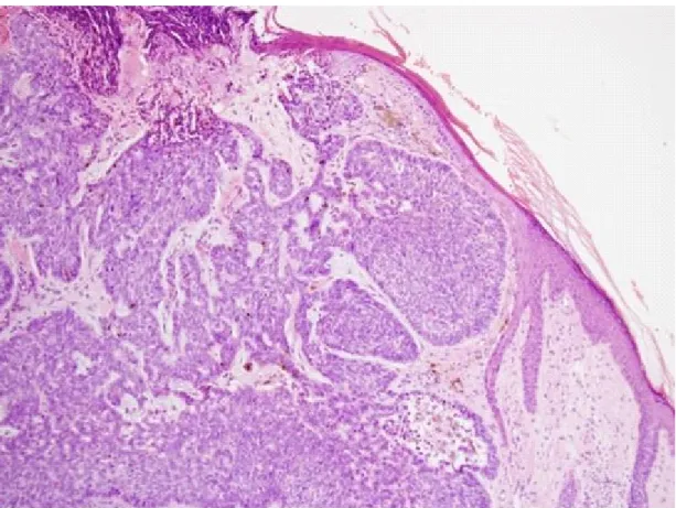
Regarding tumor site, the face was the most prev- alent site (28/35: 80%) (Fig. 1). Of the tumors on the face, 15 cases were from the nose (42.9%), 6 from the eyelid (17.1%), and 5 from the cheek (14.3%). Sites other than the face included 1 case from the anus, 1 case from the temporal area, 1 case from the clavicle, 1 case from the nasal cavity, and 2 cases from the trunk skin. Regarding gender, 15 patients were male (42.9%) and 20 pa- tients were female (53.1%), yielding a male-to-fe-
male ratio of 0.75: 1. Patients were aged between 41 and 86 years, with a mean age of 69.26 ± 173.903 years. The mean BCC size was 1.34 ± 3.853 cm, with a range of 0.3 cm to 12.0 cm. BCC arising from face skin consisted of 28 cases (80.0%). The mean tumor invasion depth from the epidermal basement membrane was 0.17 ± 0.035 cm, with a range of 0.03 cm to 1.10 cm. The mean number of frozen sections obtained from patients was 5.66
± 20.938, with a range of 0 to 19 (Table 1). Frozen sections were not obtained from five patients.
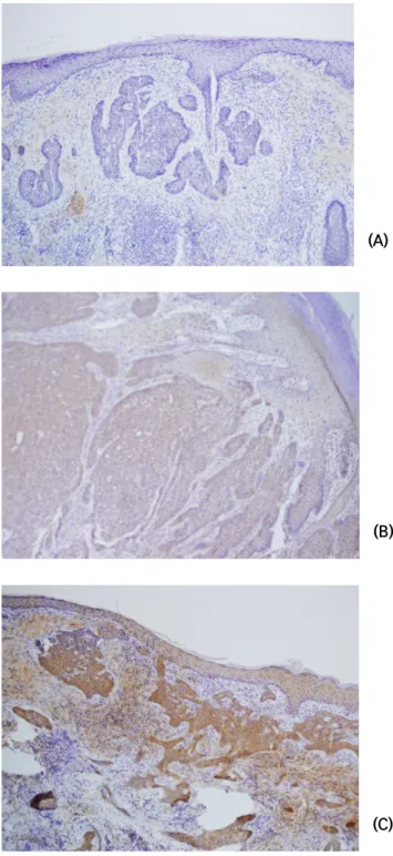
VEGF was not expressed in 14 of the 35 patients (40.0%) (Fig. 2A). However, 15 and 6 patients showed low expression (42.9%) (Fig. 2B) and high expression (17.1%) (Fig. 2C), respectively.
Fig. 1. The pathologic findings of Basal cell carcinoma of skin on the face. (H-E, x100)
(A)
(B)
(C)
Fig. 2. The expression of VEGF by immunostaining in the basal cell carcinoma (x100) (A) Negative expression of VEGF in the basal cell carcinoma
(B) Low-expression of VEGF in the basal cell carcinoma (C) High-expression of VEGF in the basal cell carcinoma
VEGF expression was significantly associated with age (P = 0.022), size (P = 0.030), tumor invasion site depth (P = 0.019), and number of frozen sec- tions(P = 0.003).
The mean age of patients with no VEGF ex- pression was 63 ± 14.486 years, whereas those of patients with low and high expression were 73.47
± 10.954 and 73.33 ± 10.875 years, respectively.
VEGF expression was significantly correlated with age (P = 0.022). In male patients, 7 cases (46.7%) had no VEGF expression. However, in female pa- tients, 7 cases (35.7%) had no VEGF expression and 10 cases (50.0%) had low VEGF expression.
VEGF expression was not associated with gender.
Mean tumor size was greater in cases with low VEGF expression compared to those with high ex-
pression (P = 0.030). Specifically, the mean tumor size was 0.710 cm in patients without VEGF ex- pression, 1.923 cm in patients with low VEGF ex- pression (VEGF-low), 1.367 cm in patients with high VEGF expression (VEGF-high), and 1.645 cm in patients with VEGF-positive. The incidence of non-face tumor sites was higher in VEGF-negative patients than in VEGF-positive patients (P = 0.013).
Tumor invasion depth was significantly deeper in VEGF-negative patients (mean: 1.21 cm) than in VEGF-low (mean: 2.21 cm) and VEGF-high (mean:
1.63 cm) patients (P = 0.019), and than VEGF-pos- itive group(mean: 1.79cm) (p = 0.000).
The mean number of frozen sections excised by the plastic surgeon was correlated with VEGF expression: 3.29 in VEGF-negative cases, 6.20 in variables total (n = 35)
(%)
Cases of VEGF expression P value
according to positivity (expression
level) negative(%)
(n = 14)
Positive reaction(n = 21) total of Positivity
(%)
low(%) (n = 15)
high(%) (n = 6)
mean age 69.26 ± 103.903 63 ± 14.486 73.41 ± 10.915 73.47 ± 10.954 73.33 ± 10.875 0.000 (0.022) gender
male 15(42.9) 7(46.7) 8(5.5) 5(33.3) 3(20.2) 0.096
(0.120)
female 20(53.1) 7(25.0) 13(75.0) 10(60.0) 3(15.0)
mean size
(cm) 1.34 ± 3.852 0.709 ± 0.289 1.645 ± 1.875 1.923 ± 2.897 1.367 ± 0.582 0.000 (0.030)
site 0.002
(0.013)
face 28(80.0) 9(32.1) 19(67.9) 14(50.0) 5(17.9)
non-face 7(20.0) 5(64.4) 2(33.6) 1(14.3) 1(14.3)
invasion
depth(mm) 1.7 ± 0.035 1.21 ± 0.092 1.79 ± 0.495 2.21 ± 0.26 1.63 ± 0.71 0.000 (0.019) numbers
of frozen section
5.66 ± 20.94 3.29 ± 2.76 8.02 ± 5.121 6.20 ± 4.13 9.83 ± 6.11 0.000 (0.003) Table 1. The association of VEGF expression with clinicopathologic variables in the basal cell carinoma
VEGF-low cases, and 9.83 in VEGF-high cases (P
= 0.003).
DISCUSSION
Angiogenesis is a crucial process in the pro- gression and metastasis of many cancers, including skin cancer.8-12 Typically, angiogenesis is required for tumors to grow beyond 1-2 mm in size and offers a pathway for tumor cells to spread to meta- static sites.8-13 Hence, tumor angiogenesis has been a challenging therapeutic target of many cut- ting-edge strategies.14 To induce angiogenesis, tu- mor cells and stromal cells in the tumor mass micro- environment must break the balance of pro- and anti-angiogenetic factors, favoring an “angiogenic switch”.15 The conversion of pro-angiogenic sig- nals to anti-angiogenic signals allows capillary sprouting by proliferation and migration of endo- thelial cells.14 Eventually, the newly formed vessels supply oxygen and nutrients to the tumor; this is required for progression.14 VEGF has been iden- tified as a potent angiogenic factor.15
VEGF mRNA levels in normal epidermis are low.
However, differentiated epidermal cell layers have been shown to have higher VEGF expression than less differentiated epidermal cells.16 In this study, distinct expression of VEGF was not identified by immunohistochemistry. In human BCCs, weak VEGF expression is typically seen in tumor cells predominantly localized to the invading front margin.17 We observed a similar expression pat-
tern, supporting this phenomenon. Bowden et al.17 reported that VEGF was expressed in 54.5% (24/44) of the examined BCCs, most of which had low expression. In this study, VEGF was not expressed in 14 of the 35 patients (40.0%), expressed at low levels in 15 of the patients (42.9%), and expressed at high levels in 6 of the patients (17.1%). In this study, 60% of the BCCs expressed VEGF and 42.9%
of the BCCs had low VEGF expression. Weninger reported that 64.3% (9/14) of the analyzed BCCs expressed VEGF mRNA.18 Thus, our results are in the range of those reported by Bowden and Weninger.
VEGF expression is low in animal skin and in- creases gradually during tumor development.19 Moreover, VEGF is suspected to be clinically upre- gulated in more aggressive tumors.7
We found that VEGF expression was significantly associated with age (P = 0.022), size (P = 0.030), site (P = 0.013), tumor invasion depth (P = 0.019), and number of frozen sections (P = 0.003). These results suggest that VEGF expression is higher in large and deeply invasive tumors (i.e., tumors that tend to become aggressive), comparable to the findings of Gaitanis.7 One study using an animal model showed that VEGF involves chemi- cally-induced tumorigenesis and that VEGF is ex- pressed rapidly at very high levels in metastatic cases.20 In the present study, one metastatic case had high VEGF expression (data not shown).
Management of BCC is dependent on a variety of factors, including lesion location, patient age, comorbidities, and tumor histologic type.3 The site
at which the lesion occurs is important because tumors that arise in cosmetically or functionally important areas are best managed with treatments that minimize the amount of tissue removed while ensuring a high chance of complete cure.5 In the elderly population, the slow growing nature of BCCs means that less invasive treatments may be favored, despite the fact that some of these meth- ods have higher recurrence rates.3 In the present study, the average patient age was 69.26 years, so patients underwent less invasive surgery per- formed by plastic surgeons and sections were fro- zen to reduce tissue loss.
Interestingly, VEGF expression was positively as- sociated with age. This unexpected result might result from the positive associations between high- er expression and larger size with increased age.
Older age is associated with increased incidence of BCCs on the face. Due to the importance of satisfactory cosmetic outcomes when tumors arise on the face, treatment decisions might differ sig- nificantly from those that would be made for BCCs arising elsewhere. Facial BCCs had more frozen sections from which the marginal status could be identified, with increased VEGF expression.
Another possible explanation is the increased tu- mor size. Regarding risk factors for BCC metastasis, one study followed ten metastatic cases for 20 years.21 The median metastatic tumor size (largest dimension) was 3.3 cm (range, 1.9-11.5 cm).
Branson13 suggested that increased tumor size, local primary tumor recurrence, aggressive histo- logic subtype, and perineural invasion were all
markers of high risk orbitofacial basal cell carcinoma. The present study included only BCCs surgically removed by plastic surgeons, with 2 cases of recurrence. While the recurrent cases showed high expression of VEGF, intraoperative con- sultation (frozen sections) was performed. Frozen sections were acquired from most (30) cases, with a mean of 5.66 sections. The number of frozen sections was significantly increased in VEGF-low cases compared to VEGF-high cases. A potential explanation for this finding is that the increased number of frozen sections may be due to tumor size and invasion depth, which lead to VEGF expression. Thus, larger size reflects more frozen sections. Tumor invasion depth was significantly deeper in patients without VEGF expression (mean:
1.21 cm) than in VEGF-low (mean: 2.21 cm) and VEGF-high (mean: 1.63 cm) patients. Interestingly, the VEGF- low-expressed group showed deeper depth of invasion than the VEGF-high-expressed group, but statistically insignificant (p=0.096). This result might be explained by small number of cases or another factors of tumor invasion, which are pericyte function for vascular sprouts and mesen- chymal transition. There are reports about dual function of VEGF in the tumor invasion that VEGF has also functions as negative regulator for peri- cytes22 and VEGF inhibits tumor cell invasion in some condition.23 Therefore, we could presume that VEGF has a crucial role in tumor invasion in early stage, however, in later stage of invasion of the basal cell carcinoma, the expression of VEGF become lower, comparing to early stage.
Cumulatively, our findings suggest that VEGF expression is a potential biomarker for aggressive BCCs with poor prognosis and that provide clini- cian with helpful information, for example, intra- operative frozen section.
A limitation of this study is the small number of cases, which hinders establishment of the clin- ical significance of VEGF expression. The next study with larger scale of cases will be needed for verifying these results.
REFERENCES
1. Rogers HW, Weinstock MA, Harris AR, Hinckley MR, Feldman SR, Fleischer AB, et al. Incidence estimate of nonmelanoma skin cancer in the United States, 2006. Arch Dermatol 2010;146:283–7.
2. Marghoob A, Kopf AW, Bart RS, Sanfilippo L, Silverman MK, Lee P, et al. Risk of another basal cell carcinoma developing after treatment of a basal cell carcinoma. J Am Acad Dermatol 1993;28:22–8.
3. Marzuka AG, Book SE. Basal cell carcinoma:
pathogenesis, epideriology, clinical features, di- agnosis, histopathology, and mangement. Yale J Biol Med 2015;88:167-79.
4. Linos E, Chren MM, Stijacic Cenzer I, Covinsky KE. Skin Cancer in U.S. Elderly Adults: Does Life Expectancy Play a Role in Treatment Decisions?
J Am Geriatr Soc 2016;64:1610-5.
5. Dvorak HF. Angiogenesis: update 2005. J Thromb Haemost 2005;3:1835–42.
6. Mueller MM, Fusenig NE. Tumor-stroma inter- actions directing phenotype and progression of epithelial skin tumor cells. Differentiation 2002;70:486–97.
7. Gaitanis G, Bassukas I. Intralesional bevacizumab as in-add adjuvant to immunocryosurery for lo- cally advanced basal cell carcinoma. J Eur Acad Dermatol Venereol 2014;28:1117-21.
8. Larcher F, Murillas R, Bolontrade M, Conti CJ, Jorcano JL. VEGF/VPF overexpression in skin of transgenic mice induces angiogenesis, vascular hyperpermeability and accelerated tumor development. Oncogene 1998;17:303–11.
9. Wilgus TA, Matthies AM, Radek KA, Dovi JV, Burns AL, Shankar R, et al. Novel function for vascular endothelial growth factor receptor-1 on epidermal keratinocytes. Am J Pathol 2005;167:1257–66.
10. Beck B, Driessens G, Goossens S, Youssef KK, Kuchnio A, Caauwe A, et al. A vascular niche and a VEGF-Nrp1 loop regulate the initiation and stemness of skin tumours. Nature 2011;478:399–
403.
11. Lichtenberger BM, Tan PK, Niederleithner H, Ferrara N, Petzelbauer P, Sibilia M. Autocrine VEGF signaling synergizes with EGFR in tumor cells to promote epithelial cancer development.
Cell 2010;140:268–79.
12. Zhu JW, Wu XJ, Luo D, Lu ZF, Cai SQ, Zheng M. Activation of VEGFr-2 signaling in response to moderate dose of ultraviolet B promotes surviv- al of normal human keratinocytes. Int J Biochem Cell Biol 2012;44:246–56.
13. Folkman J. The role of angiogenesis in tumor growth. Semin Cancer Biol 1992;3:65–7.
14. Carmeliet P. Angiogenesis in life, disease and medicine. Nature 2005;438:932–6.
15. Hanahan D, Folkman J. Patterns and emerging mechanisms of the angiogenic switch during tumorigenesis. Cell 1996;86:353–64.
16. Viac J, Palacio S, Schmitt D, Claudy A. Expression of vascular endothelial growth factor in normal epidermis, epithelial tumors and cultured keratinocytes. Arch Dermatol Res 1997;289:158–
63.
17. Bowden J, Brennan PA, Umar T, Cronin A.
Expression of vascular endothelial growth factor in basal cell carcinoma and cutaneous squamous cell carcinoma of the head and neck. J Cutan Pathol 2002;29:585–9.
18. Weninger W, Uthman A, Pammer J, Pichler A, Ballaun C, Lang IM, et al. Vascular endothelial growth factor production in normal epidermis and in benign and malignant epithelial skin tumors. Lab Invest 1996;75:647-57.
19. Larcher F, Robles AI, Duran H, Murillas R, Quintanilla M, Cano A, et al. Up-regulation of
vascular endothelial growth factor/vascular per- meability factor in mouse skin carcinogenesis correlates with malignant progression state and activated H-ras expression levels. Carcinogenesis 1996;56:5391–6.
20. Hirakawa S, Kodama S, Kunstfeld R, Kajiya K, Brown LF, Detmar M. VEGF-A induces tumor and sentinel lymph node lymphangiogenesis and promotes lymphatic metastasis. J Exp Med 2005;201:1089–99.
21. Branson SV, McClintic E, Ozgur O, Esmaeli B, Yeatts RP. Orbitofacial Metastatic Basal Cell Carcinoma: Report of 10 Cases. Ophthal Plast Reconstr Surg 2017;33:213-7.
22. Greenberg JI, Shields DJ, Barillas SG, Acevedo LM, Murphy E, Huang J, et al. A role for VEGF as a negative regulator of pericyte function and vessel maturation. Nature, 2008;456:809-13.
23. Lu KV, Chang JP, Parachoniak CA, Pandika MM, Aghi MK, Meyronet D, et al. VEGF inhibits tumor cell invasion and mesenchymal transition through a MET/VEGFR2 complex. Cancer Cell, 2012;22:21-35.