Copyright © 2014 The Korean Society for Bone and Mineral Research
This is an Open Access article distributed under the terms of the Creative Commons Attribution Non-Commercial Li-cense (http://creativecommons.org/liLi-censes/by-nc/3.0/) which permits unrestricted non-commercial use, distribu-tion, and reproduction in any medium, provided the original work is properly cited.
pISSN 2287-6375 eISSN 2287-7029
Effect of Zoledronate on the Expression of Vascular
Endothelial Growth Factor-A by Articular
Chondrocytes and Synovial Cells: An in Vitro Study
Jin Woong Yi1, Woo-Suk Lee2, Sang-Bum Kim1, Youn-Moo Heo1, Dong-Sik Chae3
1Department of Orthopedic Surgery, Konyang University Hospital, Daejeon;
2Department of Orthopedic Surgery, Yonsei University Gangnam Severance Hospital, Seoul;
3Department of Orthopedic Surgery, International St. Mary's Hospital, Catholic Kwandong University College of Medicine, Incheon, Korea
Background: The aim of this in vitro study was to determine the effect of zoledronate, which is frequently used to treat osteoporosis, on osteoarthritis by analyzing zoledro-nate-induced expression of vascular endothelial growth factor-A (VEGF-A) in
chondro-cytes and synovial cells. Methods: After chondrocytes and synovial cells were separated
and cultured, zoledronate was added, and VEGF-A and pigment epithelium-derived fac-tor (PEDF) expression were quantified by real-time polymerase chain reaction and
West-ern blotting. Results: There was no significant difference in the expression of VEGF-A
mRNA in chondrocytes between the zoledronate group and the control group on the 8th day of culture. The expression of both VEGF-A and PEDF mRNA in synovial cells was
significantly decreased in the zoledronate group (P<0.05). Conclusions: Zoledronate
decreases the expression of VEGF-A in synovial cells and may affect the development and progression of osteoarthritis.
Key Words: Chondrocytes, Osteoarthritis, Synovial cells, Vascular endothelial growth fac-tor-A, Zoledronate
INTRODUCTION
Cartilaginous joints normally do not contain blood vessels; however, angiogen-esis is observed in subchondral bone and in bony spurs of osteoarthritic bones, and is also increased in the synovial membrane.[1-3] Angiogenesis in osteoarthri-tis is thought to affect the function or homeostasis of cartilaginous joints, playing a direct or indirect role in the pathogenesis and progression of the disease.[4,5]
Vascular endothelial growth factor-A (VEGF-A) is an important factor in the pro-cess of angiogenesis. Angiogenesis at cartilaginous bone is observed during the ossification of endochondral bone in the growth phase or in growth plates, but it is not normally observed in the joints of adults.[6,7] However, granulation tissues containing blood vessels are observed in cartilaginous joints damaged by osteo-arthritis or rheumatoid osteo-arthritis. VEGF and pigment epithelium-derived factor (PEDF), an angiogenesis suppressor, are thought to be involved in the pathogenesis and progression of this disease.[8-10]
Corresponding author Woo-Suk Lee
Department of Orthopedic Surgery, College of Medicine, Yonsei University, Gangnam Severance Hospital, 211 Eonju-ro, Gangnam-gu, Seoul 135-720, Korea Tel: +82-2-2019-3417 Fax: +82-2-573-5393 E-mail: [email protected] Received: July 28, 2014 Revised: November 20, 2014 Accepted: November 21, 2014
No potential conflict of interest relevant to this article was reported.
This work was supported (in part) by Konyang University Myunggok Research Fund of 2010.
Bisphosphonate, which suppresses the proliferation and action of osteoclasts, is an agent frequently used in the treatment of osteoporosis. While the anti-inflammatory properties and protective effects of bisphosphonate on chondrocytes have been reported, its mechanism of action in cartilage has not been precisely defined.[11-13] Current theories hypothesize that bisphosphonates regulate the breakdown and reconstruction of subchondral bone to re-duce the stress put on cartilaginous joints; nonetheless, very few studies have focused on the effects of this agent on chondrocytes and synovial cells.[14,15]
In this study, chondrocytes and synovial cells obtained and cultured in vitro from patients with osteoarthritis were treated with zoledronate, and expression patterns of VEGF-A and PEDF following zoledronate treatment were analyz-ed, thereby identifying the cartilage-protecting mechanism of zoledronate via regulation of VEGF-A expression.
METHODS
1. Culture of chondrocytes and synovial cells
Cartilaginous tissues obtained from six patients under-going total knee replacement due to osteoarthritis were washed four times with phosphate buffered saline (PBS) and were cut into 2×2 mm pieces. The pieces were added to a pre-made enzyme solution (collagenase 2 mg/mL; Roche Diagnostics, Indianapolis, IN, USA) and were digest-ed for 20 hr at 37°C while being constantly stirrdigest-ed with a metal rod. The solution was then centrifuged, and the su-pernatant was removed. The isolated primary chondro-cytes in the pellet were cultured in an incubator with 5%
CO2 at 37°C in a 25 cm2 plate containing Dulbecco’s
modi-fied essential medium (DMEM) (Gibco, Paisley, Scotland, UK) and 10% bovine serum albumin, and the culture media was changed every 2 days. The chondrocytes were stored after three successive cultures, and the expression of type 2 collagen was assayed via real-time (RT) polymerase chain reaction (PCR) to identify the phenotype of the chondro-cytes. Synovial membranes were obtained from patients undergoing total knee replacement due to osteoarthritis: they were digested for 12 hr in a pre-made enzyme solu-tion (collagenase 2 mg/mL) while being stirred with a metal rod in an incubator at 37°C. The solution was then centrifuged, and the supernatant was removed and the sy-novial cells in the pellet were cultured. The study was
ap-proved by the Institutional Review Board, and all patients were provided informed consent.
2.
3-(4,5-Dimethylthiazol-2-yl)-2,5-diphenyltetrazolium bromide (MTT)
analysis and study groups
A total of 1×10⁵ cell/mL of primary-culture
chondro-cytes were plated onto 6-well plates coated with 100 mM
CaCl2, and then cultured 3-dimensionally in alginate. The
cells were cultured for 3 days without zoledronate and then
treated with 10-7 mol/L zoledronate for 48 hr. Interleukin-1
(IL-1) at 10 ng/mL was also administered for 3 days to acti-vate the chondrocytes; cells not treated with zoledronate were defined as the control group. The chondrocytes were removed on the 3rd, 5th, and 8th days of culture and stained with 0.4% trypan blue. Cells were counted, and cell viability via MTT assay at various concentrations of
zoledro-nate (10-7, 10-6, 10-5, 10-4 mol/L) was determined.
The primary cultured synovial cells were distributed to
culture plates at 1×10⁵ cells/mL and cultured in a single
layer without zoledronate. They were treated with 10-7 mol/L
of zoledronate for 3 days; cells not treated with zoledro-nate were defined as the control group. A total of six each zoledronate and control groups were created, and the study was repeated twice.
3. Analysis of VEGF-A and PEDF mRNA by
RT-PCR
Chondrocytes and synovial cells were harvested at the 3rd, 5th, and 8th days of culture, and RNA was extracted using TRIzol (Invitrogen, Penrose, Auckland, NZ) and quan-tified with a spectrophotometer. cDNA of the extracted mRNA was synthesized with a SuperSCRIPT First-Strand Synthesis RT-PCR kit (Invitrogen, Penrose, Auckland, NZ) by combining RNA, oligo (dT), and deoxynucleotide triphos-phates (dNTPs) and incubating for 5 min at 65°C, then
add-ing 10X RT Buffer, 25 Mm MgCl2, 0.1 M dithiothreitol (DTT),
RNaseOUT, and SuperScript and incubating for 50 min at 50°C and for 5 min at 85°C, then treating with RNase H for min at 37°C. Sequences of the forward and reverse primers
of VEGF-A and PEDF are given inTable 1. Five microliters of
the synthesized cDNA, 1 μL of 10 pmol forward and reverse primers, 10 μL of iQ SYBER Green Supermix (Bio-Rad, Her-cules, CA, USA), and 3 μL of nuclease-free water were mixed and subjected to 55 cycles of PCR (15 sec at 95°C, 15 sec at
56°C, and 15 sec at 72°C). The results were analyzed with Bio-Rad IQ5 software. The concentrations of mRNA of VEGF-A and PEDF were calculated with reference to a standard curve and compared to the amount of β-actin.
4. Western blot of VEGF-A and PEDF
The culture media was then stored at -70°C after chon-drocytes and synovial cells were isolated on the 3rd, 5th, and 8th days of culture. The proteins in the media were ex-tracted and subjected to Western blot for quantitative anal-ysis of VEGF-A and PEDF. The amount of protein extract so-lution (Pro-prep protein extraction soso-lution, Intron, Seon-nam, Korea) required was calculated based on the number of cells and was mixed with the media. The media was stored on ice for 10 to 20 min and was shaken vigorously every 5 min. The media was centrifuged at 13,000 rpm for 5 min, and the protein concentration was measur ed by Bradford assay (Bio-Rad Laboratories, Inc., Richmond, CA, USA).
For sodium dodecyl sulfate (SDS)-polyacrylamide gel electrophoresis (PAGE), 30 μg of the samples were mixed with an equal amount of 2X sample buffer (Laemmli sam-ple buffer, Bio-Rad Laboratories, Inc., Richmond, CA, USA), which was heated at 95°C for 5 min and then loaded onto 10% SDS-polyacrylamide gel. The gel was run at 60 volts until the sample ran into the separating gel, after which 120 volts was applied for approximately 1 hr. To transfer the protein, the gels were placed on polyvinylidene difluoride (PVDF) membrane (Amersham Biosciences, Arlington Hei-ghts, IL, USA), ensuring that no air bubble was formed. Af-ter adhering the gel to the membrane, 3 M paper wet with transfer buffer (Trizma base, glycine, and MeOH) and pads were attached at either side, and the set was loaded to the transfer kit (Mini Trans-Blot cell system, Bio-Rad Laborato-ries, Inc., Richmond, CA, USA). The transfer was performed at 300 mA for 2 hr under refrigeration, and the mem brane was washed twice with DW and then twice with TBST. The
membrane was then immersed in a blocking solution (5% skim milk with TBST: Tween 20, 1 M Tris pH 7.5, 5 M NaCl) and was shaken for 1 hr in an orbital shaker and then wash-ed with TBST (Tween 20, 1 M Tris pH 7.5, 5 M NaCl). Anti-VEGF rabbit polyclonal IgG at 200 μg/mL (Santa Cruz Bio-technology, Santa Cruz, CA, USA), anti-PEDF goat polyclonal IgG at 200 μg/mL (Santa Cruz Biotechnology), and anti-β-actin mouse monoclonal IgG at 200 μg/mL (Santa Cruz Bio-technology) were used as primary antibodies. The diluted primary antibodies were added to the membrane and in-cubated for 12 hr at 4°C. The membrane was then washed with TBST for 10 min, and the process was repeated 3 times. Donkey anti-goat IgG-horseradish peroxidase (HRP) (sc-2020, Santa Cruz Biotechnology) diluted with 5% skim milk was added to the membrane and incubated for 2 hr at room temperature to allow binding of secondary antibodies. The membrane was wash ed with TBST for 10 min, and the pro-cess was repeated 3 times. The membrane was then im-mersed in enhanced chemiluminescence (ECL) solution
(West-zolTM Plus, Intron Biotechnology, Seoul, Korea) for 5
min, and densitometric analysis (Gel Doc Gel Imaging Sys-tems, Bio-Rad Laboratories, Inc., Richmond, CA, USA) was performed.
5. Statistical analysis
The Mann-Whitney U test with SPSS version 18 (SPSS Inc., Chicago, IL, USA) was used for statistical analysis. P-values less than 0.05 were considered significant.
RESULTS
1. MTT assay and study groups
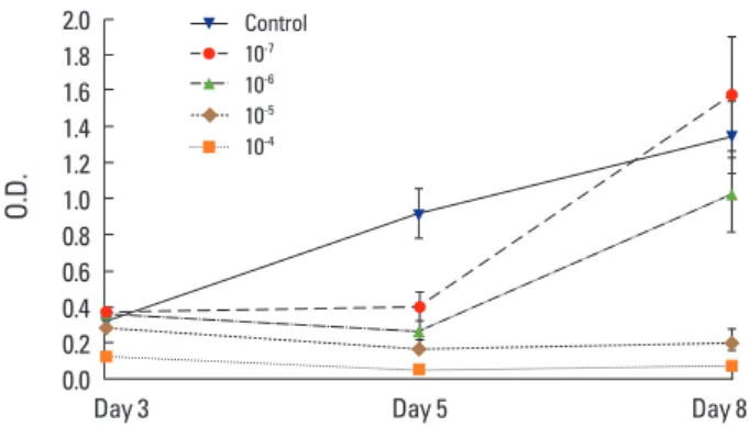
As per the MTT assay on chondrocytes, the cell survival
rate significantly decreased at 10-5 mol/L and 10-4 mol/L of
zoledronate (Fig. 1). Cell number increased in the group
treated with 10-7 mol/L of zoledronate until 3 days after
administration and then plateaued, as was the case with the control group (Fig. 2). The same concentration of zole-dronate was administered to synovial cells, and the cell number increased until 5 days after administration; the fi-nal cell number did not differ between the zoledronate and control groups (P=0.437) (Fig. 3).
2. mRNA analysis of VEGF-A and PEDF
While the expression of VEGF mRNA in chondrocytes
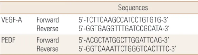
de-Table 1. Sequences of vascular endothelial growth factor-A and pig-ment epithelium-derived factor primer
Sequences VEGF-A Forward
Reverse 5’-TCTTCAAGCCATCCTGTGTG-3’ 5’-GGTGAGGTTTGATCCGCATA-3’ PEDF Forward
Reverse 5’-ACGCTATGGCTTGGATTCAG-3’ 5’-GGTCAAATTCTGGGTCACTTTC-3’ VEGF-A, vascular endothelial growth factor-A; PEDF, pigment epithelium-derived factor.
creased in the zoledronate group compared to the control group at the 8th day of culture, the difference was not sta-tistically significant (P=0.155) (Fig. 4). The expression of PEDF mRNA was also decreased in zoledronate group, but, as with VEGF, the difference was not significant (P=0.631)
(Fig. 5).
The expression of both VEGF and PEDF mRNA in synovial cells was significantly decreased in the zoledronate group compared to the control group on the 8th day of culture (P=0.022, P=0.041) (Fig. 6, 7).
Fig. 5. Expression of pigment epithelium-derived factor (PEDF) mRNA in zoledronate-treated chondrocytes was lower than in the control group at day 8, but this difference was not significant (P =0.631). ZA, zoledronate. 1.2 1.0 0.8 0.6 0.4 0.2 0 Relative ratio
Day 3 Day 5 Day 8
ZA Control
Fig. 4. Expression of vascular endothelial growth factor-A (VEGF-A) mRNA in zoledronate-treated chondrocytes was lower than in the control group at day 8, but this difference was not significant (P = 0.155). ZA, zoledronate. 1.2 1.0 0.8 0.6 0.4 0.2 0 Relative ratio
Day 3 Day 5 Day 8
ZA Control
Fig. 6. Expression of vascular endothelial growth factor-A (VEGF-A) mRNA in zoledronate-treated synovial cells was significantly lower than in the control group at day 8 (*P =0.022). ZA, zoledronate.
2.0 1.8 1.6 1.4 1.2 1.0 0.8 0.6 0.4 0.2 0 Relative ratio
Day 3 Day 5 Day 8
ZA
Control *
Fig. 3. Proliferation of synovial cells. Cell number decreased in cells treated with 10-7 mol/L zoledronate relative to the control group by day
8, but this difference was not significant for each time point (P >0.05).
4.5 4.0 3.5 3.0 2.5 2.0 1.5 1.0 0.5 0
Day 3 Day 5 Day 8
Control Zoledronate Cell number ( × 10 5 cell/mL)
Fig. 2. Proliferation of chondrocytes. Cell number increased in cells treated with 10-7 mol/L zoledronate relative to the control group by
day 3, but this difference was not significant for each time point (P > 0.05). 7 6 5 4 3 2 1 0 Cell number ( × 10 5 cell/mL)
Day 3 Day 5 Day 8
Control Zoledronate
Fig. 1. 3-(4,5-Dimethylthiazol-2-yl)-2,5-diphenyltetrazolium bromide (MTT) assay results after culture of chondrocytes in scaffolds for 3, 5, and 8 days. Significant differences are found in the 10-5 mol/L and
10-4 mol/L groups of zoledronate (P<0.001). 2.0 1.8 1.6 1.4 1.2 1.0 0.8 0.6 0.4 0.2 0.0 O.D.
Day 3 Day 5 Day 8
Control 10-7
10-6
10-5
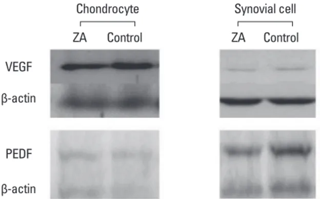
3. Western blot for VEGF-A and PEDF
The experiment was performed 6 times in both the zole-dronate and control groups. While the expression of VEGF-A and PEDF did not significantly differ between the two groups in chondrocytes (P=0.364), it was significantly de-creased in zoledronate-treated synovial cells (P=0.039, P= 0.020) (Fig. 8).
DISCUSSION
While cartilaginous joint damage, synovitis, reformation of subchondral bone and changes in bony spur formation are observed in osteoarthritis, the main pathologic chang-es are the loss of cartilage in the joints and hypertrophy and inflammatory responses of the synovial membrane. [1,3,16] While cartilaginous joints do not contain blood vessels or nerves, angiogenesis occurs in the bony carti-lage as osteoarthritis progresses, and is also increased in the synovial membrane.[5,7] Increased angiogenesis in in-traarticular tissue causes chronic synovitis and resulting pain, and also affects functional maintenance and homeo-stasis of cartilage, leading to cartilage damage. Inhibition of angiogenesis may prevent the development and progres-sion of osteoarthritis, and several studies have reported decreased inflammatory responses and angiogenesis fol-lowing administration of angiogenesis-suppressing agents. [17,18] Among the several agents known to be associated with angiogenesis, VEGF is a major player.[6,7,10] This study aimed to observe the effects of bisphosphonates on chon-drocytes and synovial cells and identify whether the drug suppresses angiogenesis in joint tissues.
VEGF is not expressed in normal cartilaginous joints, but
it is expressed in osteoarthritic joints, influencing the patho-genesis and the progression of the disease. Increased VEGF promotes not only angiogenesis, but also the invasiveness of capillaries in the joint.[6,7,10] These findings have been reported to be associated with bony spur formation in os-teoarthritis and with pathologic angiogenesis in rheuma-toid arthritis.[9,11,19]
Bisphosphonate is a potent inhibitor of osteoclasts, sup-pressing bone resorption, and is thus commonly used for the treatment of osteoporosis. While there have been re-ports on the role of bisphosphonates in providing anti-in-flammatory effects and in facilitating macrophage death in osteoarthritis, little is known regarding the effect of this agent on cartilage and synovial membranes.[20] It has been found that bisphosphonates decrease the concentration of circulating VEGF during tumor metastasis.[12,13,19,21] Re-cent studies have reported that bisphosphonates suppress the resorption of cartilage by inhibiting angiogenesis in the cartilage of growth plates; however, the underlying mechanism of its effect on cartilaginous joints has not yet been identified.[7,11,22]
In a study on the effect of bisphosphonates, including clodronate, pamidronate, and risedronate, on the survival and proliferation of chondrocytes, Van Offel et al.[23] re-ported that the survival and proliferation of cells are
sup-pressed at a bisphosphonate concentration of 10-6 mol/L.
They also reported that it is safe when used at therapeutic concentrations and that it actually suppresses steroid-in-duced death of chondrocytes. However, no studies on the
Fig. 7. Expression of pigment epithelium-derived factor (PEDF) mRNA in zoledronate-treated synovial cells was significantly lower than in the control group at day 8 (*P =0.041). ZA, zoledronate.
2.0 1.8 1.6 1.4 1.2 1.0 0.8 0.6 0.4 0.2 0 Relative ratio
Day 3 Day 5 Day 8
ZA Control
*
Fig. 8. Western blot analysis for vascular endothelial growth factor (VEGF) & pigment epithelium-derived factor (PEDF) in chondrocytes and synovial cells with or without zoledronate revealed no significant difference in chondrocytes, but a significant difference in synovial cells (P=0.039, P=0.020). VEGF, vascular endothelial growth factor; PEDF, pigment epithelium-derived factor; ZA, zoledronate.
Chondrocyte ZA Control VEGF β-actin PEDF β-actin ZA Control Synovial cell
effect of zoledronate, a more potent bone resorption sup-pressant, on the survival and proliferation of chondrocytes had been reported before now. The results of the current study revealed that zoledronate suppressed the survival and proliferation of chondrocytes at a concentration above
10-6 mol/L, similar to the results of previous study on other
bisphosphonates.[23] This suggests that the effect of zole-dronate on chondrocytes is minimal even when the drug is injected intravenously.
While the expression of VEGF-A mRNA decreased in
chon-drocytes administered 10-7 mol/L of zoledronate compared
to that in controls, the difference was not significant. This result differs from that found by Evans and Oberbauer,[11] who reported that alendronate inhibits the expression of VEGF in chondrocytes from growth plates. A direct com-parison between the two studies may be difficult since the type and the activity of the bisphophonates used in the studies are different; however, these contradictory result may be attributable to the difference in characteristics be-tween growth-plate cartilage and joint cartilage. The effect of bisphosphonates on chondrocytes is thought to be min-imal since bisphosphonates are mainly deposited in hydroxy-apatite; furthermore, the expression of PEDF, which sup-presses angiogenesis, did not change either.
Unlike in chondrocytes, the expression of VEGF-A mRNA decreased in synovial cells after treatment with zoledro-nate. While the effect of bisphosphonates on an actual joint cannot be determined from this study, it is possible that they are deposited into bone tissue adjacent to synovial cells, suppressing the expression of angiogenic factors. Mel-inte et al.[14] reported detecting the expression of VEGF in synovial cells by histological staining of tissues affected by osteoarthritis and rheumatoid arthritis. The results of this study suggest that zoledronate may decrease the expres-sion of VEGF in synovial cells and suppresses angiogenesis, reducing inflammatory changes and alleviating the resul-tant pain.
One limitation of this study is the lack of an in vivo study elucidating whether zoledronate acts on synovial cells by being incorporated directly into the synovial membrane, or by first being deposited into the bone and then affect-ing adjacent synovial cells. Nor has it been determined whe-ther its effect on synovial cells indirectly influences chon-drocytes. Regardless, this study is meaningful in that it is the first to have investigated the expression of VEGF-A and
PEDF in articular chondrocytes and synovial cells following treatment with zoledronate.
In conclusion, the effect of zoledronate on the expres-sion of VEGF-A in chondrocytes was not significant, and therefore, its action on chondrocytes seems to be minimal; however, it does decrease VEGF-A expression in synovial cells, which may impinge upon the mechanisms of inflam-mation and cartilage damage in osteoarthritis.
REFERENCES
1. Brandt KD, Dieppe P, Radin EL. Etiopathogenesis of osteo-arthritis. Rheum Dis Clin North Am 2008;34:531-59. 2. Goldring MB, Goldring SR. Articular cartilage and
subchon-dral bone in the pathogenesis of osteoarthritis. Ann N Y Acad Sci 2010;1192:230-7.
3. Krasnokutsky S, Attur M, Palmer G, et al. Current concepts in the pathogenesis of osteoarthritis. Osteoarthritis Carti-lage 2008;16 Suppl 3:S1-3.
4. Ashraf S, Mapp PI, Walsh DA. Contributions of angiogene-sis to inflammation, joint damage, and pain in a rat model of osteoarthritis. Arthritis Rheum 2011;63:2700-10. 5. Mapp PI, Walsh DA. Mechanisms and targets of
angiogen-esis and nerve growth in osteoarthritis. Nat Rev Rheuma-tol 2012;8:390-8.
6. Jansen H, Meffert RH, Birkenfeld F, et al. Detection of vas-cular endothelial growth factor (VEGF) in moderate osteo-arthritis in a rabbit model. Ann Anat 2012;194:452-6. 7. Murata M, Yudoh K, Masuko K. The potential role of
vascu-lar endothelial growth factor (VEGF) in cartilage: how the angiogenic factor could be involved in the pathogenesis of osteoarthritis? Osteoarthritis Cartilage 2008;16:279-86. 8. Broadhead ML, Akiyama T, Choong PF, et al. The patho-physiological role of PEDF in bone diseases. Curr Mol Med 2010;10:296-301.
9. Fan W, Crawford R, Xiao Y. The ratio of VEGF/PEDF expres-sion in bone marrow mesenchymal stem cells regulates neovascularization. Differentiation 2011;81:181-91. 10. Tong JP, Yao YF. Contribution of VEGF and PEDF to
choroi-dal angiogenesis: a need for balanced expressions. Clin Biochem 2006;39:267-76.
11. Evans KD, Oberbauer AM. Alendronate inhibits VEGF ex-pression in growth plate chondrocytes by acting on the mevalonate pathway. Open Orthop J 2009;3:83-8. 12. Kim MS, Kim JH, Lee MR, et al. Effects of alendronate on a
disintegrin and metalloproteinase with thrombospondin motifs expression in the developing epiphyseal cartilage in rats. Anat Histol Embryol 2009;38:154-60.
13. Ravosa MJ, Ning J, Liu Y, et al. Bisphosphonate effects on the behaviour of oral epithelial cells and oral fibroblasts. Arch Oral Biol 2011;56:491-8.
14. Melinte R, Jung I, Georgescu L, et al. VEGF and CD31 ex-pression in arthritic synovium and cartilage of human knee joints. Rom J Morphol Embryol 2012;53:911-5.
15. Zhang S, Cao W, Wei K, et al. Expression of VEGF-receptors in TMJ synovium of rabbits with experimentally induced internal derangement. Br J Oral Maxillofac Surg 2013;51: 69-73.
16. Pasternak B, Aspenberg P. Metalloproteinases and their inhibitors-diagnostic and therapeutic opportunities in or-thopedics. Acta Orthop 2009;80:693-703.
17. Choi ST, Kim JH, Seok JY, et al. Therapeutic effect of anti-vascular endothelial growth factor receptor I antibody in the established collagen-induced arthritis mouse model. Clin Rheumatol 2009;28:333-7.
18. Sekimoto T, Hamada K, Oike Y, et al. Effect of direct
angio-genesis inhibition in rheumatoid arthritis using a soluble vascular endothelial growth factor receptor 1 chimeric protein. J Rheumatol 2002;29:240-5.
19. Di Salvatore M, Orlandi A, Bagalà C, et al. Anti-tumour and anti-angiogenetic effects of zoledronic acid on human non-small-cell lung cancer cell line. Cell Prolif 2011;44:139-46. 20. Osterman T, Kippo K, Laurén L, et al. Effect of clodronate
on established adjuvant arthritis. Rheumatol Int 1994;14: 139-47.
21. Le Goff B, Heymann D. Pharmacodynamics of bisphospho-nates in arthritis. Expert Rev Clin Pharmacol 2011;4:633-41.
22. Kang JH, Choi NK, Kang SJ, et al. Alendronate affects carti-lage resorption by regulating vascular endothelial growth factor expression in rats. Anat Rec (Hoboken) 2010;293: 786-93.
23. Van Offel JF, Schuerwegh AJ, Bridts CH, et al. Effect of bispho-sphonates on viability, proliferation, and dexamethasone-induced apoptosis of articular chondrocytes. Ann Rheum Dis 2002;61:925-8.