Received: November 23, 2015.
Accepted: February 17, 2016.
Pre-published: February 17,2016.
©2016 Ferrata Storti Foundation
Check the online version for the most updated information on this article, online supplements, and information on authorship & disclosures:
www.haematologica.org/content/101/06/717
Material published in Haematologica is cov- ered by copyright. All rights reserved to the Ferrata Storti Foundation. Copies of articles are allowed for personal or internal use.
Permission in writing from the publisher is required for any other use.
Correspondence:
Ferrata Storti Foundation
EUROPEAN HEMATOLOGY ASSOCIATION
Haematologica 2016 Volume 101(6):717-723
doi:10.3324/haematol.2015.139899
The aim of the Korean Imatinib Discontinuation Study was to identify predictors for safe and successful imatinib discontinua- tion. A total of 90 patients with a follow-up of ≥12 months were analyzed. After a median follow-up of 26.6 months after imatinib dis- continuation, 37 patients lost the major molecular response. The proba- bility of sustained major molecular response at 12 months and 24 months was 62.2% and 58.5%, respectively. All 37 patients who lost major molecular response were retreated with imatinib therapy for a median of 16.9 months, and all achieved major molecular response again at a median of 3.9 months after resuming imatinib therapy. We observed newly developed or worsened musculoskeletal pain and pru- ritus in 27 (30%) patients after imatinib discontinuation. Imatinib with- drawal syndrome was associated with a higher probability of sustained major molecular response (P=0.003) and showed a trend for a longer time to major molecular response loss (P=0.098). Positivity (defined as ≥ 17 positive chambers) of digital polymerase chain reaction at screening and longer imatinib duration before imatinib discontinuation were asso- ciated with a higher probability of sustained major molecular response.
Our data demonstrated that the occurrence of imatinib withdrawal syn- drome after imatinib discontinuation and longer duration of imatinib were associated with a lower rate of molecular relapse. In addition, min- imal residual leukemia measured by digital polymerase chain reaction had a trend for a higher molecular relapse. (Trial registered at ClinicalTrials.gov: NCT01564836).
Imatinib withdrawal syndrome and longer duration of imatinib have a close association with a lower molecular relapse after treatment discontinuation: the KID study
Sung-Eun Lee,1Soo Young Choi,1Hye-Young Song,1Soo-Hyun Kim,1 Mi-Yeon Choi,1Joon Seong Park,2Hyeoung-Joon Kim,3Sung-Hyun Kim,4 Dae Young Zang,5Sukjoong Oh,6Hawk Kim,7Young Rok Do,8Jae-Yong Kwak,9 Jeong-A Kim,10Dae-Young Kim,11Yeung-Chul Mun,12Won Sik Lee,13
Myung Hee Chang,14Jinny Park,15 Ji Hyun Kwon,16 and Dong-Wook Kim1,17
1Department of Hematology, Seoul St. Mary's Hospital, The Catholic University of Korea, Seoul; 2Department of Hematology-Oncology, Ajou University School of Medicine, Suwon; 3Department of Hematology-Oncology, Chonnam National University Hwasun, Hospital; 4Department of Internal Medicine, Dong-A University College of Medicine, Busan; 5Department of Internal Medicine, Hallym University College of Medicine, Anyang; 6Division of Hematology-Oncology, Department of Internal Medicine, Kangbuk Samsung Hospital, School of Medicine, Sungkyunkwan University, Seoul; 7Division of Hematology and Cellular Therapy, Ulsan University Hospital, University of Ulsan College of Medicine; 8Division of Hematology-Oncology, School of Medicine, Keimyung
University, Daegu; 9Division of Hematology-Oncology, Chonbuk National University Medical School, Jeonju; 10Department of Hematology, St. Vincent’s Hospital, The Catholic University of Korea, Suwon; 11Department of Hematology, Asan Medical Center, University of Ulsan College of Medicine, Seoul; 12Department of Hematology, School of Medicine, Ewha Womans University, Seoul; 13Department of Internal Medicine, Inje University College of Medicine, Inje University Busan Paik Hospital; 14Department of Hematology-Oncology, National Health Insurance Service Ilsan Hospital, Ilsan;
15Department of Hematology, Gachon University Gil Hospital, Incheon; 16Department of Hematology-Oncology, Chungbuk National University Hospital, Cheongju; and 17Catholic Leukemia Research Institute, The Catholic University of Korea, Seoul, Korea
ABSTRACT
© Ferrata Storti Foundation
Introduction
Since the publication of several recent reports showing that imatinib (IM) discontinuation can be performed in patients who have had sufficient IM therapy and have undetectable molecular residual disease (UMRD), treat- ment-free remission (TFR) has emerged as a goal in the treatment of chronic myeloid leukemia (CML).1-5Although UMRD with a sensitivity of 4.5-log for at least 2 years is typically required and some factors associated with a higher rate of sustained UMRD after IM discontinuation have been reported, the precise identification of the mini- mum requirement for a safe and successful TFR and the underlying mechanism of sustained UMRD remain elu- sive.
The IM discontinuation studies have presented several predictors for TFR.1,3,4,6,7Recently, Horn et al. provided a predictive mathematical model to prognosticate the patient-specific risk of molecular relapse on treatment dis- continuation,8 and the TWISTER study tested the utility of DNA analysis by polymerase chain reaction (PCR).4 Currently, the ISAV and EURO-SKI studies are exploring the additional predictors of age, digital polymerase chain reaction (dPCR) results, and withdrawal syndromes such as musculoskeletal pain and pruritus.9,10 Our previous study of health-related profiles including quality of life (HRQOL) also demonstrated that IM withdrawal syn- drome can develop in some patients.11
This Korean multicenter prospective study (Korean Imatinib Discontinuation Study; KID Study) has been per- formed to identify predictors for safer and more successful IM discontinuation and to explore additional contributing factors for a sustained molecular response (MR) in specific patient cohorts. Herein, we analyzed the results of patients with a sufficient follow-up to confirm the signifi- cant predictors of molecular relapse using preliminary identified factors.
Methods Study patients
Patients with chronic-phase CML who were treated with first- line IM for more than 3 years and had undetectable BCR-ABL1 transcript by quantitative reverse transcription PCR (qRT-PCR) for at least 2 years were eligible for the KID study. Previous reports from the KID study included 48 patients who were enrolled from 9 October 2010 to 7 November 2012; 28 patients who had IM without transplantation, and 20 patients who received IM for post-transplant relapse.3By the data cut-off date of 31 July 2015 for this analysis, a total of 156 patients were enrolled. Among them, 90 non-transplant patients with at least 12 months of fol- low-up were included in this analysis. Written informed consent was obtained from each patient before enrollment. This study was approved by the Institutional Review Board of each participating institution and conducted in accordance with the Declaration of Helsinki. The study protocol was registered with the National Institutes of Health clinical trial registry as #NCT01564836.
Questionnaire relating to withdrawal syndrome
Patients were provided with a questionnaire composed of 43 parameters in 3 sections (IM-related adverse events, physical health, and mental health). Each parameter was rated using a five- point score at each time point and changes were calculated as dif- ferences in score from the baseline (at the time of IM discontinua-
tion).11As newly developed or worsened musculoskeletal pain and pruritus were observed in a significant proportion of patients, all patients were asked to complete an additional questionnaire for IM withdrawal syndrome (IMWS).
Evaluation of molecular response
To confirm undetectable BCR-ABL1 transcript levels by qRT- PCR for at least 2 years, duplicated qRT-PCR analyses were per- formed at more than six time points. For all screening and subse- quent follow-up samples, duplicated qRT-PCR and nested RT- PCR with at least 4.5-log sensitivity was performed in the central laboratory (Cancer Research Institute, The Catholic University of Korea, Seoul, Korea) and only qRT-PCR results with more than 50,000 ABL1 transcripts were analyzed. Major molecular response (MMR) was defined as a BCR-ABL1 transcript level of 0.1% or lower on the international scale (IS). UMRD was defined as negative PCR results in duplicated qRT-PCR assays with 5-log sensitivity.
After IM discontinuation, the molecular response was moni- tored every month for the first 6 months, every 2 months up to 12 months, and every 3 months thereafter. Loss of MMR and UMRD was defined after 2 consecutive assessments within 4 weeks, and in cases of MMR loss (molecular relapse) IM treatment was rein- troduced. After IM resumption for MMR loss, the molecular response was evaluated every month until MMR was re-achieved and every 3 months thereafter.
Digital PCR
For more sensitive quantitation of minimal residual disease, all screening samples were tested with 10-7sensitivity by digital PCR (dPCR) as previously reported.9,12 A brief method is provided in the Online Supplementary Methods. To identify the optimal cut-off
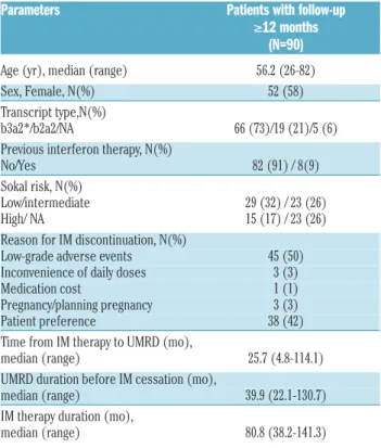
Table 1.Patient characteristics.
Parameters Patients with follow-up ≥12 months (N=90) Age (yr), median (range) 56.2 (26-82) Sex, Female, N(%) 52 (58) Transcript type,N(%) b3a2*/b2a2/NA 66 (73)/19 (21)/5 (6) Previous interferon therapy, N(%) No/Yes 82 (91) / 8(9) Sokal risk, N(%) Low/intermediate 29 (32) / 23 (26) High/ NA 15 (17) / 23 (26) Reason for IM discontinuation, N(%) Low-grade adverse events 45 (50) Inconvenience of daily doses 3 (3) Medication cost 1 (1) Pregnancy/planning pregnancy 3 (3) Patient preference 38 (42) Time from IM therapy to UMRD (mo),
median (range) 25.7 (4.8-114.1) UMRD duration before IM cessation (mo),
median (range) 39.9 (22.1-130.7) IM therapy duration (mo),
median (range) 80.8 (38.2-141.3)
IM: imatinib; NA: not available; UMRD: undetectable molecular residual disease; mo:
months.*One patient had a type of b3a2+b2a2.
© Ferrata Storti Foundation
in the 765 chambers analyzed by dPCR for predicting loss of MMR, we performed a receiver operating characteristic curve analysis and applied different cut-off levels iteratively in steps of 1 positive chamber. A cut-off of more than 17 positive chambers was chosen for a positive result of dPCR with a significantly lower probability of sustained MMR.
Statistical analysis
Time to loss of UMRD and MMR was calculated from the date of IM discontinuation to the date of the first detection of BCR-ABL1 in two consecutive analyses or, for patients who did not lose MR, was determined by default on the date of the last molecular examination. The probability of sustained MR was plotted using the Kaplan-Meier method and compared using the log-rank test.
Results
Patient characteristics
A total of 90 patients with UMRD (38 men and 52 women) with a median age of 56.2 years (range, 26-82 years) were analyzed. The percentage of patients with low, intermediate, and high Sokal risk scores was 32%, 26%, and 17%, respectively, and 26% had an unknown risk. Eight patients had a previous history of interferon treatment. The reasons for participating in this study included low-grade adverse events (n=45), inconvenience of daily doses (n=3), medication cost (n=1), pregnancy/planning pregnancy (n=3), and patient’s wishes (n=38). Prior to discontinuation, the median duration of IM therapy was 80.8 months (range, 38.2-141.3 months) and the duration of sustained UMRD was 39.9 months (range, 22.1-130.7 months). Patient characteristics are summarized in Table 1.
Outcomes after discontinuation of IM therapy
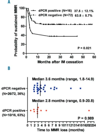
After a median follow-up of 26.6 months (range, 12.6- 58.1 months) after IM discontinuation, 45 patients (50%) lost UMRD. Eight patients who lost UMRD but not MMR exhibited different patterns of BCR-ABL1 kinetics: 7 patients spontaneously re-achieved UMRD after a median time of 1.2 months (range, 0.7-5.4 months) and 1 patient showed fluctuation of BCR-ABL1 transcript under the level of 0.1% on IS for 20.6 months. The other 37 patients lost MMR in 2 consecutive analyses. The overall 12- month and 24-month probability of sustained MMR was 62.2% and 58.5%, respectively (Figure 1A). The overall 12-month and 24-month probability of sustained UMRD was 50.0% and 50.0%, respectively. Among the 37 patients with molecular relapse, the median time to MMR loss was 3.3 months (range, 0.9-20.8 months) after IM dis- continuation (Figure 1B) and the median time from loss of UMRD to loss of MMR was 1.0 month (range, 0.0-13.6 months) (Table 2).
Molecular kinetics after IM resumption
All 37 patients who lost MMR were retreated with IM therapy for a median of 16.9 months (range, 4.6-47.4 months). All patients re-achieved MMR at a median of 3.9 months (range, 0.5-11.1 months) after resuming IM thera- py, and 32 re-achieved UMRD at a median of 7.2 months (range, 3.3-14.7 months). The other five patients are still on MMR for a median of 6.3 months (range 4.6-23.2) of follow-up duration.
Association of baseline minimal residual disease with molecular kinetics
Among 88 patients with available dPCR data at screen- ing, 16 (18%) patients had a positive result of dPCR based on a cut-off of 17 positive chambers. Ten out of 16 (63%) patients with positivity of dPCR and 26 out of 72 (36%) patients with negativity of dPCR had molecular relapse.
Patients in the dPCR-negative group had a higher proba- bility of sustained MMR than patients in the dPCR-posi- tive group (63.8% vs. 37.5%, P=0.021) (Figure 2A). Among 37 patients with molecular relapse, the median time to MMR loss was not different according to positivity of dPCR at baseline (3.6 months in the dPCR negative group vs. 2.8 months in the dPCR positive group, P=0.989) (Figure 2B).
Association of withdrawal syndrome with molecular kinetics
Changes in musculoskeletal pain and pruritus after IM discontinuation were carefully evaluated for all 90 patients. Interestingly, aggravation or new development of musculoskeletal pain and/or pruritus after IM discontinu- ation was presented in 27 (30%) patients, which was defined as IMWS. Online Supplementary Tables S1 and S2 present the characteristics of patients with aggravation or new development of musculoskeletal pain and pruritus after IM discontinuation, respectively. Musculoskeletal pain seemed to continue for quite a while, with a median duration of 6 months (range 1-36 months), and pruritus continued for a median of 3 months (range 2-54 months).
In addition, there was no patient who restarted IM due to IMWS. Five patients in the IMWS group (18%) lost MMR compared with 32 (51%) patients in the no IMWS group.
Patients with IMWS had a higher probability of sustained Figure 1. Probability of sustained MMR (A) and time to MMR loss (B) in patients with follow-up ≥ 12 months (N=90).
A
B
© Ferrata Storti Foundation
MMR than patients without IMWS (79.5% vs. 49.2%, P=0.003) (Figure 3A). Among 37 patients with molecular relapse, the median time to MMR loss was longer in the patients with IMWS than in those without IMWS (3.0 months vs. 7.6 months, P=0.098) (Figure 3B). In addition creatine phosphokinase (CPK) was not noted in associa- tion with the IMWS (Online Supplementary Figure S1). The elevation of erythrocyte sedimentation rate (ESR) and C- reactive protein (CRP) were noted in 5 and 4 of 8 evaluable patients with IMWS, respectively, whereas both were nor- mal in 9 evaluable patients without IMWS (data not shown).
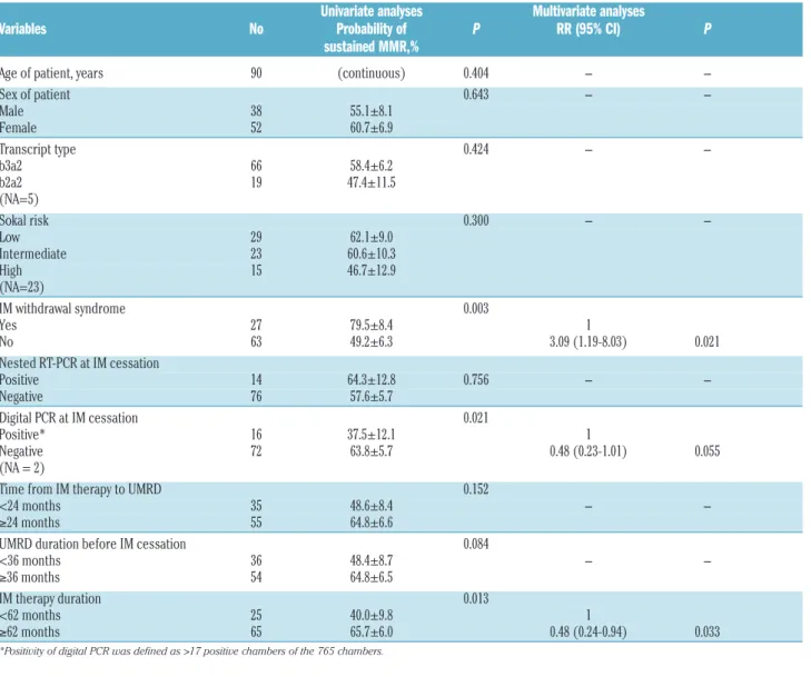
Overall predictive factors affecting sustained MMR Univariate and multivariate analyses for predictive fac- tors of sustained MMR were performed (Table 3). In addi- tion to IMWS (P=0.003), negativity of digital PCR at screening (P=0.021) and longer duration of IM therapy (P=0.013) were identified as potential factors for sustained MMR. Longer duration of UMRD (P=0.084) showed a trend for a higher rate of sustained MMR. Multivariate analyses showed that the presence of IMWS (P=0.021) and longer duration of IM therapy (P=0.033) were inde- pendent factors for sustained MMR, and negativity of dPCR at screening (P=0.055) maintained a trend for sus- tained MMR. Combining models according to the number of predictive factors for sustained MMR allows better dis- crimination (Figure 4).
Discussion
Although the results of prospective clinical trials indicat- ed the possibility of TFR in 30-50% of patients after IM discontinuation, because the variables for a sustained molecular response have changed and relapse rates have increased with longer follow-up, many clinical questions regarding safe and successful IM discontinuation remain.2,
13-15We previously reported results from 48 patients show-
ing that the 12-month probability of sustained MMR and UMRD was 79.9% and 80.8%, respectively (64.4% and 66.3% among 28 non-transplant patients).3In this further analysis, the 12-month probability of sustained MMR and UMRD was 62.2% and 50.0%, respectively. This rate was similar to that reported by the A-STIM trial (median IM duration 79 months; median UMRD duration 41 months)2 and slightly higher than that reported by the TWISTER trial (median IM duration 70 months; median UMRD duration 36 months).4 In our study, the median duration of
IM therapy and UMRD before IM discontinuation was 80.8 months and 39.9 months, respectively. We employed stringent PCR sensitivity criteria for accurate measure- ment of BCR-ABL1 transcript levels prior to discontinua- tion and examined BCR-ABL1 kinetics in response to IM resumption and after IM cessation in detail.
The confirmation of predictive factors for durable TFR is a key issue. Increased IM therapy duration was reported to be strongly associated with a higher probability of sus- tained UMRD in previous studies.1,3,6,7Sokal risk score,1,4,6 sex,1prior interferon treatment,4,7UMRD duration before IM discontinuation,3,7 time to UMRD,6 age,9 and dPCR9 were also reported as potential predictive factors, albeit with conflicting data. To identify additional predictors and to validate predictors explored in our previous study,3 we performed further analysis with more patients who had at least 12 months of follow-up. This analysis was based on additional clinical observations: first, we observed an interesting phenomenon in some patients who developed musculoskeletal pain with or without pruritus after dis- continuation of IM, implying a possible link to autoimmu- nity;11and second, detectable minimal residual leukemia by the more sensitive approach of dPCR may have a sig- nificant positive predictive value for molecular relapse.9
In this study, we observed that most IM-related adverse events such as nausea, indigestion, peripheral edema, and skin whitening and fragility were rapidly resolved, where- as musculoskeletal pain and pruritus were newly devel- oped or worsened in some patients after IM discontinua- tion. These observations are similar to the findings of the Table 2.Outcomes after imatinib discontinuation.
Patients with follow-up ≥ 12 months (N=90) Follow-up duration (mo), median (range) 26.6 (12.6-58.1) Loss of UMRD, N (%) 45* (50) Loss of MMR, N (%) 37 (41) Time to loss of UMRD (mo) median (range) 2.0 (0.9-9.5) Time to loss of MMR (mo), median (range) 3.3 (0.9-20.8) Time to from loss of UMRD to loss of MMR (mo), median (range) 1.0 (0-13.6)
mo: months; UMRD: undetectable molecular residual disease; MMR: major molecular response. *Among 8 patients who lost UMRD but not MMR, 7 patients spontaneously re- achieved UMRD and 1 patient showed fluctuation of BCR-ABL1 transcript under the level of 0.1% on IS.
Figure 2. Probability of sustained MMR (A)and time to MMR loss (B) according to positivity of dPCR (defined as > 17 positive chambers) at baseline.
A
B
© Ferrata Storti Foundation
EURO-SKI study,10in which 15 (30%) out of 50 patients reported musculoskeletal pain evolving gradually from 1 to 6 weeks after TKI discontinuation, and may suggest that the newly developed or worsened musculoskeletal
pain and pruritus were due to IMWS. Interestingly, our study found an association between IMWS and sustained MMR: the patients with IMWS showed a higher rate of sustained MMR and a longer time to MMR loss.
Table 3. Univariate and multivariate analyses of variables affecting the probability of sustained MMR in the patients with follow-up ≥12 months (N=90).
Univariate analyses Multivariate analyses
Variables No Probability of P RR (95% CI) P
sustained MMR,%
Age of patient, years 90 (continuous) 0.404 - -
Sex of patient 0.643 - -
Male 38 55.1±8.1
Female 52 60.7±6.9
Transcript type 0.424 - -
b3a2 66 58.4±6.2
b2a2 19 47.4±11.5
(NA=5)
Sokal risk 0.300 - -
Low 29 62.1±9.0
Intermediate 23 60.6±10.3
High 15 46.7±12.9
(NA=23)
IM withdrawal syndrome 0.003
Yes 27 79.5±8.4 1
No 63 49.2±6.3 3.09 (1.19-8.03) 0.021
Nested RT-PCR at IM cessation
Positive 14 64.3±12.8 0.756 - -
Negative 76 57.6±5.7
Digital PCR at IM cessation 0.021
Positive* 16 37.5±12.1 1
Negative 72 63.8±5.7 0.48 (0.23-1.01) 0.055
(NA = 2)
Time from IM therapy to UMRD 0.152
<24 months 35 48.6±8.4 - -
≥24 months 55 64.8±6.6
UMRD duration before IM cessation 0.084
<36 months 36 48.4±8.7 - -
≥36 months 54 64.8±6.5
IM therapy duration 0.013
<62 months 25 40.0±9.8 1
≥62 months 65 65.7±6.0 0.48 (0.24-0.94) 0.033
*Positivity of digital PCR was defined as >17 positive chambers of the 765 chambers.
Figure 3. Probability of sustained MMR (A)and time to MMR loss (B)according to the presence of IM withdrawal syndrome.
A B
© Ferrata Storti Foundation
Considering the role of inflammatory cytokines in muscu- loskeletal pain,16 whether IMWS is linked to immunologic background warrants further investigation. Briefly, we observed that ESR and CRP were increased in some patients with IMWS (data not shown) but CPK were not noted in association with the musculoskeletal symptoms.
The importance of immunologic status for successful TFR is illustrated by the beneficial effect of natural killer (NK) cell activity17,18and interferon treatment on a higher TFR rate,4,7 indicating that the re-activation of autologous immunity may contribute to the elimination of MRD after IM discontinuation via the emergence of NK cells or cyto- toxic T lymphocytes (CTLs) against CML-associated anti- gens.19-21Although we analyzed the frequency and func- tion of NK cells after IM discontinuation in some patients, no significant association was found.
In our previous study, we showed that 24 out of 32 PCR-negative samples as assayed by conventional qRT- PCR showed detectable BCR-ABL1 in dPCR. More importantly, dPCR with preamplification showed a greater sensitivity with 2-3-log improvement and a con- tinuous decrease in BCR-ABL1 upon IM treatment was evident in the follow-up samples analyzed by dPCR.12 These results suggest that the duration of UMRD on con- tinued IM treatment will be an important predictor of sustained molecular remission and that the detection of very low levels of BCR-ABL1 by dPCR may help to select candidates for a trial of TFR. In this study, positivity of dPCR at baseline was confirmed as an important deter- minant of molecular relapse. However, there are conflict- ing data regarding the association of the depth of MRD at baseline. In A-STIM, which included patients with intermittent low-level positive qRT-PCR tests during the 2 years prior to IM discontinuation, there was no differ- ence in the rate of loss of MMR according to MRD.2In
the TWISTER study, which used a highly sensitive (6-log below baseline) patient-specific assay for BCR-ABL1 DNA, the presence or absence of MRD was not inform- ative with regard to the subsequent loss of MMR, although this approach did enable earlier detection of MMR loss than qRT-PCR.4 In addition to the persistence of BCR-ABL1 genomic DNA in non-cycling CML stem cell clones, this discrepancy may be caused by the diverse factors affecting the sustained MR, including the biologic nature of high-risk disease and host immune response. Therefore, further studies are warranted to val- idate the utility of dPCR in the TFR setting and to deter- mine concomitant immunobiologic factors.
In terms of BCR-ABL1 transcript kinetics after IM dis- continuation, in our study, among the 37 patients who lost MMR, 20 lost MMR within 3 months of discontinuing IM but the latest loss of MMR was at 20.8 months. This implies that qRT-PCR monitoring should be continued to safeguard patients with delayed relapse. However, the fact that most patients re-achieved MMR at approximately 4 months after resuming IM therapy supported the safety of IM discontinuation under intensive follow-up.
Our data demonstrated that the occurrence of IMWS after discontinuation of long-term IM therapy was closely associated with a lower molecular relapse, and longer duration of IM provided a predictive factor for successful IM discontinuation. In addition, minimal residual leukemia measured by dPCR had a trend for a higher molecular relapse.
Funding
This study was supported by a grant from the National R&D Program for Cancer Control, Ministry of Health & Welfare, Republic of Korea (1020400) and the Korea Leukemia Bank (NRF-2013M3A9B8031236).
Figure 4. Probability of sustained MMR according to the number of predictive factors (negative dPCR, presence of IMWS, and longer than 62 months of IM therapy duration).
References
1. Mahon FX, Rea D, Guilhot J, et al.
Discontinuation of imatinib in patients with chronic myeloid leukaemia who have maintained complete molecular remission for at least 2 years: the prospective, multi- centre Stop Imatinib (STIM) trial. Lancet Oncol. 2010;11(11):1029-1035.
2. Rousselot P, Charbonnier A, Cony- Makhoul P, et al. Loss of major molecular response as a trigger for restarting tyrosine kinase inhibitor therapy in patients with chronic-phase chronic myelogenous leukemia who have stopped imatinib after durable undetectable disease. J Clin Oncol.
2014;32(5):424-430.
3. Lee SE, Choi SY, Bang JH, et al. Predictive factors for successful imatinib cessation in
chronic myeloid leukemia patients treated with imatinib. Am J Hematol. 2013;88(6):
449-454.
4. Ross DM, Branford S, Seymour JF, et al.
Safety and efficacy of imatinib cessation for CML patients with stable undetectable minimal residual disease: results from the TWISTER study. Blood. 2013;122(4):515- 522.
5. Thielen N, van der Holt B, Cornelissen JJ, et
© Ferrata Storti Foundation
al. Imatinib discontinuation in chronic phase myeloid leukaemia patients in sus- tained complete molecular response: a ran- domised trial of the Dutch-Belgian Cooperative Trial for Haemato-Oncology (HOVON). Eur J Cancer. 2013;49(15):3242- 3246.
6. Yhim HY, Lee NR, Song EK, et al. Imatinib mesylate discontinuation in patients with chronic myeloid leukemia who have received front-line imatinib mesylate thera- py and achieved complete molecular response. Leuk Res. 2012;36(6):689-693.
7. Takahashi N, Kyo T, Maeda Y, et al.
Discontinuation of imatinib in Japanese patients with chronic myeloid leukemia.
Haematologica. 2012;97(6):903-906.
8. Horn M, Glauche I, Muller MC, et al.
Model-based decision rules reduce the risk of molecular relapse after cessation of tyro- sine kinase inhibitor therapy in chronic myeloid leukemia. Blood. 2013;121(2):378- 384.
9. Mori S, Vagge E, le Coutre P, et al. Age and dPCR can predict relapse in CML patients who discontinued imatinib: The ISAV study. Am J Hematol. 2015;90(10):910-914.
10. Richter J, Soderlund S, Lubking A, et al.
Musculoskeletal pain in patients with chronic myeloid leukemia after discontinu-
ation of imatinib: a tyrosine kinase inhibitor withdrawal syndrome? J Clin Oncol.
2014;32(25):2821-2823.
11. Park JS, Lee SE, Jeong SH, et al. Change of health-related profiles after Imatinib cessa- tion in chronic phase chronic myeloid leukemia patients. Leuk Lymphoma. 2015 Jun 12:1-7. [Epub ahead of print].
12. Goh HG, Lin M, Fukushima T, et al.
Sensitive quantitation of minimal residual disease in chronic myeloid leukemia using nanofluidic digital polymerase chain reac- tion assay. Leuk Lymphoma.
2011;52(5):896-904.
13. Ross DM, Hughes TP. How I determine if and when to recommend stopping tyrosine kinase inhibitor treatment for chronic myeloid leukaemia. Br J Haematol.
2014;166(1):3-11.
14. Breccia M, Alimena G. Discontinuation of tyrosine kinase inhibitors and new approaches to target leukemic stem cells:
treatment-free remission as a new goal in chronic myeloid leukemia. Cancer Lett.
2014; 347(1):22-28.
15. Mahon F-X, Rea D, Guilhot J, et al.
Discontinuation of Imatinib in Patients with Chronic Myeloid Leukemia Who Have Maintained Complete Molecular Response:
Update Results of the STIM Study. ASH
Annual Meeting Abstracts 2011;118(21):603.
16. Marchand F, Perretti M, McMahon SB. Role of the immune system in chronic pain. Nat Rev Neurosci. 2005;6(7):521-532.
17. Mizoguchi I, Yoshimoto T, Katagiri S, et al.
Sustained upregulation of effector natural killer cells in chronic myeloid leukemia after discontinuation of imatinib. Cancer Sci.
2013;104(9):1146-1153.
18. Ohyashiki K, Katagiri S, Tauchi T, et al.
Increased natural killer cells and decreased CD3(+)CD8(+)CD62L(+) T cells in CML patients who sustained complete molecular remission after discontinuation of imatinib.
Br J Haematol. 2012;157(2):254-256.
19. Molldrem JJ, Lee PP, Wang C, et al. Evidence that specific T lymphocytes may participate in the elimination of chronic myelogenous leukemia. Nat Med. 2000;6(9):1018-1023.
20. Kanodia S, Wieder E, Lu S, et al. PR1-specif- ic T cells are associated with unmaintained cytogenetic remission of chronic myeloge- nous leukemia after interferon withdrawal.
PLoS One. 2010;5(7):e11770.
21. Burchert A, Wolfl S, Schmidt M, et al.
Interferon-alpha, but not the ABL-kinase inhibitor imatinib (STI571), induces expres- sion of myeloblastin and a specific T-cell response in chronic myeloid leukemia.
Blood. 2003;101(1):259-264. © Ferrata Storti Foundation