pISSN 2383-7837 © 2020 The Korean Society of Pathologists/The Korean Society for Cytopathology
TP53 is a tumor suppressor gene that encodes the protein p53, which is involved in cell cycle arrest in damaged cells that require DNA repair or in cases of damage beyond repair, trig-gering apoptosis. A defect in TP53 is a crucial step in carcino-genesis. Previous studies noted that either a defect of the TP53 gene itself or of a gene upstream or downstream of TP53 was found in virtually all human cancers [1-3]. In gastric cancer (GC), p53 overexpression has been reported in 37.8%–54% of cases [4-6]. According to those studies, overexpression of p53 was generally associated with worse overall survival (OS) as well as well-known prognostic factors such as vascular invasion and lymph node metastasis.
In 2014, The Cancer Genome Atlas (TCGA) Research
Net-work Group proposed a molecular classification of GC [6]. The four subgroups were Epstein-Barr virus (EBV)–positive, micro-satellite instability, genomic stability, and chromosomal insta-bility. TP53 alteration is a characteristic of the chromosomal instability group. In the following year, the Asian Cancer Research Group (ACRG) presented a different molecular classification that considered the three factors of microsatellite instability, ep-ithelial-mesenchymal transition, and TP53 mutation [7]. The four groups classified by those factors exhibited different prog-noses. However, one of the limitations of those two studies was that the methodology used requires high-end and high-cost technologies such as next-generation gene sequencing. Differ-ent groups have attempted to develop a more practical
imple-Prediction of
TP53 mutations by p53 immunohistochemistry and their
prognostic significance in gastric cancer
Hye Jung Hwang1, Soo Kyung Nam1, Hyunjin Park2, Yujun Park1, Jiwon Koh3,
Hee Young Na1, Yoonjin Kwak3, Woo Ho Kim3, Hye Seung Lee1
1Department of Pathology, Seoul National University Bundang Hospital, Seoul National University College of Medicine, Seongnam; 2Department of Pathology, Gangnam Severance Hospital, Yonsei University College of Medicine, Seoul;
3Department of Pathology, Seoul National University Hospital, Seoul National University College of Medicine, Seoul, Korea
Background: Recently, molecular classifications of gastric cancer (GC) have been proposed that include TP53 mutations and their
func-tional activity. We aimed to demonstrate the correlation between p53 immunohistochemistry (IHC) and TP53 mutations as well as their clinicopathological significance in GC. Methods: Deep targeted sequencing was performed using surgical or biopsy specimens from 120 patients with GC. IHC for p53 was performed and interpreted as strong, weak, or negative expression. In 18 cases (15.0%) with discrepant TP53 mutation and p53 IHC results, p53 IHC was repeated. Results: Strong expression of p53 was associated with TP53 missense mutations, negative expression with other types of mutations, and weak expression with wild-type TP53 (p<.001). The sensi-tivity for each category was 90.9%, 79.0%, and 80.9%, and the specificity was 95.4%, 88.1%, and 92.3%, respectively. The TNM stage at initial diagnosis exhibited a significant correlation with both TP53 mutation type (p=.004) and p53 expression status (p=.029). The Kaplan-Meier survival analysis for 109 stage II and III GC cases showed that patients with TP53 missense mutations had worse overall survival than those in the wild-type and other mutation groups (p=.028). Strong expression of p53 was also associated with worse overall survival in comparison to negative and weak expression (p=.035). Conclusions: Results of IHC of the p53 protein may be used as a simple surrogate marker of TP53 mutations. However, negative expression of p53 and other types of mutations of TP53 should be carefully interpreted because of its lower sensitivity and different prognostic implications.
Key Words: Gastric cancer; p53; TP53; Next-generation sequencing; Immunohistochemistry
Received: March 30, 2020 Revised: May 22, 2020 Accepted: June 1, 2020
Corresponding Author: Hye Seung Lee, MD, PhD, Department of Pathology, Seoul National University Bundang Hospital, Seoul National University College of Medicine, 82 Gumi-ro 173beon-gil, Bundang-gu, Seongnam 13620, Korea
mentation of the molecular classification of GC in clinical set-tings based on the biomarkers of TCGA and ACRG studies [8-10]. The immunohistochemistry (IHC) of p53 was used to practi-cally predict the mutation status of TP53, but interpretation of p53 IHC was varied and has yet to be confirmed. Köbel et al. [11] demonstrated that optimal p53 IHC can accurately predict the mutation status of TP53 in ovarian cancer, which can be very useful in diagnosis of high-grade serous carcinoma. This tech-nique has yet to be validated for GC.
In this study, we aimed to measure the sensitivity, specificity, and accuracy of p53 IHC as a representation of TP53 mutation status and to investigate the correlation between clinicopatho-logic features and p53 IHC or TP53 mutations in GC. There-fore, we performed next-generation sequencing (NGS) and p53 IHC in 120 GC cases, and the TP53 mutation statuses were compared with the p53 IHC results.
MATERIALS AND METHODS
Characterization of patients and sample acquisition
The study population was composed of 120 patients treated at Seoul National University Bundang Hospital (Seongnam, Korea) from 2009 to 2019. The median age was 60 years (range, 34 to 82 years), and 85 patients (70.8%) were men. Thirty-eight of the 120 cases (31.7%) were stage II at initial diagnosis, 71 (59.2%) cases were stage III, and 11 (9.2%) were stage IV. Among them, 109 stage II and III patients (90.8%) underwent curative radical resection (R0 resection) without preoperative chemotherapy or radiotherapy. In the 11 stage IV cases, endo-scopic biopsy specimen was collected in one case, metastatectomy specimens in four cases, conversion surgery specimens after che-motherapy in five patients, and gastrectomy specimen in one case for the experiments. Analysis according to the World Health Organization (WHO) classification [12] revealed that tubular adenocarcinoma accounted for 54.2% (65 cases) of diagnoses, mucinous adenocarcinoma for 3.3% (4 cases), papillary adeno-carcinoma for 3.3% (4 cases), poorly cohesive adeno-carcinoma for 30.0% (36 cases), and other minor histologic types for 9.2% (11 cases). For survival analysis, 109 patients with stage II and III GC were followed up from the date of surgery to the date of death or final follow-up. The median follow-up period was 42.2 months (range of 5.4-87.7 months).
Next-generation sequencing
Targeted sequencing of 170 cancer-related gene panels was performed using formalin-fixed, paraffin-embedded tissue (FFPE)
samples as previously described [13]. All FFPE materials had a short cold ischemic time not exceeding 2 hours, fixation time ranging from 8 to 72 hours, and were aged between 0 and 9 years.
In brief, approximately 3 µg of genomic DNA was extracted from FFPE tumor tissues, and the sequencing library was pre-pared using an Agilent SureSelect Target Enrichment Kit (Agi-lent Technologies, Santa Clara, CA, USA) following the manu-facturer’s guidelines. High-throughput sequencing was performed using the HiSeq 2500 system (Illumina, San Diego, CA, USA) (Macrogen Inc., Seoul, Korea). After quality control of the FASTQ files, sequencing reads were aligned to the reference ge-nome (GRCh37/hg19) using Burrows-Wheeler Aligner-MEM (BWA-MEM) [14]. Single nucleotide variants and small inser-tions and deleinser-tions (INDELs) were detected using the MuTect2 algorithm [15]. SnpEff and SnpSift v4.3i [16] with dbNSFP v2.9.3 [17] were used for variant annotation with various data-bases including the OncoKB [18] and ClinVar archives [19].
IHC staining
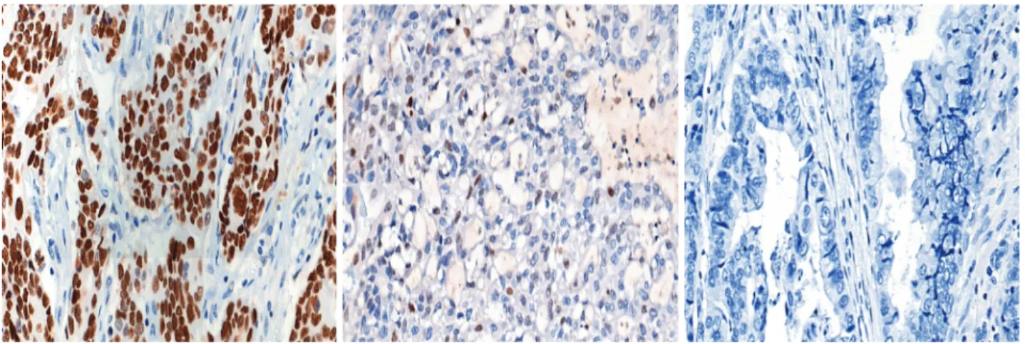
Immunohistochemical (IHC) staining for p53 (DO7, mouse monoclonal, Dako, Agilent Technologies) was performed on 3-μm-thick slides using an automated immunostainer (Bench-Mark XT, Ventana Medical Systems, Tucson, AZ, USA) follow-ing the manufacturer’s protocol. The p53 IHC was interpreted in three tiers: strong nuclear staining in more than 10% of the tumor cells was considered strong positivity, samples without any nuclear staining of tumor cells (complete absence) were in-terpreted as negativity, and cases exhibiting weak, scattered, or patchy positivity were regarded as weak positivity. Representa-tive images for each category are shown in Fig. 1. Cut-offs of 20% and 30% nuclear positivity were additionally applied for validation of the results.
For cases where gene mutation and protein expression status did not match (18 cases or 15.0%), p53 IHC was repeatedly per-formed and interpreted. In most cases (17 out of 18 or 94.4%), repeated immunohistochemical assays did not alter the initial interpretation. Tumor heterogeneity accounted for the change in one case. Initially, strong nuclear expression of p53 was observed in some areas of the tumor (<10%) but was not sufficient to be classified as strong expression. Subsequent IHC was performed on another section of the same tumor, exhibiting overall strong expression of p53.
EBV in-situ hybridization
The EBV status was tested using EBV in-situ hybridization as previously described [20]. A fluorescein-conjugated EBV
en-coded small RNA (EBER) oligonucleotide probe (INFORM EBVencoded RNA probe, Ventana Medical Systems) was used, and positive cases were defined as diffuse nuclear reactivity for EBER in tumor cells.
Microsatellite instability analysis
Representative tumor tissues and matched normal gastric mucosal tissues were selected for microsatellite instability (MSI) testing. Five NCI markers (BAT-26, BAT-25, D5S346, D17S250, and S2S123) amplified through polymerase chain reaction were analyzed using an automated sequencer (ABI 3731 Genetic An-alyzer; Applied Biosystems, Foster City, CA, USA). MSI-high was defined as two or more markers with unstable peaks, MSI-low was defined as one unstable marker, and microsatellite stable was defined as no unstable marker.
Statistical analyses
Chi-square or Fisher exact tests were used to assess significant differences in the distribution of TP53 mutations and p53 ex-pression. For univariate survival analysis, Kaplan-Meier survival curves were plotted in 109 patients with stage II and III GC cases. The survival differences were compared using the log-rank test. For multivariate survival analysis, the Cox regression model was used. All statistical analyses were performed using SPSS Sta-tistics ver. 22.0 (IBM Corp., Armonk, NY, USA).
RESULTS
Gene mutation and protein expression correlation
Table 1 summarizes the p53 IHC results according to TP53 mutations. TP53 mutations were present in 52 cases (43.3%), of which missense mutations were the most common (33 of 52
cases, 63.5%). Strong expression was observed in 34 cases (28.3%) and negative expression was observed in 27 cases (22.5%). When TP53 mutations were compared with p53 IHC, 30 of the 33 missense mutation cases (90.9%) exhibited strong p53 expres-sion, but negative expression of p53 was the dominant pattern (15 cases, 78.9%) among the 19 cases of other types of muta-tions (p<.001). Based on clinical significance, 37 cases (30.1%) had pathogenic or likely pathogenic TP53 mutations, of which 22 cases (59.5%) exhibited strong expression of p53, 13 cases (36.1%) negative expression, and two cases (5.4%) weak
ex-Table 1. Comparison between TP53 genetic mutations and p53 immunohistochemistry
TP53 mutation p53 expression by IHC Total value p-Strong Negative Weak
Mutation status <.001 Wild-type 1 (2.9) 12 (44.4) 55 (93.2) 68 (56.7) Mutation present 33 (97.1) 15 (55.6) 4 (6.8) 52 (43.3) Variant summary <.001 Wild-type 1 (2.9) 12 (44.4) 55 (93.2) 68 (56.7) Missense 30 (88.2) 0 3 (5.1) 33 (27.5) Other 3 (8.9) 15 (55.6) 1 (1.7) 19 (15.8) Stop-gained 2 (5.9) 3 (11.1) 1 (1.7) 6 (5.0) Splice region 0 5 (18.5) 0 5 (4.2) Frameshift 0 7 (25.9) 0 7 (5.8) In-frame deletion 1 (2.9) 0 0 1 (0.8) Clinical significancea <.001 Wild-type 1 (2.9) 12 (44.4) 55 (93.2) 68 (56.7) Pathogenic or likely pathogenic 22 (64.7) 13 (48.1) 2 (3.4) 37 (30.1) Uncertain significance 5 (14.7) 2 (7.4) 1 (1.7) 8 (6.7) Conflicting interpretation 6 (17.6) 0 1 (1.7) 7 (5.8) Total 34 27 59 120
Values are presented as number (%).
aAccording to the ClinVar and OncoKB databases accessed on March 18,
2020.
pression (p<.001). Nevertheless, most cases of uncertain signif-icance (62.5%) and conflicting interpretations (85.7%) also showed strong expression of p53 by IHC.
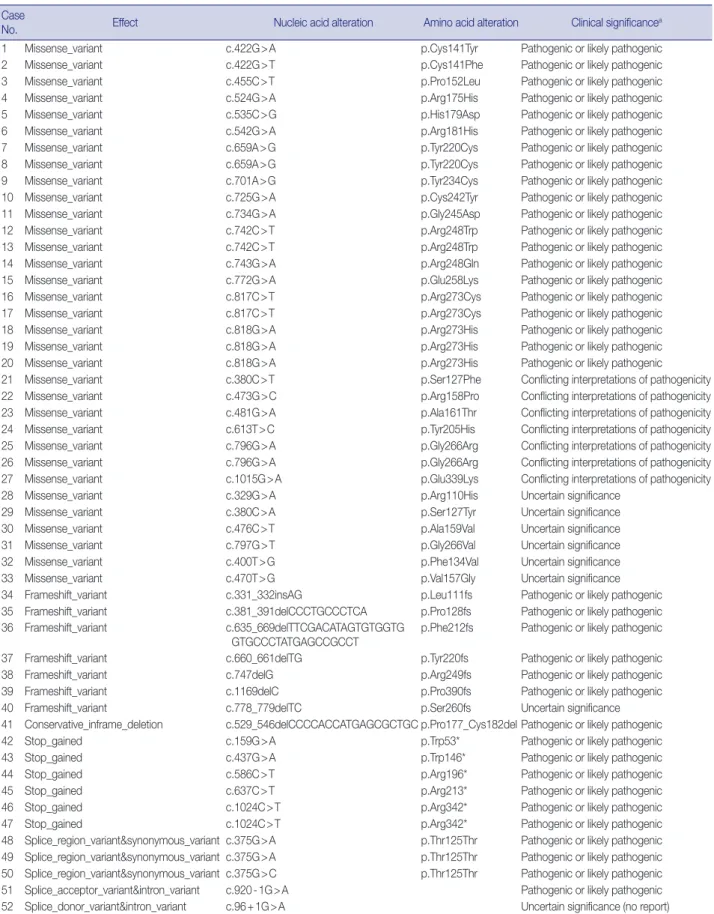
Detailed information about TP53 mutations and p53 expres-sion status is shown in Table 2. Two mutations were observed in three cases, of which one representative mutation was included in this table. One among seven cases with TP53 mutations of conflicting interpretations regarding pathogenicity had weak expression of p53 (case No. 27 in Table 2). There have been reports suggestive of the “likely benign” and “uncertain significance” nature of this mutation. The mutations c.659A>G, c.742C>T, c.817C>T, c.796G>A, c.1024C>T, and c.375G>A were found in two cases, and c.818G>A mutation was found in three cases. The IHC results matched in cases with the same muta-tion. In 44 cases with single nucleotide polymorphism, C:G to T:A conversion was observed in 32 (72.7%), C:G to A:T in four (9.1%), C:G to G:C in two (4.5%), T:A to C:G in four (9.1%), and T:A to G:C in two (4.5%).
Sensitivity, specificity, and accuracy of p53 IHC for predict-ing TP53 mutations
In general, nonsynonymous mutations detected using NGS were related to strong p53 expression in IHC. Similarly, all other types of mutations tended to show negative expression, of p53 while cases with wild-type TP53 exhibited weak protein expres-sion. The sensitivity of strong expression of p53 by IHC for predicting nonsynonymous TP53 mutations was 90.9%, sensi-tivity of negative expression for other types of mutations was 79.0%, and the sensitivity of weak expression for wild-type TP53 was 80.9% (Table 3). The specificity for each category was 95.4%, 88.1%, and 92.3%, respectively. The accuracy for each category was 94.2%, 86.7%, and 85.8%, respectively. In addi-tion, the sensitivity, specificity, and accuracy of p53 IHC at 20% and 30% cut-offs are shown in Supplementary Table S1. The sensitivity of strong expression of p53 for nonsynonymous TP53 mutations was highest at the 10% cut-off.
Clinicopathological variables and protein expression correlations
The correlation between clinicopathological characteristics and TP53 mutations or p53 expression status is summarized in Table 4. TNM stage at initial diagnosis was the only variable that showed significant correlation with both TP53 mutation type and p53 expression status (p=.004 and p=.029, respec-tively). Of the 38 stage II gastric cancer cases, 27 (71.1%) did not exhibit any detectable mutations in the TP53 gene, but five
nonsynonymous (13.2%) and six other types of mutations (15.8%) were found. Strong p53 expression was found in seven of the 38 stage II cases (18.4%). Among the stage III cases, which accounted for 71 cases, the proportions of nonsynonymous gene mutations and strong expression of p53 mutations increased to 39.4% (28 cases) and 38.0% (27 cases), respectively. On the other hand, the proportions of wild-type TP53 cases and weak expression cases decreased from 71.0% to 45.0% and from 55.2% to 43.6%, respectively.
TP53 mutations were more frequently observed in intestinal-type GC (25 of 45 cases, 55.6%) compared to the non-intestinal type (27 of 75 cases, 36.0%), but with borderline statistical sig-nificance (p=.065). Other clinicopathological variables such as sex, age, tumor location, and WHO classification were not sta-tistically significant.
Survival analysis
One hundred nine patients with stage II and III GC at initial diagnosis were selected for survival analysis. The patients under-went curative surgery followed by adjuvant chemotherapy. Patients with any TP53 mutations tended to have worse OS compared to those without mutations, although the difference was not statis-tically significant (p=.227). When OS was analyzed based on TP53 mutation type, patients with nonsynonymous mutations had the worst OS, and the wild-type and other types of muta-tions exhibited similar OS (p=.074) (Fig. 2A). This trend be-came statistically significant when the nonsynonymous muta-tion group was compared to the combined wild-type and other mutation groups (p=.028) (Fig. 2B). The expression pattern of p53 was not significantly associated with patient OS (p=.107) (Fig. 2C), but it was statistically significant when strong expres-sion of p53 was compared to the combined negative and weak expression cases (p=.035) (Fig. 2D). Patients with abnormal— negative and strong expression—expression did not exhibit a statistically significant survival difference compared to patients with weak expression (p=.208). The Kaplan-Meier survival curves of p53 expression status at 20% and 30% cut-offs were additionally plotted in Supplementary Fig. S1. The difference in survival was largest at the 30% cut-off. Multivariate Cox re-gression analysis showed that strong expression of p53 was as-sociated with patient OS independent of stage with borderline significance (p=.070, data not shown). The presence of nonsyn-onymous missense mutations of TP53 was not an independent prognostic factor in multivariate analysis (p=.130).
Table 2. Detailed information of TP53 mutation and p53 expression status in gastric cancer patients with any TP53 mutation
Case
No. Effect Nucleic acid alteration Amino acid alteration Clinical significancea
1 Missense_variant c.422G>A p.Cys141Tyr Pathogenic or likely pathogenic
2 Missense_variant c.422G>T p.Cys141Phe Pathogenic or likely pathogenic
3 Missense_variant c.455C>T p.Pro152Leu Pathogenic or likely pathogenic
4 Missense_variant c.524G>A p.Arg175His Pathogenic or likely pathogenic
5 Missense_variant c.535C>G p.His179Asp Pathogenic or likely pathogenic
6 Missense_variant c.542G>A p.Arg181His Pathogenic or likely pathogenic
7 Missense_variant c.659A>G p.Tyr220Cys Pathogenic or likely pathogenic
8 Missense_variant c.659A>G p.Tyr220Cys Pathogenic or likely pathogenic
9 Missense_variant c.701A>G p.Tyr234Cys Pathogenic or likely pathogenic
10 Missense_variant c.725G>A p.Cys242Tyr Pathogenic or likely pathogenic
11 Missense_variant c.734G>A p.Gly245Asp Pathogenic or likely pathogenic
12 Missense_variant c.742C>T p.Arg248Trp Pathogenic or likely pathogenic
13 Missense_variant c.742C>T p.Arg248Trp Pathogenic or likely pathogenic
14 Missense_variant c.743G>A p.Arg248Gln Pathogenic or likely pathogenic
15 Missense_variant c.772G>A p.Glu258Lys Pathogenic or likely pathogenic
16 Missense_variant c.817C>T p.Arg273Cys Pathogenic or likely pathogenic
17 Missense_variant c.817C>T p.Arg273Cys Pathogenic or likely pathogenic
18 Missense_variant c.818G>A p.Arg273His Pathogenic or likely pathogenic
19 Missense_variant c.818G>A p.Arg273His Pathogenic or likely pathogenic
20 Missense_variant c.818G>A p.Arg273His Pathogenic or likely pathogenic
21 Missense_variant c.380C>T p.Ser127Phe Conflicting interpretations of pathogenicity
22 Missense_variant c.473G>C p.Arg158Pro Conflicting interpretations of pathogenicity
23 Missense_variant c.481G>A p.Ala161Thr Conflicting interpretations of pathogenicity
24 Missense_variant c.613T>C p.Tyr205His Conflicting interpretations of pathogenicity
25 Missense_variant c.796G>A p.Gly266Arg Conflicting interpretations of pathogenicity
26 Missense_variant c.796G>A p.Gly266Arg Conflicting interpretations of pathogenicity
27 Missense_variant c.1015G>A p.Glu339Lys Conflicting interpretations of pathogenicity
28 Missense_variant c.329G>A p.Arg110His Uncertain significance
29 Missense_variant c.380C>A p.Ser127Tyr Uncertain significance
30 Missense_variant c.476C>T p.Ala159Val Uncertain significance
31 Missense_variant c.797G>T p.Gly266Val Uncertain significance
32 Missense_variant c.400T>G p.Phe134Val Uncertain significance
33 Missense_variant c.470T>G p.Val157Gly Uncertain significance
34 Frameshift_variant c.331_332insAG p.Leu111fs Pathogenic or likely pathogenic
35 Frameshift_variant c.381_391delCCCTGCCCTCA p.Pro128fs Pathogenic or likely pathogenic
36 Frameshift_variant c.635_669delTTCGACATAGTGTGGTG
GTGCCCTATGAGCCGCCT
p.Phe212fs Pathogenic or likely pathogenic
37 Frameshift_variant c.660_661delTG p.Tyr220fs Pathogenic or likely pathogenic
38 Frameshift_variant c.747delG p.Arg249fs Pathogenic or likely pathogenic
39 Frameshift_variant c.1169delC p.Pro390fs Pathogenic or likely pathogenic
40 Frameshift_variant c.778_779delTC p.Ser260fs Uncertain significance
41 Conservative_inframe_deletion c.529_546delCCCCACCATGAGCGCTGC p.Pro177_Cys182del Pathogenic or likely pathogenic
42 Stop_gained c.159G>A p.Trp53* Pathogenic or likely pathogenic
43 Stop_gained c.437G>A p.Trp146* Pathogenic or likely pathogenic
44 Stop_gained c.586C>T p.Arg196* Pathogenic or likely pathogenic
45 Stop_gained c.637C>T p.Arg213* Pathogenic or likely pathogenic
46 Stop_gained c.1024C>T p.Arg342* Pathogenic or likely pathogenic
47 Stop_gained c.1024C>T p.Arg342* Pathogenic or likely pathogenic
48 Splice_region_variant&synonymous_variant c.375G>A p.Thr125Thr Pathogenic or likely pathogenic 49 Splice_region_variant&synonymous_variant c.375G>A p.Thr125Thr Pathogenic or likely pathogenic 50 Splice_region_variant&synonymous_variant c.375G>C p.Thr125Thr Pathogenic or likely pathogenic 51 Splice_acceptor_variant&intron_variant c.920-1G>A Pathogenic or likely pathogenic
52 Splice_donor_variant&intron_variant c.96+1G>A Uncertain significance (no report)
IHC, immunohistochemistry.
DISCUSSION
TP53 is the most well-known tumor suppressor gene, and p53 IHC is a method used in daily practice as a surrogate marker in various cancer patients. In this study, we performed targeted deep sequencing for detecting various TP53 mutations and IHC for p53 using a commercially available and validated primary antibody with an automatic immunostainer. Strong expression
of p53 could predict nonsynonymous missense mutations of TP53 with a sensitivity of 90.9%, specificity of 95.4%, and accu-racy of 94.2%. However, weak expression of p53 was less spe-cific (80.9%) for predicting wild-type TP53, and negative expres-sion was less sensitive (79.0%) for predicting other mutations of TP53. These results suggest that p53 IHC can be used as a sur-rogate marker in predicting TP53 mutations, especially for strong expression, to predict nonsynonymous mutations. There
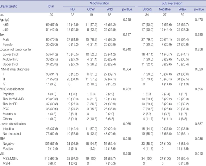
Table 4. Clinicopathologic characteristics according to TP53 mutation and p53 expression status
Characteristic Total TP53 mutation p53 expression
NS Other Wild p-value Strong Negative Weak p-value
No. 120 33 19 68 34 27 59 Age (yr) 0.248 0.470 <65 69 (57.5) 15 (45.5) 11 (57.9) 43 (63.2) 17 (50.0) 15 (55.6) 37 (62.7) ≥65 51 (42.5) 18 (54.5) 8 (42.1) 25 (36.8) 17 (50.0) 12 (44.4) 22 (37.3) Sex 0.117 0.285 Male 85 (70.8) 27 (81.8) 15 (78.9) 43 (63.2) 27 (79.4) 20 (74.1) 38 (64.4) Female 35 (29.2) 6 (18.2) 4 (21.1) 25 (36.8) 7 (20.6) 7 (25.9) 21 (35.6)
Location of tumor center 0.940 0.856
Lower third 53 (44.2) 15 (45.5) 10 (52.6) 28 (41.2) 16 (47.1) 11 (40.7) 26 (44.1) Middle third 33 (27.5) 9 (27.3) 4 (21.1) 20 (29.4) 7 (20.6) 8 (29.6) 18 (30.5) Upper third 34 (28.3) 9 (27.3) 5 (26.3) 20 (29.4) 11 (32.4) 8 (29.6) 15 (25.4) TNM at initial diagnosis 0.004 0.029 II 38 (31.7) 5 (15.2) 6 (31.6) 27 (39.7) 7 (20.6) 10 (37.0) 21 (35.6) III 71 (59.2) 28 (84.8) 11 (57.9) 32 (47.1) 27 (79.4) 13 (48.1) 31 (52.5) IV 11 (9.2) 0 � 2 (10.5) 9 (13.2) 0 � 4 (14.8) 7 (11.9) WHO classification 0.733 0.596 Papillary 4 (3.3) 1 (3.0) 1 (5.3) 2 (2.9) 1 (2.9) 2 (7.4) 1 (1.7) Tubular WD/MD 28 (23.3) 10 (30.3) 6 (31.6) 12 (17.6) 10 (29.4) 6 (22.2) 12 (20.3) Tubular PD 37 (30.8) 9 (27.3) 7 (36.8) 21 (30.9) 10 (29.4) 8 (29.6) 19 (32.2) PCC 36 (30.0) 8 (24.2) 3 (15.8) 25 (36.8) 7 (20.6) 7 (25.6) 22 (37.3) Mucinous 4 (3.3) 2 (6.1) 0 � 2 (2.9) 2 (5.9) 1 (3.7) 1 (1.7) Others 11 (9.2) 3 (9.1) 2 (10.5) 6 (8.8) 4 (11.7) 3 (11.1) 4 (6.8) Lauren classification 0.065 0.587 Intestinal 45 (37.5) 14 (42.4) 11 (57.9) 20 (29.4) 15 (44.1) 10 (37.0) 20 (33.9) Non-intestinal 75 (62.5) 19 (57.6) 8 (42.1) 48 (70.6) 19 (55.9) 17 (63.0) 39 (66.1) EBV 0.215 0.036 Negative 105 (87.5) 31 (93.9) 18 (94.7) 56 (82.4) 30 (88.2) 27 (100) 48 (81.4) Positive 15 (12.5) 2 (6.1) 1 (5.3) 12 (17.6) 4 (11.8) 0 � 11 (18.6) MSI 0.258 0.010 MSS/MSI-L 112 (93.3) 32 (97.0) 19 (100) 61 (89.7) 34 (100) 27 (100) 51 (86.4) MSI-H 8 (6.7) 1 (3.0) 0 � 7 (10.3) 0 � 0 � 8 (13.6)
Values are presented as number (%).
NS, nonsynonymous; Other, other type mutation; wild, wild-type; WHO, World Health Organization; WD, well-differentiated; MD, moderately differentiated; PD, poorly differentiated; PCC, poorly cohesive carcinoma; EBV, Epstein-Barr virus; MSI, microsatellite instability; MSS, microsatellite stable; MSI-L, microsat-ellite instability-low; MSI-H, microsatmicrosat-ellite instability-high.
Table 3. The sensitivity, specificity, and accuracy of p53 immunohistochemistry for predicting TP53 mutation, cut-off 10%
TP53 mutation Sensitivity (%) Specificity (%) Accuracy (%)
Nonsynonymous mutation by p53 strong expression 90.9 95.4 94.2
Other type mutation by negative expression of p53 79.0 88.1 86.7
have been recent attempts to use IHC for molecular classifica-tions, and their clinicopathological significance has been increas-ingly important in GC [8,10]. Our results will be helpful for these new molecular classifications although negative expres-sion should be cautiously interpreted.
Based on the Kaplan-Meier survival analysis, strong expres-sion of p53 was significantly associated with worse OS compared to weak and negative expression of p53 in this study. Previous studies that investigated any relationship between p53 overex-pression and survival reported p53 overexoverex-pression as a poor prog-nostic factor [21-23], similar to the findings from our study. Some studies did not reveal the prognostic significance of p53
overex-pression in GC, but a meta-analysis demonstrated that it is a poor prognostic factor [24]. In those studies, the median cut-off value was 10% [24]. Therefore, we applied a cut-off value of 10% for defining strong expression of p53. In addition to the 10% cut-off, we applied 20% and 30% cut-offs in this study. Although the survival difference was largest at the 30% cut-off, the sensi-tivity of strong expression of p53 for predicting nonsynony-mous TP53 mutation was highest at the 10% cut-off. There-fore, further studies are needed to validate various cut-offs.
For interpretation of p53 IHC, Köbel et al. [11,25] proposed a three-tiered scoring system, including overexpression, complete absence, and normal or wild-type pattern in ovarian cancer. The 1.0 0.8 0.6 0.4 0.2 0.0 1.0 0.8 0.6 0.4 0.2 0.0 1.0 0.8 0.6 0.4 0.2 0.0 1.0 0.8 0.6 0.4 0.2 0.0 Cumulative survival Cumulative survival Cumulative survival Cumulative survival Time (mo) Time (mo) Time (mo) Time (mo) 0 20 40 60 80 100 0 20 40 60 80 100 0 20 40 60 80 100 0 20 40 60 80 100 Other Other WT NS NS Neg Neg+ Weak Weak Strong Strong A C B D
Fig. 2 . Kaplan-Meier survival curves of cumulative survival rate versus follow-up months after surgery according to mutation status (A, B) and immunohistochemistry results (C, D). (A) Nonsynonymous mutations (NS, beige) versus wild-type (WT, green) versus other types of mu-tations (other, green) (p=.074). (B) Nonsynonymous mutations (NS, green) versus combined wild-type and other types of mutations (other, blue) (p=.028). (C) Strong (beige) versus weak (green) versus negative expression (Neg) (p=.107). (D) Strong (green) versus combined weak and negative expression (Neg+Weak, blue) (p=.035).
scoring system exhibited good correlation with TP53 mutation status: overexpression with nonsynonymous mutation; com-plete absence with stop gain, frameshift, and splicing muta-tions; and a normal pattern with the wild-type TP53 gene [11]. Shin et al. [26] investigated the prognostic roles of p53 expres-sion status in patients with GC. They defined group 0 as com-plete absence, group 1 as weak staining in <50%, group 2 as strong staining in 50%–90%, and group 3 as strong staining in >90%. When the Kaplan-Meier survival analysis was performed, group 1 was associated with better survival than groups 0, 2, and 3, but with borderline statistical significance. Our results showed a similar relationship between p53 IHC and TP53 mu-tation status to those of previous studies. If weak expression in this study was defined as a normal or wild-type pattern, the Ka-plan-Meier survival curves did not show a significant difference between normal and abnormal expression patterns. Further-more, patients with GC having nonsynonymous TP53 muta-tions had significantly worse prognosis compared to patients with other types of mutations and the wild-type TP53 group. Similarly, strong expression of p53, which was related to non-synonymous TP53 mutations, was shown to be a poor prognostic factor. The complete absence of p53 expression or other types of TP53 mutations might not be significant for predicting prog-nosis.
There were 17 cases (14.2%) with discrepant results between p53 IHC and TP53 mutations. Most discrepant cases had nega-tive expression of p53 and wild-type TP53. Weak expression of p53 was observed in four missense mutation cases and one stop gain mutation case. These findings might be due to tumor het-erogeneity or tissue quality issues, such as specimen ischemic time or archival age. In addition, there was one case with weak p53 expression and a missense mutation of conflicting pathogenic interpretation. Considering this case was conflicting between uncertain significance and likely benign significance, one of the possible reasons for the discrepancy is a non-pathogenic muta-tion. To discriminate functional and nonfunctional p53, Nenutil et al. [27] performed IHC for p53, Ki67, MDM2, and p21 in human cancers, and overexpressed p53 without increased MDM2 indicated inactivating mutations in their study. p21, a tran-scriptional target of p53, was considered to reflect p53 activity and could decrease false-positive results of p53 expression [28]. The ACRG considered a TP53-activity signature using p21 and MDM2 genes [7]. Therefore, in addition to p53 IHC, IHC for p21 and MDM2 would be helpful for evaluating the functional status of p53. The need for these additional analyses reflects a potential limitation of this study, necessitating further research.
In a total of 120 gastric cancer cases, 52 (43.3%) had TP53 mutations, including nonsynonymous missense, frameshift, stop gain, in-frame deletion, and splice region mutations. Hot spot mutations within the central core (R175, G245, R248, R273, and R282) were observed in a minority of cases (10 out of 120 or 8.3%). In accordance with our results, a previous study reported hot spot mutations in 6.2% of gastric cancer cases [29]. Therefore, sequencing (Sanger or NGS) is suggested as a suitable method for detecting TP53 mutations in gastric cancer.
In summary, we investigated the relationship between p53 expression and TP53 mutation status to predict TP53 muta-tions by p53 IHC and reveal their prognostic significance. TP53 mutations were observed in 43.3% of cases. Strong p53 expres-sion could predict nonsynonymous missense mutations with high sensitivity and specificity, but only half of the p53 negative cases (55.6%) exhibited other types of TP53 mutations. Protein overexpression and nonsynonymous genetic mutations of TP53 significantly predicted worse OS. p53 IHC could be regarded as a simple surrogate marker of TP53 mutations, but negative ex-pression of p53 and other types of TP53 mutations should be cautiously considered in daily practice or scientific research. Over-all, our study results will be informative for simple molecular classification of patients with GC.
Supplementary Information
The Data Supplement is available with this article at https://doi.org/10.4132/ jptm.2020.06.01.
Ethics Statement
This study was approved by the Institutional Review Board of Seoul Na-tional University Bundang Hospital (IRB number: B-2001/591-105). Writ-ten informed consent was waived by the IRB.
ORCID
Hye Jung Hwang https://orcid.org/0000-0002-3251-0270 Soo Kyung Nam https://orcid.org/0000-0002-4372-1516 Hyunjin Park https://orcid.org/0000-0001-7193-9849 Yujun Park https://orcid.org/0000-0002-2335-7606 Jiwon Koh https://orcid.org/0000-0002-7687-6477 Hee Young Na https://orcid.org/0000-0002-2464-0665 Yoonjin Kwak https://orcid.org/0000-0001-5314-2465 Woo Ho Kim https://orcid.org/0000-0003-0557-1016 Hye Seung Lee https://orcid.org/0000-0002-1667-7986
Author Contributions
Conceptionalization: WHK, HSL. Data curation: HJH, HSL. Formal analy-sis: HJH, HSL. Funding Acquisition: HSL. Investigation: HJH, YP, HP, JK, HYN, YK, HSL. Methodology: HJH, SKN, HP, HSL. Supervision: HSL. Writing—original draft: HJH, HSL. Writing—review &editing: HJH, WHK, HSL. Approval of final manuscript: all authors.
Conflicts of Interest
W.H.K. and H.S.L., contributing editors of the Journal of Pathology and Translational Medicine, were not involved in the editorial evaluation or de-cision to publish this article. All remaining authors have declared no con-flicts of interest.
Funding Statement
This work was supported by a National Research Foundation of Korea (NRF) grant funded by the Korean government (Ministry of Science and ICT) (No. 2019R1A2C1086180).
References
1. Junttila MR, Evan GI. p53: a Jack of all trades but master of none. Nat Rev Cancer 2009; 9: 821-9.
2. Bartkova J, Horejsi Z, Koed K, et al. DNA damage response as a candidate anti-cancer barrier in early human tumorigenesis. Nature 2005; 434: 864-70.
3. Starzynska T, Bromley M, Ghosh A, Stern PL. Prognostic signifi-cance of p53 overexpression in gastric and colorectal carcinoma. Br J Cancer 1992; 66: 558-62.
4. Kakeji Y, Korenaga D, Tsujitani S, et al. Gastric cancer with p53 overexpression has high potential for metastasising to lymph nodes. Br J Cancer 1993; 67: 589-93.
5. Maehara Y, Tomoda M, Hasuda S, et al. Prognostic value of p53 protein expression for patients with gastric cancer: a multivariate analysis. Br J Cancer 1999; 79: 1255-61.
6. Cancer Genome Atlas Research Network. Comprehensive molecu-lar characterization of gastric adenocarcinoma. Nature 2014; 513: 202-9.
7. Cristescu R, Lee J, Nebozhyn M, et al. Molecular analysis of gastric cancer identifies subtypes associated with distinct clinical outcomes. Nat Med 2015; 21: 449-56.
8. Setia N, Agoston AT, Han HS, et al. A protein and mRNA expression-based classification of gastric cancer. Mod Pathol 2016; 29: 772-84. 9. Ahn S, Lee SJ, Kim Y, et al. High-throughput Protein and mRNA
Expression-based classification of gastric cancers can identify clini-cally distinct subtypes, concordant with recent molecular classifica-tions. Am J Surg Pathol 2017; 41: 106-15.
10. Koh J, Lee KW, Nam SK, et al. Development and validation of an easy-to-implement, practical algorithm for the identification of mo-lecular subtypes of gastric cancer: prognostic and therapeutic im-plications. Oncologist 2019; 24: e1321-30.
11. Kobel M, Piskorz AM, Lee S, et al. Optimized p53 immunohisto-chemistry is an accurate predictor of TP53 mutation in ovarian car-cinoma. J Pathol Clin Res 2016; 2: 247-58.
12. Fukayama M, Rugge M, Washington MK. Tumours of the stomach. In: Lokuhetty D, White VA, Watanabe R, Cree IA, eds. WHO classi-fication of tumours. Lyon: International Agency for Research on Cancer (IARC), 2019; 85-103.
13. Koh J, Nam SK, Roh H, et al. Somatic mutational profiles of stage II and III gastric cancer according to tumor microenvironment im-mune type. Genes Chromosomes Cancer 2019; 58: 12-22.
14. Martin M. Cutadapt removes adapter sequences from high-through-put sequencing reads. EMBnet J 2011; 17: 10-2.
15. Cibulskis K, Lawrence MS, Carter SL, et al. Sensitive detection of somatic point mutations in impure and heterogeneous cancer sam-ples. Nat Biotechnol 2013; 31: 213-9.
16. Cingolani P, Patel VM, Coon M, et al. Using Drosophila melano-gaster as a model for genotoxic chemical mutational studies with a new program, SnpSift. Front Genet 2012; 3: 35.
17. Liu X, Wu C, Li C, Boerwinkle E. dbNSFP v3.0: a one-stop data-base of functional predictions and annotations for human nonsyn-onymous and splice-site SNVs. Hum Mutat 2016; 37: 235-41. 18. Chakravarty D, Gao J, Phillips SM, et al. OncoKB: a precision
oncol-ogy knowledge base. JCO Precis Oncol 2017; 2017: 10.1200/PO.17. 00011.
19. Landrum MJ, Lee JM, Benson M, et al. ClinVar: public archive of in-terpretations of clinically relevant variants. Nucleic Acids Res 2016; 44: D862-8.
20. Park Y, Koh J, Na HY, et al. PD-L1 testing in gastric cancer by the combined positive score of the 22C3 PharmDx and SP263 assay with clinically relevant cut-offs. Cancer Res Treat 2020 Jan 10 [Epub]. https://doi.org/10.4143/crt.2019.718.
21. Song KY, Jung CK, Park WS, Park CH. Expression of the antiapop-tosis gene survivin predicts poor prognosis of stage III gastric ade-nocarcinoma. Jpn J Clin Oncol 2009; 39: 290-6.
22. Mrena J, Wiksten JP, Kokkola A, Nordling S, Ristimaki A, Haglund C. COX-2 is associated with proliferation and apoptosis markers and serves as an independent prognostic factor in gastric cancer. Tumour Biol 2010; 31: 1-7.
23. Zha Y, Cun Y, Zhang Q, Li Y, Tan J. Prognostic value of expression of Kit67, p53, TopoIIa and GSTP1 for curatively resected advanced gas-tric cancer patients receiving adjuvant paclitaxel plus capecitabine chemotherapy. Hepatogastroenterology 2012; 59: 1327-32.
24. Yildirim M, Kaya V, Demirpence O, Gunduz S, Bozcuk H. Prognos-tic significance of p53 in gastric cancer: a meta- analysis. Asian Pac J Cancer Prev 2015; 16: 327-32.
25. Kobel M, Reuss A, du Bois A, et al. The biological and clinical value of p53 expression in pelvic high-grade serous carcinomas. J Pathol 2010; 222: 191-8.
26. Shin YJ, Kim Y, Wen X, et al. Prognostic implications and interac-tion of L1 methylainterac-tion and p53 expression statuses in advanced gastric cancer. Clin Epigenetics 2019; 11: 77.
27. Nenutil R, Smardova J, Pavlova S, et al. Discriminating functional and non-functional p53 in human tumours by p53 and MDM2 im-munohistochemistry. J Pathol 2005; 207: 251-9.
28. Watanabe G, Ishida T, Furuta A, et al. Combined immunohisto-chemistry of PLK1, p21, and p53 for predicting TP53 status: an inde-pendent prognostic factor of breast cancer. Am J Surg Pathol 2015; 39: 1026-34.
29. Tahara T, Shibata T, Okamoto Y, et al. Mutation spectrum of TP53 gene predicts clinicopathological features and survival of gastric cancer. Oncotarget 2016; 7: 42252-60.