INTRODUCTION
Drug resistance is a major obstacle in the successful treat- ment and an important cause of death in acute leukemia. Such resistance may be present before begining treatment or may develop during chemotherapy. Drug resistance that extends to structurally and functionally unrelated drugs is termed multidrug resistance (MDR) (1).
Several molecular biological mechanisms have been identi- fied as being associated with MDR (2). P-glycoprotein (PGP), also named P-170, is a product of the multidrug resistance 1 gene (MDR1) and is an ATP-dependent pump capable of expelling drugs out of cancer cells (3, 4). PGP is a transmem- brane glycoprotein conferring cross-resistance to a variety of mechanistically and structurally unrelated cytotoxic drugs, such as anthracyclines, taxanes, vinca alkaloids and epipodo- phyllotoxins (5). Another protein, the multidrug resistance- related protein (MRP), is structurally similar to PGP and be- longs to the same transmembrane transporter superfamily (6). The substrate specificity of MRP is similar to but more
limited than that of PGP, and its normal physiological role may be detoxification of intracellular oxidants (5). In addition to these two proteins, a 110 kDa protein has been identified in a PGP-negative MDR lung cancer cell line. This protein was termed the lung resistance protein (LRP) and acts as a major vault protein in humans (7). Despite the identification of these proteins, the pathways that result in drug resistance in leukemic cells remain largely uncharacterized. New infor- mation regarding drug resistance mechanisms is likely to in- crease the chances of cure either through development of new drugs or by means of strategies that may modulate or reverse resistance (8). While drug resistance gene expression has been studied in acute leukemia (9-12), the value of MDR1, MRP and LRP gene expression as independent predictors of treat- ment success is still controversial.
In the present study, we investigated whether outcomes in acute leukemia patients were associated with MDR1, MRP and LRP mRNA expression.
Hee Jin Huh, Chan-Jeoung Park*, Seongsoo Jang*, Eul-Ju Seo*, Hyun-Sook Chi*, Je-Hwan Lee�, Kyoo-Hyung Lee�, Jong Jin Seo�, Hyung Nam Moon�, Thad Ghim�
Department of Laboratory Medicine*, Internal Medicine�and Pediatrics�, College of Medicine, University of Ulsan and Asan Medical Center, Seoul;
Department of Laboratory Medicine, College of Medicine, Dogguk University, Seoul, Korea
Address for correspondence Chan Jeoung Park, M.D.
Department of Laboratory Medicine, Asan Medical Center, 388-1 Pungnap-dong, Songpa-gu, Seoul 138-736, Korea
Tel : +82.2-3010-4508, Fax : +82.2-478-0884 E-mail : [email protected]
253
Prognostic Significance of Multidrug Resistance Gene 1 (MDR1), Multidrug Resistance-related Protein (MRP) and Lung Resistance Protein (LRP) mRNA Expression in Acute Leukemia
The prognostic significance of multidrug resistance (MDR) gene expression is con- troversial. We investigated whether multidrug resistance gene 1 (MDR1), multidrug resistance-related protein (MRP) and lung resistance protein (LRP) mRNA expres- sion are associated with outcomes in acute leukemia patients. At diagnosis we exam- ined MDR1, MRP and LRP mRNA expression in bone marrow samples from 71 acute leukemia patients (39 myeloid, 32 lymphoblastic) using nested RT-PCR. The expression of each of these genes was then expressed as a ratio in relation to - actin gene expression, and the three genes were categorized as being either 0, 1+, 2+ or 3+. MDR1, MRP and LRP mRNA expression was detected in 23.9%, 83.1%
and 45.1 %, respectively. LRP mRNA expression was significantly associated with resistance to induction chemotherapy in acute leukemia patients, and in the AML proportion (p=0.02 and p=0.03, respectively). MRP and high MDR1 mRNA expres- sion was associated with poorer 2-yr survival (p=0.049 and p=0.04, respectively).
Patients expressing both MRP and LRP mRNA had poorer outcomes and had worse 2-yr survival. The present data suggest that MDR expression affects complete remis- sion and survival rates in acute leukemia patients. Thus, determination of MDR gene expression at diagnosis appears likely to provide useful prognostic information for acute leukemia patients.
Key Words :Genes, MDR; Multidrug Resistance Gene 1; P-Glycoprotein Multidrug Resistance-related Pro- tein 1; lung resistance protein; Prognosis; Leukemia
Received : 18 March 2005 Accepted : 5 October 2005
Patients
The study involved 71 patients (34 males and 37 females) diagnosed with acute leukemia between January 1998 and January 1999. The median age of the patients was 30 yr (ran- ge, 3 months - 75 yr). Diagnosis and classification of acute leukemia was made according to the French-American-British (FAB) criteria and immunophenotype analyses. There were 39 acute myeloid leukemia (AML) and 32 acute lymphoblas- tic leukemia (ALL) patients. Among 39 AML patients, eleven patients had AML M1, 11 M2, 7 M3, 6 M4, 2 M5, 1 M6 and 1 M7. The patients were classified as being good progno- sis group, intermediated prognosis group and poor progno- sis group according to cytogenetic results. AML patients with t(8;21), t(15;17) and inv (16) were defined as “good progno- sis group”, patients with 5-, 7-, 5q-, 7q-, hyperdiploidy and multiple chromosomal abnormality were defined as “poor prognosis group”, and the others were defined as the “interme- diate prognosis group”. Among AML patients, eight patients (t(8;21), 1; t(15;17), 7) were in good prognosis group, 20 (nor- mal karyotype, 19; t(11;19), 1) in intermediate prognosis group and 12 ( trisomy 8, 2; other complex abnormality, 10) in poor prognosis group. Among ALL patients, two were in good prognosis group, 11 in intermediate prognosis group and 19 in poor prognosis group. The induction chemother- apy regimen were cytarabine plus idarubicin (AI regimen) in adult AML patients and vincristine, prednisone, daunorubi- cin, L-asparaginase (VPDL regimen) in adult ALL patients.
Newly diagnosed childhood ALL patients were treated accord- ing to Children’s Cancer Group (CCG) 1881, 1882, 1891 or 1901 protocols. Childhood AML patients were treated accord- ing to BFM (Frankfurt Munster group) 83 protocols, includ- ing cyclophosphamide and prednisone. Clinical information was obtained by reviewing charts.
Definition of disease phase and response
Complete remission (CR) was defined as a cellularity of more than 20% with fewer than 5% blasts in the bone mar- row (BM) after induction chemotherapy, and relapse as more than 5% blasts in the BM. The CR rate was calculated only using patients that underwent bone marrow analysis follow- ing initial diagnosis (64 cases).
Semiquantitative RT-PCR
Seventy one bone marrow aspirates collected in EDTA were obtained at the initial diagnosis. A COR-L23/R cell line was used as a positive control for MDR1 and LRP mRNA expres- sion, and the HL60/Adr cell line for MRP mRNA expression.
Negative controls included deionized RNAase-free water, and bone marrow aspirates obtained from patients with lympho-
plification was used as an internal control.
Mononuclear cells were separated by density gradient cen- trifugation using Ficoll-Hypaque within 3 hr after bone mar- row aspiration. Total cellular RNA was extracted using the RNeasy kit (Qiagen Inc., Valencia, CA, U.S.A.) according to the manufacturer’s instructions.
cDNA was synthesized using the First Strand cDNA Syn- thesis Kit for RT-PCR (Behringer Mannheim Inc, Germany).
Total RNA (1 g) was reverse transcribed in 20 L reaction mixture (2 L 10X Reaction buffer [100 mM Tris, 500 mM KCL, pH 8.3 and 25 mM MgCl2], 10 mM each of four dNTPs, 1.6 g/ L random primers, 50 U/ L RNase inhibitor and 1 L AMV Reverse Transciptase). The reaction was performed at 70℃for 2 min and 42℃for 60 min. MDR1, MRP and LRP mRNA amplifications were performed after heating the reaction mixture to 99℃for 5 min. The first round of PCR reactions involved 30 cycles of: 1 min denaturing at 94℃, 1 min annealing at 64℃and 2 min extension at 72℃using a thermocycler (Perkin Elmer-Cetus, Wellesley, PA, U.S.A.).
The PCR reactions were carried out in a final volume of 50 L containing 2 g cDNA, 10 pg/ L of each primer, 0.4 U/ L taq polymerase, 200 M of each dNTP, 5 L 10X buffer con- taining 500 mM KCl, 100 mM Tris (pH 8.3) and 25 mM MgCl2. The first round of PCR was followed by a second ro- und of 20 cycles. Amplification of -actin mRNA (20 cycle PCR) was performed and the data obtained used to normalize any variations between samples (e.g. differences in concen- tration or quality of extracted RNA). PCR primer sequences
Target Direction Sequences of primer
MDR1 External Sense 5′-CCAGTGGTGTTTTTAGGGTCATCA-3′ primers
Antisense 5′-CCAAAGAGGCTCTGGATGAAAG- TA-3′
Internal Sense 5′-TGTTGTTGGTGTATTTTGTGCCATT-
primers AT-3′
Antisense 5′-GTATCGGAGCCGCTTGGTGAGA-3′ MRP External Sense 5′-TACACCGTGCTGCTGTTTGTCACT-3′
primers
Antisense 5′-GTCTTGGTCATCGCCATCACA-3′ Internal Sense 5′-ACTTCCACATCTGCTTCGTCAGTG-3′ primers
Antisense 5′-ATTCAGCCACAGGAGGTAGAGA- GC-3′
LRP External Sense 5′-GTCTTCGGGCCTGAGCTGGTGT-
primers CG-3′
Antisense 5′-CTTGGCCGTCTCTTGGGGGTCC- TT-3′
Internal Sense 5′-TTCTGGATTTGGTGGACG-3′ primers
-actin Antisense 5′-ACTTCTCTCCCTTGACCA-3′ Sense 5′-GTGGGGCGCCCCAGGCACCA -3′ Antisense 5′-GTCCTTAATGTCACGCACGATTTC-3′ Table 1.RT-PCR primer sequences for MDR1, MRP, LRP and
-actin mRNA amplification
. .
are shown in Table 1. PCR products were electrophoretically separated on 2% agarose gels and visualized using ethidium bromide staining. The gel was photographed with Polaroid film (Fig. 1) and the film scanned into a computer (Sharp JX- 330, Japan) for analysis. For semiquantitative measurements of mRNA, the relative ratios of MDR1, MRP or LRP mRNA expression to -actin mRNA expression was calculated using ImageMaster software (Pharmacia, Sweden). Following the determination of this ratio, MDR genes were classified as either not expressed (0), weakly expressed (ratio 0-0.7=1+), moderately expressed (ratio 0.7-1=2+), or strongly expressed (ratio >1=3+).
Statistics
Data were analyzed using chi square or Fisher exact tests.
Survival curves were plotted using the Kaplan-Meier method, and differences were analyzed using log-rank tests. A p value
<0.05 was deemed to indicate a significant difference. All statistical analyses were performed using SPSS version 11.5 (SPSS, Chicago, IL., U.S.A.).
RESULTS
Expression rates, levels of MDR mRNA and cytogenetic results
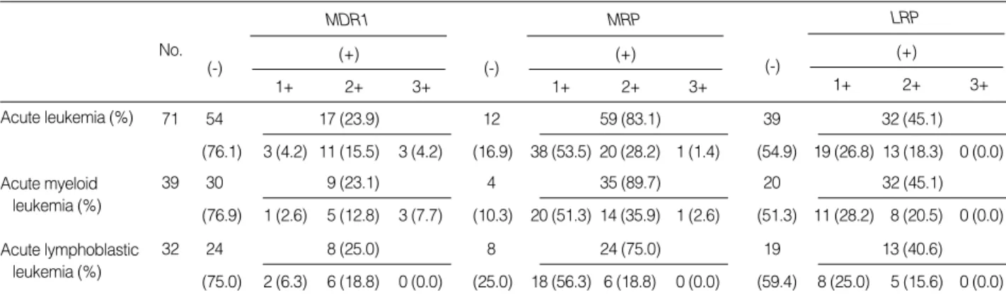
We found that the proportion of acute leukemia patients expressing MDR1, MRP and LRP mRNA was 23.9%, 83.1
% and 45.1%, respectively (Table 2). In terms of leukemia categories, MDR1, MRP and LRP mRNA was expressed in 23.1%, 89.4% and 48.7% respectively of AML patients, and in 25.0%, 75.0% and 40.6% respectively of ALL patients.
While fewer patients expressed MDR1 mRNA compared to MRP and LRP mRNA, the rate of MDR1 mRNA expression (3+) was higher than the rate of either MRP or LRP mRNA high expression (3+) (Table 2). Univariate analysis showed no correlation between cytogenetic results and MDR1, MRP and LRP mRNA expression in AML and ALL patients.
Association of MDR1, MRP and LRP mRNA expression at diagnosis with the rate of complete remission, relapse and survival time
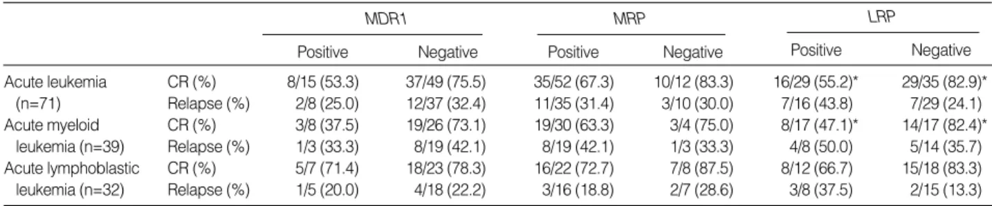
LRP mRNA expression at initial diagnosis was associated with a lower CR rate after induction chemotherapy, with only 55.2% (16/29) of LRP-positive acute leukemia patients achi- eving CR, compared to 82.9% (29/35) of LRP-negative pa- tients achieving CR (p=0.02). Similarly, for AML patients, 47.1% (8/17) of LRP-positives achieved CR, while 82.4%
(14/17) of LRP-negatives achieved CR (p=0.03). MDR1 and MRP mRNA expression appeared to have statistically no sig- nificant effect on patient outcome following induction chemo- therapy. However, MDR1 or MRP mRNA-positive patients showed the tendency of lower CR rates than MDR1 or MRP- negative patients (Table 3). MDR1, MRP or LRP mRNA expression had no effect on relapse rates. The multivariate logistic regression analysis of age, MDR mRNA expression and cytogenetic results indicated that the MDR mRNA ex- pression did not give significant effects on relapse and contin- uous CR after induction chemotherapy.
Kaplan-Meier analysis showed that the median survival for
Fig. 1.RT-PCR products following MDR1, MRP and LRP mRNA amplification. Lane S, size marker; lane 1, MDR1-positive cell line (band at 243 bp); lane 2, DW; lane 3, normal bone marrow; lane 4, MDR1-positive patient; lane 5, MRP-positive line (bands at 575 and 420 bp); lane 6, DW; lane 7, normal bone marrow; lane 8, s-positive patient; lane 9, LRP-positive cell line (band at 239 bp); lane 10, DW; lane 11, normal bone marrow; lane 12, LRP-positive patient;
lane 13, -actin (internal control, band at l66 bp). (DW=deionized water (negative control)).
S 1 2 3 4 5 6 7 8 9 10 11 12 13 S
575 bp 420 bp 243 bp 166 bp
No.
Table 2.Semiquantitative MDR gene expression results in acute leukemia patients MDR1
1+ 2+ 3+
(-) (+)
MRP
1+ 2+ 3+
(-) (+)
LRP
1+ 2+ 3+
(-) (+)
(76.9) 1 (2.6) 5 (12.8) 3 (7.7)
39 30 9 (23.1)
(10.3) 20 (51.3) 14 (35.9) 1 (2.6)
4 35 (89.7)
(51.3) 11 (28.2) 8 (20.5) 0 (0.0)
20 32 (45.1)
(76.1) 3 (4.2) 11 (15.5) 3 (4.2)
71 54 17 (23.9)
(16.9) 38 (53.5) 20 (28.2) 1 (1.4)
12 59 (83.1)
(54.9) 19 (26.8) 13 (18.3) 0 (0.0)
39 32 (45.1)
(75.0) 2 (6.3) 6 (18.8) 0 (0.0)
32 24 8 (25.0)
(25.0) 18 (56.3) 6 (18.8) 0 (0.0)
8 24 (75.0)
(59.4) 8 (25.0) 5 (15.6) 0 (0.0)
19 13 (40.6)
Acute leukemia (%)
Acute myeloid leukemia (%)
Acute lymphoblastic leukemia (%)
MRP-positive acute leukemia patients was 600 days, while MRP-negative patients did not reach the median survival time.
There was a significant difference in the 2-yr survival between these two groups (p=0.049) (Fig. 2). There was statistically no prognostic significance for MDR1 (p=0.05) or LRP mRNA (p=0.08) expression in acute leukemic patients in total, nor for AML or ALL patients when analyzed separately.
High level MDR1 mRNA expression was significantly associated with poorer 2-yr survival in acute leukemia patients (p=0.04) (Fig. 3), with all three such patients dying within 2 yr (20 days, 228 days and 405 days) of diagnosis. Among these patients, two were diagnosed with adult AML and one with childhood AML. Only one of these three patients achie- ved CR after induction chemotherapy.
While the survival time appeared to decrease with increas- ing MRP and LRP mRNA expression levels, this observation was not supported by statistical analysis.
Outcome in acute leukemia patients expressing two MDR genes
Double positivity for MDR1 and MRP mRNA was detect- ed in 17 samples (23.4%), MDR1 and LRP mRNA in 15 (21.1%) and MRP and LRP mRNA in 31 (43.7%). MDR1 and LRP positive patients also showed positivity for MRP.
While 57.1% (16/28) of acute leukemia patients expressing both MRP and LRP mRNA achieved CR, 80.6% (29/36) of those not expressing both genes achieved CR (p=0.04). Ex- pressing both MDR1 and MRP mRNA, or both MDR1 and LRP mRNA had no significant influence on the CR rate after induction chemotherapy. The median survival of MRP and LRP-double positive patients was 405 days; the other patients did not reach median survival. There was a significant differ- ence in the 2-yr survival between the two groups (p=0.04).
Expressing both MDR1 and MRP mRNA, or MDR1 and LRP mRNA had no negative impact on 2-yr survival.
DISCUSSION
The present study indicates that MDR gene expression can affect outcomes in acute leukemia patients. LRP gene expres- sion at diagnosis appeared to be associated with resistance to induction chemotherapy in acute leukemia patients. This was not the case for MDR1 or MRP gene expression. For AML patients, while over 80% of LRP-negative subjects achieved CR, less than half of the LRP-positive subjects achieved CR (p=0.03). Therefore, LRP mRNA expression was particular- ly influential in outcomes following induction chemotherapy in AML patients. This correlation between LRP expression
Positive Negative
MDR1 MRP
Positive Negative
LRP
Positive Negative
*p value <0.05; CR, complete remission.
Days since initial diagnosis
0 200 400 600 800
Probability
1.1 1.0 0.9 0.8 0.7 0.6 0.5 0.4 0.3 0.2 0.1 0.0
p=0.049
Fig. 2.Relationship between MRP mRNA expression and 2-yr sur- vival in acute leukemia patients.
negative
positive
Days since initial diagnosis
0 200 400 600 800
Probability
1.2
1.0
0.8
0.6
0.4
0.2
0.0
p=0.004
Fig. 3.Relationship between MDR1 mRNA expression levels and 2-yr survival in acute leukemia patients.
0
1+or 2+
3+
Acute leukemia CR (%) 8/15 (53.3) 37/49 (75.5) 35/52 (67.3) 10/12 (83.3) 16/29 (55.2)* 29/35 (82.9)*
(n=71) Relapse (%) 2/8 (25.0) 12/37 (32.4) 11/35 (31.4) 3/10 (30.0) 7/16 (43.8) 7/29 (24.1) Acute myeloid CR (%) 3/8 (37.5) 19/26 (73.1) 19/30 (63.3) 3/4 (75.0) 8/17 (47.1)* 14/17 (82.4)*
leukemia (n=39) Relapse (%) 1/3 (33.3) 8/19 (42.1) 8/19 (42.1) 1/3 (33.3) 4/8 (50.0) 5/14 (35.7) Acute lymphoblastic CR (%) 5/7 (71.4) 18/23 (78.3) 16/22 (72.7) 7/8 (87.5) 8/12 (66.7) 15/18 (83.3) leukemia (n=32) Relapse (%) 1/5 (20.0) 4/18 (22.2) 3/16 (18.8) 2/7 (28.6) 3/8 (37.5) 2/15 (13.3)
and treatment outcome is consistent with suggestions that chemosensitive leukemic cells have low LRP mRNA levels in AML (10, 13).
Interestingly, it was reported that LRP mRNA expression more strongly correlated with drug resistance than LRP pro- tein expression (14). Sauerbrey et al. (15) suggested that LRP overexpression might be a mechanism involved in childhood ALL drug resistance. While the present data appeared to sug- gest LRP-positive ALL patients showed a lower probability for first remission, this observation was not supported statis- tically.
In AML patients, PGP/MDR1 expression was found to be an independent prognostic variable associated with reduced probability for achieving remission following conventional anthracene-containing induction regimens, and in some re- ports this expression was linked to inferior leukemia-free and overall survivals (16-18). However, while our data appeared to suggest that MDR1-positive AML and ALL patients had low CR rates and poor 2-yr survivals, this observation was not supported by statistical analysis. It may be that analysis of a larger number of patients would provide the statistical power for significance. The different sensitivities of the detec- tion techniques may be responsible for the discrepant statis- tical significance. However, three patients with strong MDR1 mRNA expression died within 2 yr, and high levels of MDR1 mRNA were significantly associated with poor 2-yr survival in acute leukemia patients. Therefore, we need to consider that patients with high MDR1 mRNA expression may have poorer outcomes.
The reported proportion of patients positive for MDR gene expression varies widely and appears to be related to the study method employed and the patient characteristics of any par- ticular study. In the present study, 83.1% of patients expre- ssed MRP mRNA, and 2-yr survival in these patients was less than that in MRP-negative patients (p=0.049). Although some studies show MRP expression lacked prognostic power (19, 20), others found it was predictive of poorer outcome (21).
In the present study, MRP and LRP-double positive patients showed reduced remission rates after induction chemothera- py and poorer 2-yr overall survival compared to patients not expressing both of these genes. Only two studies have previ- ously attempted to determine the impact of simultaneous expression of two MDR genes, and the conclusions drawn by these studies were not consistent with each other (21, 22).
The present study demonstrates that outcomes in acute leukemia patients can vary depending upon expression of MDR1, MRP and LRP mRNA. Simultaneous expression of MRP and LRP mRNA correlated with a low CR rate and poor survival. These results suggest that analysis of MDR gene expression at diagnosis of acute leukemia may provide useful prognostic information. Such data is also likely to assist in determining the mechanisms underlying drug resistance.
REFERENCES
1. Riordan JR, Ling V. Genetic and biochemical characterization of multidrug resistance. Pharmacol Ther 1985; 28: 51-75.
2. McKenna SL, Padua RA. Multidrug resistance in leukaemia. Br J Haematol 1997; 96: 659-74.
3. Shen DW, Cardarelli C, Hwang J, Cornwell M, Richert N, Ishii S, Pastan I, Gottesman MM. Multiple drug-resistant human KB carci- noma cells independently selected for high-level resistance to colchicine, adriamycin, or vinblastine show changes in expression of specific proteins. J Biol Chem 1986; 261: 7762-70.
4. Lee NY, Bae HG, Kwon EH, Heo WB, Shin SH, Suh JS. Relation among the tests and comparison of positivity of tests for multi-drug resistance in newly diagnosed acute leukemia. Korean J Lab Med 2003; 23: 143-50.
5. Chauncey TR. Drug resistance mechanisms in acute leukemia. Curr Opin Oncol 2001; 13: 21-6.
6. Kruh GD, Chan A, Myers K, Gaughan K, Miki T, Aaronson SA. Ex- pression complementary DNA library transfer establishes mrp as a multidrug resistance gene. Cancer Res 1994; 54: 1649-52.
7. Scheper RJ, Broxterman HJ, Scheffer GL, Kaaijk P, Dalton WS, van Heijningen TH, van Kalken CK, Slovak ML, de Vries EG, van der Valk P. Overexpression of a M (r) 110,000 vesicular protein in non- P-glycoprotein-mediated multidrug resistance. Cancer Res 1993; 53:
1475-9.
8. Szabo D, Keyzer H, Kaiser HE, Molnar J. Reversal of multidrug re- sistance of tumor cells. Anticancer Res 2000; 20: 4261-74.
9. Kakihara T, Tanaka A, Watanabe A, Yamamoto K, Kanto K, Kata- oka S, Ogawa A, Asami K, Uchiyama M. Expression of multidrug resistance-related genes does not contribute to risk factors in newly diagnosed childhood acute lymphoblastic leukemia. Pediatr Int 1999;
41: 641-7.
10. List AF, Spier CS, Grogan TM, Johnson C, Roe DJ, Greer JP, Wolff SN, Broxterman HJ, Scheffer GL, Scheper RJ, Dalton WS. Overex- pression of the major vault transporter protein lung-resistance pro- tein predicts treatment outcome in acute myeloid leukemia. Blood 1996; 87: 2464-9.
11. Dhooge C, De Moerloose B, Laureys G, Kint J, Ferster A, De Bac- quer D, Philippe J, Benoit Y. P-glycoprotein is an independent pro- gnostic factor predicting relapse in childhood acute lymphoblastic leukaemia: results of a 6-year prospective study. Br J Haematol 1999; 105: 676-83.
12. Gurbuxani S, Zhou D, Simonin G, Raina V, Arya LS, Sazawal S, Marie JP, Bhargava M. Expression of genes implicated in multidrug resistance in acute lymphoblastic leukemia in India. Ann Hematol 1998; 76: 195-200.
13. Xu D, Arestrom I, Virtala R, Pisa P, Peterson C, Gruber A. High levels of lung resistance related protein mRNA in leukaemic cells from patients with acute myelogenous leukaemia are associated with inferior response to chemotherapy and prior treatment with mitoxantrone. Br J Haematol 1999; 106: 627-33.
14. Laurencot CM, Scheffer GL, Scheper RJ, Shoemaker RH. Increased LRP mRNA expression is associated with the MDR phenotype in intrinsically resistant human cancer cell lines. Int J Cancer 1997;
analysis of the multidrug resistance related protein (MRP1) and the lung resistance protein (LRP) in de novo and relapsed childhood acute lymphoblastic leukemia. Leuk Lymphoma 2002; 43: 875-9.
16. Guerci A, Merlin JL, Missoum N, Feldmann L, Marchal S, Witz F, Rose C, Guerci O. Predictive value for treatment outcome in acute myeloid leukemia of cellular daunorubicin accumulation and P-gly- coprotein expression simultaneously determined by flow cytometry.
Blood 1995; 85: 2147-53.
17. Mahadevan D, List AF. Targeting the multidrug resistance-1 trans- porter in AML: molecular regulation and therapeutic strategies.
Blood 2004; 104: 1940-51.
18. Schaich M, Soucek S, Thiede C, Ehninger G, Illmer T. MDR1 and MRP1 gene expression are independent predictors for treatment out- come in adult acute myeloid leukaemia. Br J Haematol 2005; 128:
324-32.
19. Borg AG, Burgess R, Green LM, Scheper RJ, Yin JA. Overexpres- sion of lung-resistance protein and increased P-glycoprotein func-
103: 1083-91.
20. den Boer ML, Pieters R, Kazemier KM, Rottier MM, Zwaan CM, Kaspers GJ, Janka-Schaub G, Henze G, Creutzig U, Scheper RJ, Veerman AJ. Relationship between major vault protein/lung resis- tance protein, multidrug resistance-associated protein, P-glycopro- tein expression, and drug resistance in childhood leukemia. Blood 1998; 91: 2092-8.
21. Legrand O, Simonin G, Beauchamp-Nicoud A, Zittoun R, Marie JP.
Simultaneous activity of MRP1 and Pgp is correlated with in vitro resistance to daunorubicin and with in vivo resistance in adult acute myeloid leukemia. Blood 1999; 94: 1046-56.
22. Valera ET, Scrideli CA, Queiroz RG, Mori BM, Tone LG. Multiple drug resistance protein (MDR-1), multidrug resistance-related pro- tein (MRP) and lung resistance protein (LRP) gene expression in childhood acute lymphoblastic leukemia. Sao Paulo Med J 2004;
122: 166-71.