VOL.23 NO.3 : 091-100 (2010)
하고초 열수추출물이 대식세포 면역만응에 미치는 영향
경원대학교 한의과대학 부인과학교실 차지혜, 김윤상, 임은미
ABSTRACT
Effects of P runellae Spica Water Extract on Immune Response in Macrophage Cells
Ji-Hea Cha, Yoon-Sang Kim, Eun-Mee Lee
Dept. of Gynecology, College of Oriental Medicine, Kyung-Won University Purpose: The purpose of this study was to investigate the effects of Prunellae Spica Water Extract(PSE) on immune response in macrophage cells.
Methods: We had devided two group the one is normal group; not treated with PSE, and the other is experimental group; treated with PSE. We measured the cell viability of PSE on RAW 264.7 cells and investigated production of nitric oxide(NO) and cytokines such as interleukin(IL)-1β, IL-6 and tumor necrosis factor (TNF)-α with sample PSE.
Results:
1. Cell viability of PSE on RAW 264.7 cells was significantly decreased in both 24 hr and 48 hr incubation.
2. NO production of PSE on RAW 264.7 cells was significantly increased in both 24 hr and 48 hr incubation.
3. IL-1β production of PSE on RAW 264.7 cells was significantly increased under concentration over 50 ㎍/㎖ in 24 hr incubation.
4. IL-6 production of PSE on RAW 264.7 cells was significantly increased under concentration over 50 ㎍/㎖ in 24 hr incubation.
5. TNF-α production of PSE on RAW 264.7 cells was significantly increased under concentration over 50 ㎍/㎖ in 24 hr incubation.
Conclusion: NO, IL-1β, IL-6 and TNF-α production of PSE on RAW 264.7 cells was significantly increased. This study suggest that PSE stimulates the macrophage and enhances the immune response.
Key Words: Prunellae Spica , macrophage, immune response, NO, cytokine
7 )
“이 연구는 2010년도 경원대학교 지원에 의한 결과임.”
교신저자(김윤상) : 인천시 중구 용동 117번지 경원대학교 부속 길한방병원 한방부인과 전화 : 032-770-1227 이메일 :
[email protected]
Ⅰ. 서 론
여성에게 陰血은 생리적․병리적인 측 면에서 모두 중요하며, 陰血이 부족하면 衝任이 영양을 받지 못하고 內熱이 발생 하여 질병 치료 시 우선적으로 고려되므 로 補血하는 本草들은 부인과적 질환에 다용된다 1) . 이와 같은 맥락에서 본 연구 의 대상이 되는 하고초(夏枯草 : Prunellae Spica ) 역시 淸熱解毒하는 효능 2) 이외에 補養血脈하는 효능 3) 을 중심으로 여성의 陰血을 다스림으로서 부인과 질환의 면 역력 증강에 효능이 있을 것으로 사료되 어 본 연구를 진행하게 되었다.
하고초는 꿀풀과(脣形科 : Labiatae) 에 속한 다년초 본초인 꿀풀(Prunella vulgaris var. lilacina Nakai)의 꽃이삭으 로 淸熱瀉火藥에 속하여 일반적으로 淸 火, 明目, 散結 및 消腫하는 작용을 가지 고 있으나 2) 이 외에 補養血脈하는 효능 도 있다 3) .
기존의 연구를 통해 하고초는 대식세 포의 반응산소중간물질의 생성능과 대식 세포의 탐식능을 유의하게 증가시켜 세 포성 및 체액성 면역과 선천적 면역능을 증가시키는 것으로 보고된 바 있어 4) 본 연구에서는 마우스 대식세포를 이용하여 하고초가 세포생존률 및 면역매개물질 생성에 미치는 영향을 조사하였다.
하고초 열수추출물( Prunellae Spica Water Extract, PSE)로 마우스 대식세 포에 전처리한 다음 cell viability 및 nitric oxide(NO) 생성과 interleukin(IL) -1β, IL-6 및 tumor necrosis factor(TNF)-α 등의 cytokine 생성에 미치는 영향을 관 찰하여 유의한 결과를 얻었기에 補養血
脈하는 효능을 중심으로 한 하고초에 관 한 본 연구를 통해 부인과 질환의 면역 력 증강 효능을 제시하는 바이다.
Ⅱ. 연구방법
1. 재 료
1) 약재 및 시료
실험에 사용된 하고초(옴니허브, Korea) 는 2009년 2월에 구입(NO : 2009-02-0011) 하였고, 사용 전 ultrasonic cleaner를 이 용하여 불순물을 제거하였다. 하고초 50 g을 정확하게 측정한 뒤 환류추출기에 1 차 증류수 2,000 ㎖와 함께 넣은 후 끓는 시점으로부터 2시간 동안 가열하여 추출 한 다음 추출액을 filter paper로 감압 여 과하고, rotary vacuum evaporator로 농 축시켜 농축액을 얻었다. 이 농축액을 동결건조기를 이용하여 건조시켜 건조 추출물 8 g을 얻었으며, 수율은 16%였다.
2) 시약 및 기기 (1) 시 약
본 실험을 위해서 ethyl alcohol(Samchun
chemical, Korea), methyl alcohol(Samchun
chemical, Korea), fetal bovine serum
(FBS), dimethyl sulfoxide(DMSO, Sigma,
USA), Dulbecco's modied essential medium
(DMEM, Sigma, USA), 1×phosphate
buffered saline(PBS, Sigma, USA),
ethylenediaminetetraacetic acid(EDTA,
Sigma, USA), acetic acid(Sigma, USA),
isopropanol(Sigma, USA), trypsin-EDTA
(Sigma, USA), MTT(Sigma, USA), NO
assay kit(Oxford biomedical research, USA)
및 Bio-Plex cytokine assay kit(Panomics,
USA) 등이 사용되었다.
(2) 기 기
본 실험에 사용된 기기는 filter paper (Advantec No.2, Japan), carbon dioxide (CO2) incuvator(NUAIRE, USA), flask (Falcon, USA), pulverizer(Rong tsong, Taiwan), rotary vacuum evaporator(Eyela, Japan), air compressor(Tamiya, Japan), homogenizer(Omni, USA), research microscope (Becton dickinson, USA), centriuge(Hanil, Korea), fume hood(Hanil, Korea), clean bench(Jeio thec, Korea), ultrasonic cleaner (Branson, USA), microplate reader(Molecular devices, USA), microplate reader(Bio-rad, USA), thermo aluminum bath(Fine PCR, USA), vortex mixer(Vision scientific co, Korea), water bath(iNtRON biotech, Korea), ice-maker(Vision scientific co, Korea) 및 Bioplex-200(Bio-rad, USA) 등이다.
2. 실험방법 1) 세포배양
실험에 사용된 대식세포는 mouse macrophage(Raw 264.7 cell line)로서, 한국 세포주 은행(KCLB, Korea)에서 구입하였다. Raw 264.7 cells는 37℃, 5%
CO 2 조건에서 10% FBS, penicillin(100 U/㎖) 및 streptomycin(100 ㎍/㎖) 등이 첨가된 DMEM 배지에서 배양되었다.
Cells는 75 ㎠ flask에서 충분히 증식된 후 배양 3일 간격으로 배양세포 표면을 PBS 용액으로 씻어준 뒤 50 ㎖ flask 당 1 ㎖의 0.25% trypsin-EDTA 용액을 넣 고 실온에서 1분간 처리한 다음 trypsin 용액을 버리고 37℃에서 5분간 보관하여 세포를 탈착하여 계대 배양하였다. 탈착 된 세포는 10% FBS가 첨가된 DMEM
배양액 10 ㎖에 부유시킨 다음 새로운 배양용기(50 ㎖ culture flask)에 옮겨 1 : 2의 split ratio로 CO 2 배양기(37℃, 5% CO 2 )에서 배양하였다.
2) MTT assay
PSE의 세포독성 유발 효과를 알아보 기 위하여 Mosmann 등 5) 의 방법을 응용 하여 MTT assay를 실시하였다. 96 well plate에 1×10 4 cells/well의 cells을 100 ㎕ 씩 넣고 37℃, 5% CO 2 incubator에서 24 시간 동안 배양한 후 배지를 버린 뒤 배 양세포 표면을 1×PBS 용액으로 씻어 주 었다. 같은 양의 배지와 0, 50, 100 및 200 ㎍/㎖ 등 다양한 농도의 PSE를 각 well에 처리하고 24 혹은 48시간 동안 배 양하였다. 배양이 끝난 후 PBS에 녹인 1
㎎/㎖ MTT를 100 ㎕씩 각 well에 처리 하여 알루미늄호일로 차광시킨 후 2시간 동안 같은 조건에서 배양하였다. 배양액 을 모두 제거한 후 DMSO를 100 ㎕ 처리 하고 37℃에서 2시간 방치 후 microplate reader를 이용하여 490 ㎚에서 흡광도를 측정하였다. Cell viability는 다음 공식 으로 계산되었다.
Cell Viability(%) = 100×AT/AC AC - absorbance of control
AT - absorbance of tested extract solution.
3) NO 생성 측정
PSE의 NO 생성 효과를 알아보기 위 하여 Weissman 등 6,7) 의 방법을 응용하여 다음과 같이 실험을 실시하였다. 세포배 양액 중에 존재하는 NO의 농도를 추정 하기 위해 microplate reader를 이용, 540
㎚에서 흡광도를 측정 비교하였다. 이를
위해 0, 50, 100 및 200 ㎍/㎖ 등 다양한
농도의 PSE를 배지에 담아 각 well에 처
리하고, 24 혹은 48시간 동안 37℃, 5%
CO 2 incubator에서 배양한 후 세포배양 상등액 60 ㎕을 채취하여 여기에 Griess 시약 100 ㎕을 혼합하여 15분 동안 반응 시킨 다음 microplate reader를 이용하여 540 ㎚에서 흡광도를 측정하였다. 세포의 NO 생성은 다음 공식으로 계산되었다.
Productions of NO(%) = 100×AT/AC AC - absorbance of control
AT - absorbance of tested extract solution.
4) Cytokine 생성 측정
PSE의 cytokine 생성 효과를 알아보 기 위하여 Politch 등 8) 의 방법을 응용하 여 다음과 같이 실험을 실시하였다. 96 well plate에 1×10⁵ cells/㎖의 cell을 100
㎕씩 넣고 37℃, 5% CO 2 incubator에서 24시간 동안 배양한 후 배지를 버리고 배 양세포 표면을 1×PBS 용액으로 씻어준 뒤 각 well에 0, 50, 100 및 200 ㎍/㎖ 등 다양한 농도의 PSE와 함께 배지에 담아 처리하고 24시간 동안 배양하였다. 배양 이 끝나면 상등액을 채취하여 Bio-Plex suspension assay system을 이용, quantitative multiplexed cytokine/chemokine assay를 실시하여 마우스 대식세포의 IL-1β, IL-6 및 TNF-α 등의 cytokine 생성을 측정하였다.
5) 통계처리
본 실험에서 얻은 결과에 대해서는 mean±SD로 나타내었으며, 정상군과 각 실험군과의 평균의 차이는 Student t-test 로 분석하여 p -value 값이 0.05 미만일 때 통계적으로 유의한 차이가 있는 것으 로 판정하였다.
Ⅲ. 결 과
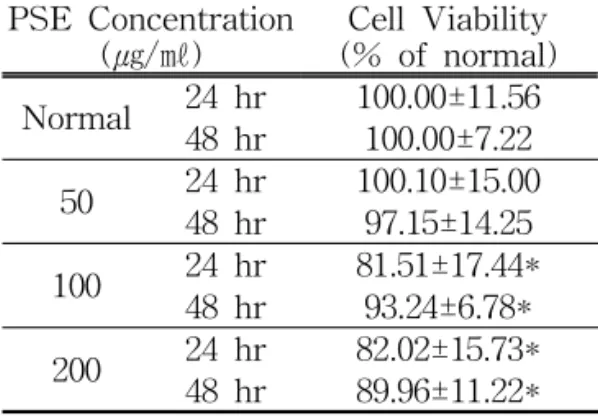
1. 세포생존율의 변화
PSE가 마우스 대식세포의 생존율에 미치는 영향을 살펴본 결과 24시간 배양 시 정상군에 비하여, 실험군 50, 100 및 200 ㎍/㎖ 등은 각각 100.10±15.00, 81.51
±17.44 및 82.02±15.73% 등의 세포생존 율을 보였고, 48시간 배양 시 정상군에 비하여, 50, 100 및 200 ㎍/㎖ 등은 각각 97.15±14.25, 93.24±6.78, 89.96±11.22% 등 의 세포생존율을 보였다(Table 1, Fig. 1).
PSE Concentration
(㎍/㎖) Cell Viability (% of normal) Normal 24 hr 100.00±11.56
48 hr 100.00±7.22 50 24 hr 100.10±15.00
48 hr 97.15±14.25 100 24 hr 81.51±17.44*
48 hr 93.24±6.78*
200 24 hr 82.02±15.73*
48 hr 89.96±11.22*
Values are the mean±SD of the three independent experiments.
Normal : Not treated with PSE.
* represents p < 0.05 compared to the normal.
Table 1. Effects of PSE on Cell Viability in RAW 264.7 Cells for 24, 48 hr Incubation
* * * *
Fig. 1. Effects of PSE on cell viability in RAW 264.7 cells for 24, 48 hr incubation
Values are the mean±SD of the three independent experiments.Normal : Not treated with PSE.
* represents p < 0.05 compared to the normal.
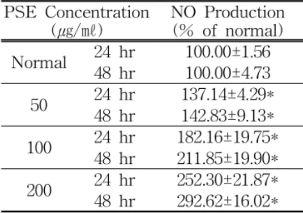
2. NO 생성의 변화
PSE가 마우스 대식세포의 NO 생성에 미치는 영향을 살펴본 결과 24시간 배양 시 정상군에 비하여, 실험군 50, 100 및 200 ㎍/㎖ 등은 각각 137.14±4.29, 182.16
±19.75 및 252.30±21.87 등으로 나타났고, 48시간 배양 시 정상군에 비하여, 실험군 50 , 100 및 200 ㎍/㎖ 등은 각각 142.83
±9.13, 211.85±19.90 및 292.62±16.02 등으 로 나타나 모든 실험군에서 정상군에 비해 NO 생성이 유의하게 증가하였다(Table 2, Fig. 2).
PSE Concentration
(㎍/㎖) NO Production (% of normal) Normal 24 hr 100.00±1.56
48 hr 100.00±4.73 50 24 hr 137.14±4.29*
48 hr 142.83±9.13*
100 24 hr 182.16±19.75*
48 hr 211.85±19.90*
200 24 hr 252.30±21.87*
48 hr 292.62±16.02*
Values are the mean±SD of the three independent experiments.
Normal : Not treated with PSE.
* represents p < 0.05 compared to the normal.
Table 2. Effects of PSE on NO Production of RAW 264.7 Cells for 24, 48 hr Incubation
*
*
* *
* *
Fig. 2. Effects of PSE on NO production of RAW 264.7 cells for 24, 48 hr incubation
Values are the mean±SD of the three independent experiments.
Normal : Not treated with PSE.
* represents p < 0.05 compared to the normal.
3. IL-1β 생성의 변화
PSE가 마우스 대식세포의 IL-1β 생 성에 미치는 영향을 살펴본 결과 24시간 배양 시 정상군이 4.30±0.50인데 비하여, 실험군 50, 100 및 200 ㎍/㎖ 등은 각각 5.8±0.50, 10.0±0.82 및 15.8±3.33 등으로 나타나 모든 실험군에서 정상군에 비해 IL-1β 생성이 유의하게 증가하였다(Table 3, Fig. 3).
PSE Concentration
(㎍/㎖) IL-1β Production (pg /㎖) Normal 4.3±0.50 50 5.8±0.50*
100 10.0±0.82*
200 15.8±3.33*
Values are the mean±SD of the three independent experiments.
Normal : Not treated with PSE.
* represents p < 0.05 compared to the normal.
Table 3. Effects of PSE on IL-1β Production of RAW 264.7 Cells for 24 hr Incubation
*
*
*
Fig. 3 Effects of PSE on IL-1β production of RAW 264.7 cells for 24 hr incubation
Values are the mean±SD of the three independent experiments.Normal : Not treated with PSE.
* represents p < 0.05 compared to the normal.
4. IL-6 생성의 변화
PSE가 마우스 대식세포의 IL-6 생성 에 미치는 영향을 살펴본 결과 24시간 배양 시 정상군이 395.8±74.47인데 비하 여, 실험군 50, 100 및 200 ㎍/㎖ 등은 496.5±19.12, 754.3±88.18 및 1442.8±202.11 등으로 나타나 모든 실험군에서 정상군 에 비해 IL-6 생성이 유의하게 증가하였 다(Table 4, Fig. 4).
PSE Concentration
(㎍/㎖) IL-6 Production (pg /㎖) Normal 395.8±74.47
50 496.5±19.12*
100 754.3±88.18*
200 1442.8±202.11*
Values are the mean±SD of the three independent experiments.
Normal : Not treated with PSE.
* represents p < 0.05 compared to the normal.
Table 4. Effect of PSE on IL-6 Production of RAW 264.7 Cells for 24 hr Incubation
*
*
*
Fig. 4. Effects of PSE on IL-6 production of RAW 264.7 cells for 24 hr incubation
Values are the mean±SD of the three independent experiments.Normal : Not treated with PSE.
* represents p < 0.05 compared to the normal.
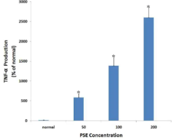
5. TNF-α 생성의 변화
PSE가 마우스 대식세포의 TNF-α 생 성에 미치는 영향을 살펴본 결과 24시간 배양 시 정상군이 14.3±3.4인데 비하여, 실험군 50, 100 및 200 ㎍/㎖ 등은 584.4
±101.57, 1386.3±215.47 및 2601.3±250.19 등으로 나타나 모든 실험군에서 정상군 에 비해 TNF-α 생성이 유의하게 증가 하였다(Table 5, Fig. 5).
PSE Concentration
(㎍/㎖) TNF-α(pg /㎖) Normal 14.3±3.40
50 584.4±101.57*
100 1386.3±215.47*
200 2601.3±250.19*
Values are the mean±SD of the three independent experiments.
Normal : Not treated with PSE.
* represents p < 0.05 compared to the normal.
Table 5. Effects of PSE on TNF-α Production of RAW 264.7 Cells for 24 hr Incubation
*
*
*
Fig. 5, Effects of PSE on TNF-α production of RAW 264.7 cells for 24 hr incubation
Values are the mean±SD of the three independent experiments.Normal : Not treated with PSE.
* represents p < 0.05 compared to the normal.