J o u r n a l o f R h e u m a t i c D i s e a s e s V o l . 2 1 , N o . 5 , O c t o b e r , 2 0 1 4
http://dx.doi.org/10.4078/jrd.2014.21.5.253 □ Case Report □
253
<Received:August 23, 2013, Revised (1st: October 1, 2013, 2nd: October 14, 2013), Accepted:October 16, 2013>
Corresponding to:In Seol Yoo, Department of Internal Medicine, Chungnam National University School of Medicine, 640, Deasa-dong, Jung-gu, Daejeon 301-721, Korea. E-mail:[email protected]
pISSN: 2093-940X, eISSN: 2233-4718
Copyright ⓒ 2014 by The Korean College of Rheumatology
This is a Free Access article, which permits unrestricted non-commerical use, distribution, and reproduction in any medium, provided the original work is properly cited.
Localized Mesenteric Vasculitis in a Patient with Polymyalgia Rheumatica
Seung Taek Song, Young Kim, Chan Keol Park, Su Jin Yoo, Jin Hyun Kim, Seong Wook Kang, In Seol Yoo
Department of Internal Medicine, Chungnam National University School of Medicine, Daejeon, Korea
Polymyalgia rheumatica (PMR) is an uncommon disorder characterized by bilateral pain and stiffness in the should- er and pelvic girdles. Polymyalgia rheumatica and giant cell arteritis (GCA) occur in the same patient population and share a common pathogenesis. Giant cell arteritis pre- dominantly affects the cranial arteries and rarely involves the gastrointestinal tract. Moreover, giant cell arteritis has
rarely been reported in Asians. Here, we present a case with 62-year-old Asian woman who developed polymyalgia rheumatica with localized vasculitis in the mesenteric arteries.
Key Words. Gastrointestinal tract, Mesenteric vasculitis, Polymyalgia rheumatica
Introduction
Polymyalgia rheumatica (PMR) is an inflammatory condition of unknown etiology characterized by bilateral myalgia of the hip and shoulder girdles with accompanying morning stiffness that lasts for more than 1 hour. Giant-cell arteritis (GCA) is an inflammatory vasculopathy that affects large and me- dium-size arteries. PMR and GCA occur in the same patient population and share common risk factors and pathogenic pathways (1). Approximately 10% of patients with PMR de- velop GCA, and approximately 50% of patients with GCA de- velop PMR symptoms. GCA predominantly affects the cranial arteries and rarely involves other sites, including the mesen- teric arteries (2). A recent study found that Asians were 20 times less likely to present with GCA than Caucasians (3).
Moreover, the cases of GCA involving the mesenteric arteries have not been reported in Asians (4). Herein, we present the case of an Asian woman who developed PMR with localized mesenteric vasculitis.
Case Report
A 62-year-old Korean woman was admitted to the hospital because of diffuse abdominal pain and diarrhea for 3 days.
Five months before admission, proximal myalgia of the neck and the shoulder and pelvic girdles developed; this was ac- companied by morning stiffness that lasted for more than 1 hour. At that time, abdominal pain and diarrhea preceded the musculoskeletal symptoms. Two months before admission, the woman was admitted to our hospital with sustained symptoms and was diagnosed with PMR based on the facts that she was over the age of 50; showed bilateral aching and stiffness in- volving the neck, shoulder, and hip; and showed an elevated erythrocyte sedimentation rate (113 mm/hr); Subdeltoid bursi- tis and biceps tendinitis on sonography. The patient was treat- ed with 20 mg of prednisone per day, and her symptoms im- proved within 3 days. At that time, she was also diagnosed with type 2 diabetes mellitus.
At the time of admission, the pain and stiffness of the neck, shoulder, and hip had recurred, and had not been resolved with 15 mg of prednisone per day. The initial vital signs may be summarized as follows: body temperature 36.4°C, pulse rate 72/minute, respiration rate 20/minute, blood pressure 100/60 mm Hg. On physical examination, she had a diffusely tender abdomen, especially in the periumbilical region and in the right lower quadrant. Bowel sounds were increased. There
254 Seung Taek Song et al.
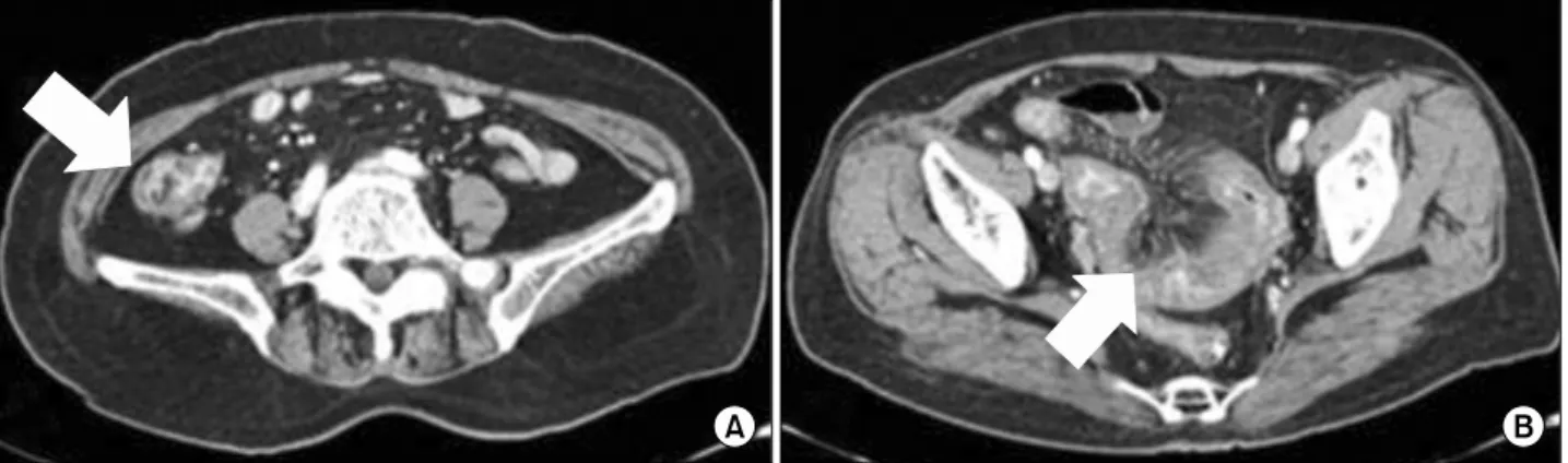
Figure 1. Abdomen and pelvis CT. (A) Diffuse severe edematous wall thickening in distal ileum and ileocecal region with luminal collapse (arrow). (B) Vascular congestion seen in the mesenteric vessels (arrow), suggesting ischemic bowel disease due to vasculitis.
Figure 2. (A, B) Follow-up CT scan after Steroid and MTX therapy shows improved bowel wall thickening and engorged vasa recta of adjacent small bowel mesentery.
was no skin lesion, oral ulcer and arthritis except shoulder and hip joints. The CBC results may be summarized as follows:
WBC 13,270 cells/μL (neutrophil 74.9%, lymphocyte 13.0%, monocyte 11.0%, eosinophil 0.1%), platelet count 220,000 cells/μL, hemoglobin 11.8 g/dL, Hct 35.3%. The erythrocyte sedimentation rate (ESR) and the C-reative protein (CRP) lev- el were elevated (ESR 113 mm/hr, CRP 4.2 mg/dL). Other laboratory tests, including urine analysis, AST, ALT, ALP, LDH, CPK, ANCA, Rheumatoid factor and Anti-Nuclear anti- body were normal except elevated serum glucose (150 mg/dL). No organisms were found in blood and urine cultures.
The stool examination results showed hemoglobin 697 ng/mL (0∼100) and stool WBC many /HPF. Parasites were not found and the stool culture was negative. Abdominal pain and diarrhea were not resolved with antibiotics. An abdominal and pelvic computerized tomography (CT) scan showed diffuse se- vere edematous wall thickening in the distal ileum and ileoce- cal region with luminal collapse (Figure 1A) and engorged va-
sa recta of adjacent small bowel mesentery suggesting vasculi- tis (Figure 1B). The colonoscopic examination showed linear ulceration and erythematous lesions in the terminal ileum and the cecum. Also, colonoscopic biopsy showed ileocolitis with erosion and no evidence of tumor. The gross finding and biop- sy results of colonoscopy were compatible with ischemic colitis. On the third day of admission, we raised the dose of prednisone to 30 mg per day, and then, the patient’s abdomi- nal and musculoskeletal symptoms slightly improved. On the seventh day of admission, her neck and shoulder pain wors- ened and the ESR and CRP levels were still high (ESR 103 mm/hr, CRP 4.4 mg/dL); therefore, we treated the patient with 55 mg (1 mg/kg) of prednisone daily and 15 mg of methotrex- ate (MTX) weekly. After that, the patient’s symptoms im- proved with resolution of abdominal tenderness and muscu- loskeletal symptoms within several days. Also, the follow-up CT scan and aorta CT showed improvement of inflammation in previous noted ischemic bowel and mesenteric arteries
PMR with Mesenteric Vasculitis 255
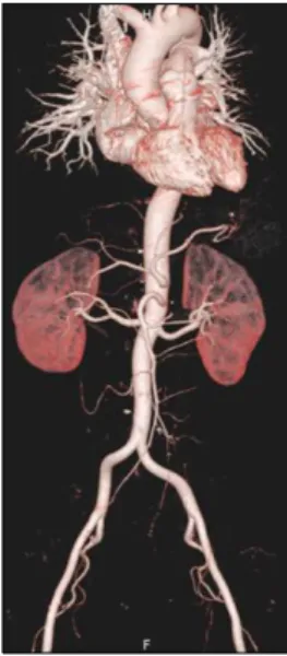
Figure 3. Aorta CT. There was no evidence of vasculitis in aorta and its major branches.
(Figures 2 and 3). But there was no evidence of vasculitis in the aorta or its major branches (Figure 3). We have checked the patient’s status in the outpatient department monthly.
There have been no signs of recurrence in her physical and laboratory findings up to now. The level of acute phase re- actants were within normal limits (ESR 15 mm/hr, CRP 0.1 mg/dL) on latest visit. So we tapered the dose of prednisone to 5 mg per day and the dose of MTX to 10 mg per week.
Discussion
Gastrointestinal (GI) manifestations of vasculitis are consid- ered rare, and the presentation is often nonspecific. However, if there is significant involvement of the major vessels of the gastrointestinal system, life-threatening sequelae, including perforation and bowel ischemia, may occur. GI involvement frequently occurs in Henoch–Schönlein purpura, Polyarteritis nodosa (PAN), ANCA-associated vasculitis (Wegener’s gran- ulomatosis, Churg–Strauss syndrome and Microscopic poly- angiitis), and Takayasu arteritis (5). Less commonly, GI in- volvement occurs in GCA (4). Vasculitis of the GI tract may occur in isolation, and may represent a form of single-organ vasculitis (SOV) (5). Focal SOV tends to have a good prog- nosis, and excision of the vasculitic lesion can be curative, although SOV can also progress to a systemic illness (6).
However, localized vasculitis of the GI tract (LVGT) with se- vere ischemic or hemorrhagic complications may have a sig- nificant impact on morbidity and premature mortality in the affected patients (5). There have been a few reports on GCA patients with mesenteric artery involvement (4). Twelve pa- tients have been identified to have mesenteric ischemia attrib-
uted to GCA. Concomitant cranial and abdominal symptoms were present in 7 of the 12 patients, and cranial symptoms were absent in 5 patients who presented with abdominal complaints. Bowel involvement was the initial presentation in two patients. Burke et al. (7) reported a large series of patients with GI vasculitis, including the clinical and pathological find- ings of 63 patients presenting with vasculitis of the GI tract.
When followed over time, 6 of 23 patients with isolated poly- arteritis of GI tract subsequently developed systemic vasculitis. Similar with these rare cases, our patient showed no signs or symptoms of cranial involvement. Since there were not severe ischemic or hemorrhagic complications, we did not perform a mesenteric biopsy. However, the abdominal CT scan and colonoscopy suggested LVGT, and the patient’s condition improved with high doses of corticosteroid (1 mg/kg/day). Only a few case series of LVGT have been re- ported, and in those studies, there was no patient who devel- oped PMR (5,8-12).
Summary
This is the first case of PMR presenting as LVGT in an Asian patient. Although the incidence of GCA in Asians is very low, patients with PMR may develop GCA. Therefore, a long-term follow up period is needed to differentiate our pa- tient from those who go on to develop a systemic vasculitis.
References
1. Salvarani C, Cantini F, Boiardi L, Hunder GG.
Polymyalgia rheumatica and giant-cell arteritis. N Engl J Med 2002;347:261-71.
2. Weyand CM, Fulbright JW, Hunder GG, Evans JM, Goronzy JJ. Treatment of giant cell arteritis: interleukin-6 as a biologic marker of disease activity. Arthritis Rheum 2000;43:1041-8.
3. Pereira LS, Yoon MK, Hwang TN, Hong JE, Ray K, Porco T, et al. Giant cell arteritis in Asians: a comparative study. Br J Ophthalmol 2011;95:214-6.
4. Annamalai A, Francis ML, Ranatunga SK, Resch DS.
Giant cell arteritis presenting as small bowel infarction.
J Gen Intern Med 2007;22:140-4.
5. Salvarani C, Calamia KT, Crowson CS, Miller DV, Broadwell AW, Hunder GG, et al. Localized vasculitis of the gastrointestinal tract: a case series. Rheumatology (Oxford) 2010;49:1326-35.
6. Hernández-Rodríguez J, Molloy ES, Hoffman GS.
Single-organ vasculitis. Curr Opin Rheumatol 2008;20:
40-6.
7. Burke AP, Sobin LH, Virmani R. Localized vasculitis of the gastrointestinal tract. Am J Surg Pathol 1995;19:
338-49.
8. Gonzalez-Gay MA, Vazquez-Rodriguez TR, Miranda-Filloy JA, Pazos-Ferro A, Garcia-Rodeja E. Localized vasculitis
256 Seung Taek Song et al.
of the gastrointestinal tract: a case report and literature review. Clin Exp Rheumatol 2008;26(3 Suppl 49):S101-4.
9. Garcia-Porrua C, Gutierrez-Duque O, Soto S, Garcia- Rodeja E, Gonzalez-Gay MA. Localized vasculitis of the gastrointestinal tract. Semin Arthritis Rheum 2006;35:
403-6.
10. Will U, Gerlach R, Wanzar I, Urban H, Manger T, Meyer F. Isolated vasculitis of the stomach: a novel or rare dis- ease with a difficult differential diagnosis. Endoscopy
2006;38:848-51.
11. Adajar MA, Painter T, Woloson S, Memark V. Isolated celiac artery aneurysm with splenic artery stenosis as a rare presentation of polyarteritis nodosum: a case report and review of the literature. J Vasc Surg 2006;44:647-50.
12. Vlahos K, Theodoropoulos GE, Lazaris ACh, Agapitos E, Christakopoulos A, Papatheodorou D, et al. Isolated colon- ic leukocytoclastic vasculitis causing segmental megacolon:
report of a rare case. Dis Colon Rectum 2005;48:167-71.