증 례
건강하던 24세 남자 환자가 1개월 전부터 시작된 호흡
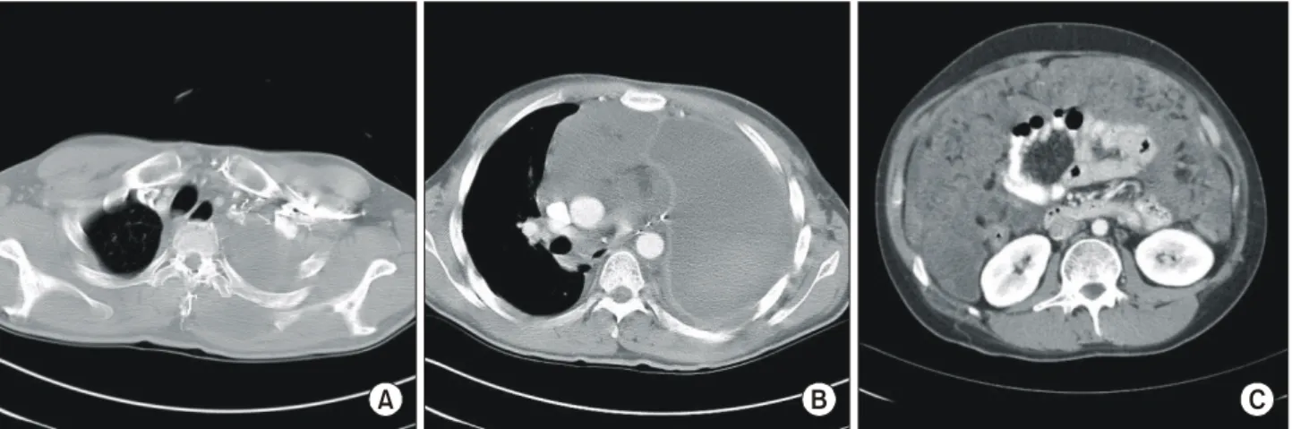
곤란과 흉부 불편감으로 타 병원을 방문하여 검사한 흉부 단순촬영 및 전산화단층촬영에서 좌측 혈흉이 의심되어 좌측 흉관삽관술을 시행받았다(Fig. 1A, B). 혈성 흉수가 Chun Soo Park, M.D.*, Young Tae Kim, M.D.*, Sook Whan Sung*, Joo Hyun Kim, M.D.*
Mediastinum is a very rare primary site of liposarcoma. In general, wide surgical excision with adequate resection margin is the treatment of choice for liposarcoma. We experienced a case of liposarcoma in a 24 year-old male who complained of dyspnea and chest discomfort. Symptoms had been developed a month before admission, and the intensity had been gradually increased. He visited another general hospital, and there he received left closed thoracostomy because hemothorax was suspected. Afterwards, he was transferred to our hospital without a specific diagnosis, on review of outside chest computed tomography film, mass shadow was detected in the mediastinum.
For the further evaluation, we checked the chest sonography and chest magnetic resonance imaging. MRI showed 10 cm sized mass contacted with pulmonary artery trunk and left main pulmonary artery. The radiologist strongly suggested sarcoma. On the 4th day after admission, we performed emergent exploratory left thoracotomy for hematoma evacuation because mediastinal shifting progressed and heart rate was increased. Biopsy confirmed that the evacuated materials were extraskeletal myxoid chondrosarcoma, so we performed extrapleural left pneumo- nectomy including diaphragm and a part of the pericardium. The final pathologic diagnosis was myxoid/round cell liposarcoma. He was discharged without complication and systemic chemotherapy was scheduled to begin 2 month later. During chemotherapy, local recurrence and peritoneal metastasis developed, and he died 10 month after the surgical excision. We report this case with reviewal of literature.
(Korean J Thorac Cardiovasc Surg 2004;37:286-291) ꠏ
Key words: 1. Liposarcoma
2. Mediastinal neoplasm 3. Pneumonectomy 4. Chemotherapy
*서울대학교병원 흉부외과, 서울대학교 의과대학 흉부외과학교실, 서울대학교병원 임상의학연구소
Department of Thoracic and Cardiovascular Surgery, Seoul National University Hospital, Seoul National University College of Medicine, Seoul National University Hospital Clinical Research Institute
논문접수일:2003년 9월 8일, 심사통과일:2003년 12월 6일
책임저자 : 김영태 (110-744) 서울특별시 종로구 연건동 28번지, 서울대학교병원 흉부외과 (Tel) 02-760-3161, (Fax) 02-765-7117, E-mail: [email protected]
본 논문의 저작권 및 전자매체의 지적소유권은 대한흉부외과학회에 있다.
배액되었으며, 세포병리검사에서 악성 세포는 관찰되지 않았다. 이후 시행한 흉부 단순촬영에서 좌측 흉수의 감 소가 관찰되지 않고, 발열, 호흡곤란 지속되어, 본원으로 전원되었다. 입원 후 흉관을 재삽입하였고, 당시 점액성의 혈성 흉수가 배액되었고, 이에 대해 시행한 세포병리검사 에서 특이소견은 없었다. 입원 후 4일째 38.5oC의 발열과 호흡곤란이 진행되었으며, 빈맥 및 중심정맥압이 상승하
였고, 흉부 단순촬영에서 종격동의 우측 편위가 관찰되어 응급수술을 시행하였다. 비디오 흉강경을 이용하여 흉곽 을 관찰하였으나 시야 확보가 여의치 않았고, 다량의 점 액성 흉수에 대한 배액이 어려워, 좌측 개흉술로 전환하 여 점액성 혈성 흉수의 배액을 시행하였다. 수술 후 환자 의 증상, 활력증후 및 흉부 단순촬영 소견은 호전되었다 (Fig. 1C). 배액된 점액성 혈성 흉수에서 골격계외 점액성
Fig. 1. Preoperative chest images. (A) simple
chest radiography at first visit; near total haziness of left lung, (B) chest computed tomography at first visit; mass shadow in anterior mediastinum, (C) simple chest radio- graphy after emergent drainage; improved hazi- ness of left hemithorax, (D) chest computed tomography after emergent drainage; more defitie mass shadow, (E) and (F) magnetic res- onance image after emergent drainage.A B
C D
E F
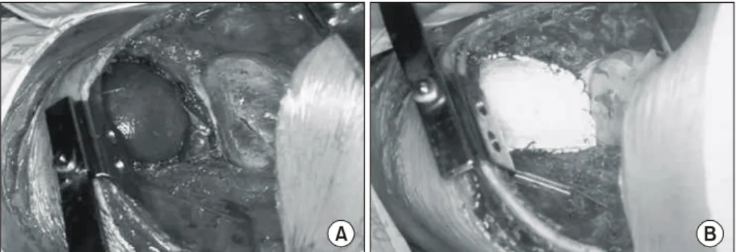
연골육종(extraskeletal myxoid chondrosarcoma)으로 진단되 었고, 이후 시행한 흉부 전산화단층촬영과 자기공명영상 에서 심장과 대혈관의 좌측 종격동에 심낭기원으로 생각 되는 최대 직경 10 cm 가량의 종괴가 관찰되어(Fig. 1D, E), 수술적 절제를 계획하였다. 입원 19일째 좌측 개흉술 을 시행하여 5번째 늑골을 절제 후 흉막 외 공간으로 박 리를 시행하였다. 종괴는 전종격동에 위치하고 있었으며, 좌주폐동맥과 좌상폐정맥을 압박하고 있었고, 폐의 좌상 엽을 침범하고 있는 양상이었다. 종양은 후측방으로 파열 되어 점액성 혈성액체가 배액되고 있었다. 폐문은 종괴의 위치상 위쪽에서 박리가 어려워 앞쪽에서 심낭을 열고 박 리하였으며, 심낭의 일부 및 횡격막의 일부를 포함한 흉 막외 전폐적출술을 시행하였다. 또, 응급수술 시의 절개부 위 피부를 포함한 흉벽 절제술 및 흉관을 삽입했던 자리 의 피부를 포함한 흉벽절제술을 시행하고 심낭결손부위 와 횡격막결손부위는 인조첩포를 이용하여 막아주었다 (Fig. 2). 조직검사에서는 종격동기원으로 흉막을 넘어 폐
의 일부를 침범하는 점액성/원형세포(myxoid/round cell) 지방육종으로 진단되었고(Fig. 3), 모든 절단면(resection margin)에서는 악성세포가 관찰되지 않았다. 수술 시 절제 한 종격동 림프절 및 기관지 주위 림프절에서는 악성세포 가 관찰되지 않았다. 환자는 술 후 11일째부터 약 2주간 원인불명의 발열이 있었으나 해소되었고 술 후 37일째 퇴 원하였다(Fig. 4). 술 후 2개월째 1회의 항암화학요법 (Ifosfamide+Adriamycin: IA) 후 1개월째 좌측 견갑골 부위 에 종괴가 발견되어 시행한 조직검사에서 재발한 점액성/
원형세포 지방육종으로 진단되었고, 당시 시행한 흉부 전 산화단층촬영에서 종격동에 9 cm 직경의 종괴와, 좌측 배 부의 종괴와 종격동 임파선비대 소견을 보여 재발에 합당 하였다(Fig. 5A). 이에 약제를 바꾸어(Etoposide+Ifosfamide +Cisplatin: VIP) 항암화학요법을 시행하였고, 3회차와 6 회차 후 시행한 흉부전산화단층촬영에서는 종양 크기가 크게 감소하는 양상이었다. 술 후 9개월째 8회차까지 시 행 후 복부팽만과, 체중증가로 시행한 복수천자 및 복부
Fig. 3. Gross and microscopic find-
ings of tumor. (A) gross specimen, (B) microscopic specimen (H&E stan- ing, ×400).A B
전산화단층촬영에서 복강전이로 진단되었고(Fig. 5B), 술 후 10개월째 사망하였다.
고 찰
지방육종은 성인에서 발생하는 육종 중 가장 흔하고, 주로 사지나 후복막강에 발생한다. 그러나 종격동에 원발 성으로 생기는 경우는 매우 드물어서 현재까지 전 세계적 으로 100여 예 정도가 보고되었으며, 우리나라에서는 지 금까지 4예가 보고되었다[1].
종격동에 발생하는 지방육종은 주로 전종격동에 있는 흉선과 관련된 지방조직에서 발생하게 되지만 후종격동
에서도 발생할 수 있고[2,3], 발견되기 전까지 상당한 크기 로 자라서 인접한 늑막강으로까지 범위를 확장할 수 있다 [4]. 증상과 징후는 주로 종양 자체에 의한 덩이효과(mass effect)나, 침범한 주위구조물에 의한 것으로, 호흡곤란, 빈 호흡 등의 호흡기계 증상이나, 흉통 등이 대부분이며, 증 상 없이 방사선 영상에 의해 우연히 발견되는 경우도 있 다. 본 증례에서는 종괴의 덩이효과로 인한 호흡곤란과, 흉부 불편감이 발생하였고, 이후 종양의 파열로 인한 다 량의 흉수로 증상 및 징후가 급격히 악화되었다.
진단으로는 질환의 희귀성 때문에 일단 그 질환을 의심 하는 것이 무엇보다 중요하겠고, 흉부단순촬영이나, 흉부 전산화단층촬영, 그리고 주위 연조직으로의 침범여부 등
Fig. 4. Postoperative simple radio-
graphy and chest computed tomog- raphy.A B
Fig. 5. Follow-up images. (A) and (B) chest computed tomography at 2 months after operation; low attenuated lesion in left back
muscle (A) and mass shadow in anterior mediastinum (B), (C) abdominal computed tomography at 9 month after operation shows peritoneal seeding.A B C
행하여 여기서 얻어진 조직을 통해 조직학적 진단이 가능 하였다.
종양의 생물학적인 행태나 재발, 전이에 있어서 종양의 크기, 위치, 조직학적 형태가 중요하고[4], 이 중 조직학적 형태가 그 예후에 있어서 매우 중요하다. 종양은 일반적 으로 4가지 조직학적 형태인 1) 고분화성, 2) 점액성, 3) 원형세포성, 4) 다형성으로 분류된고, 이 중 점액성이 가 장 흔하며, 다음으로 다형성의 순이다[7]. 본 증례의 경우 처음 응급수술 후 배액된 점액성 물질에 대한 조직검사에 서 골격외점액성연골육종(extraskeletal myxoid chondrosar- coma)으로 진단되었으나, 이는 지방아세포가 관찰되지 않 았으며, 연골형성부위가 많았기 때문이었고, 종양절제술 이후 조직검사에서는 점액성 아형과 원형세포성의 아형 이 혼재된 지방육종으로 진단되었다.
종양의 치료로는 광범위한 수술적 절제술이 추천되지 만, 상당한 경우에서 완전한 절제가 불가능한데[8], 이는 종격동에 발생하는 지방육종이 명확한 경계를 짓는 경우 가 적고, 주위에 중요한 흉곽 내 구조물들과 인접해 있기 때문이다[9]. 그러나 완전절제가 불가능한 경우라도 부분 절제로 부피를 줄여줌으로써(debulking) 증상의 완화를 기 대할 수 있다[10]. 종격동 임파선으로의 전이는 매우 드물 게 발생하기 때문에 수술 시 종격동 임파선 절제술은 추 천되지 않는다[4]. 비록 그 효과에 있어서 여러 다른 의견 들이 있지만 수술적 절제가 가능한 경우 종양의 완전한 절제 후에 방사선 치료를 추가함으로써 국소재발의 위험 성을 줄일 수 있고, 수술만 단독으로 시행한 경우에 비해 높은 5년 생존율을 보이며, 재발한 경우나 수술이 불가능 한 경우에 있어서 방사선 치료 단독으로 효과를 볼 수 있 다는 보고가 있다[11,12]. 본 증례에서는 종양의 범위가 넓 었고, 술 전 종양의 파열이 있어 방사선 조사 범위의 한계 가 있어 술 후 방사선 치료를 시행하지 못했다. 항암화학 요법의 경우 고도의 조직학적 아형인 경우나 완전한 절제
형의 경우에는 12.5%의 5년 생존율을 보인다[15]. 종격동 에 발생하는 육종은 종양의 완전한 절제여부가 생존에 중 요한 영향을 미친다고 알려져 있고, 다른 부위의 육종에 비해 예후가 좋지 않은 이유는 생물학적 행태나 위치 때 문에 완전절제가 불가능한 경우가 많기 때문이다. 원격전 이의 경우에도 조직학적 분화가 나쁘고, 종양의 크기가 클 수록 그 가능성이 높고[12], 주로 폐나 뼈로 잘 전이되며, 이외에 후복막강이나, 척추, 심장 등으로 전이될 수 있다.
저자들은 종격동에 발생한 지방육종의 수술적 치료 후 항암화학요법을 시행하여 추적관찰하였기에 문헌고찰과 함께 보고한다.
참 고 문 헌
1. Kim YH, Lee HW, Park SI, Kim DK, Sohn KH, Gong GY.
Primary mediastinal liposarcoma:1case report. Korean J
Thorac Cardiovasc Surg 1996;29:125-8.2. Grewal RG, Prager K, Austin JHM, Rotterdam H. Long-term
survival in non-encapsulated primary liposarcoma of the mediastinum. Thorax 1993;48:1276-7.
3. Eisenstat R, Bruce D, Williams LE, Katz DS. Primary
liposarcoma of the mediastinum with coexistent mediastinal lipomatosis. Am J Roentgenol 2000;174:572-3.
4. Enzinger FM, Weiss SW, editors. Liposarcoma. Soft tissue
tumors. 3
rded. St. Louis, MO: Mosby. 1995;431-66.
5. Barile A, Zugaro L, Catalucci A, et al. Soft tissue lipo-
sarcoma: histological subtypes, MRI and CT findings.
Radiologia Medica 2002;104:140-9.
6. Attal H, Jensen J, Reyes CV. Myxoid liposarcoma of the
anterior mediastinum: Diagnosis by fine needle aspiration biopsy. Acta Cytol 1995;39:511-3.
7. Ezinger FM, Winslow DJ. Liposarcoma: A study of 103
cases. Virchows Arch Pathol Anat 1962;335:367-88.
8. Crist WM, Raney RB, Newton W, Lawrence W, Tefft M, Foulkes MA. Interthoracic soft tissue sarcomas in children.
Cancer 1982;50:598-604.
9. Plukker JTM, Joosten HJM, Rensing JBM, Van Haelst UJGM. Primary liposarcoma of the mediastinum in a child.
J Surg Oncol 1988;37:257-63.
10. Schweitzer DL, Aguam AS. Primary liposarcoma of the
mediastinum. Report of a case and review of the literature.
J Thorac Cardiovasc Surg 1977;74:83-97.
11. Evans HL. Liposarcoma. Am J Surg Pathol 1979;3:507-23.
12. Razzuk MA, Urschel HC, Race GJ, Kingsley WB, Paulson DL. Liposarcoma of the mediastinum. Case report and
review of the literature. J Thorac Cardiovasc Surg 1971;61:
819-26.
13. Antman KA, Blum RH, Wilson RE, et al. Survival of
patients with localized high-grade soft tissue sarcoma with multimodality therapy. Cancer 1983;51:396-401.
14. Sawamura K, Hashimoto T, Nanjo S, et al. Primary lipo-
sarcoma of the lung: report of a case. J Surg Oncol 1982;
19:243-6
15. Enterline HT, Culberson JD, Rochlin DB, Brady LW.
Liposarcoma: a clinical and pathological study of 53 cases.
Cancer 1960;13:932-50.
=국문 초록=
원발성 지방육종은 종격동에서 매우 드물게 발생하는 악성 종양으로 수술적 절제가 가장 효과적인 치료로 알려져 있다. 24세 남자 환자가 1개월 전부터 시작된 호흡곤란과 흉부 불편감으로 타 병원에 서 좌측 혈흉을 의심하여 좌측 흉관삽관술을 시행받았다. 본원 입원 후 흉부 단순촬영에서 종격동 편 위소견을 보였고 빈맥이 진행하였으며, 중심정맥압이 상승하는 양상보여 진단 및 혈괴제거를 위해 응급으로 좌측 개흉술을 시행하여 다량의 점액성의 혈성 흉수를 제거하였고, 조직검사 결과 골격계 외 점액성 연골육종으로 진단되었다. 2주 후 좌측 개흉술로 횡격막 및 심낭의 일부를 포함하는 흉막 외 좌측 전폐적출술과 함께 종격동 종양을 절제하였다. 조직검사에서 점액성/원형세포 지방육종으로 진단되었으며, 입원 54일만에 합병증 없이 퇴원하였다. 술 후 2개월째 1회의 항암화학요법(Ifosfamide +Adriamycin: IA) 후 종격동, 좌측 견갑골 부위 및 종격동 임파선에 재발하여, 약제를 바꾸어 (Etoposide+Ifosfamide+Cisplatin: VIP) 항암화학요법을 시행하였으나 복강전이로 술 후 10개월째 사 망하였다. 종격동에 발생하는 원발성 지방육종은 매우 드문 질환으로 수술적 절제 및 항암요법 후 추 적관찰한 1예를 문헌고찰과 함께 보고하는 바이다.
중심 단어:1. 지방육종 2. 종격동 신생물 3. 전폐적출술 4. 항암화학요법