160
A 38-year-old female presented with fever, shortness of breath and loss of appetite. Physical examination and electro- cardiogram were unremarkable. Chest X-ray revealed pleural effusion and wide mediastinum.
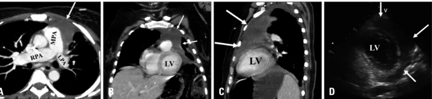
Contrast-enhanced computed tomography (CT) showed a large heterogeneous anterior mediastinal mass invading the me- diastinal structures and left anterior chest wall with encircle- ment and compression of the main and left pulmonary arteries.
The mass invaded the pericardium and was inseparable from the ventricular walls (Fig. 1A, B, and C). CT guided biopsy was
obtained and histopathology was consistent with a high-grade diffuse large B-cell lymphoma.
The patient underwent transthoracic echocardiogram (TTE) as part of routine work-up before chemotherapy. It uncovered normal left ventricular (LV) size with moderate systolic dysfunc- tion and a large mass encircling the anterior and lateral LV walls causing akinesis (Fig. 1D, Supplementary movie 1). Cardiac magnetic resonance (CMR) confirmed the findings of TTE. The mass was hyperintense on T2-weighted and isointense on T1- weighted sequences with no evidence of perfusion. Additionally
pISSN 1975-4612/ eISSN 2005-9655 Copyright © 2014 Korean Society of Echocardiography www.kse-jcu.org http://dx.doi.org/10.4250/jcu.2014.22.3.160
Unusual Cardiac Infiltration in Diffuse Large B-Cell Lymphoma
Sherif Moustafa, MBBCh1,2, David J. Patton, MD3, Nanette Alvarez, MD4, Timothy Prieur, MD4, Michael S. Connelly, MBBS4,
Mohammed Alnasser, BSc5, and Farouk Mookadam, MD2
1Department of Cardiovascular Diseases, Prince Salman Heart Center, King Fahad Medical City, Riyadh, Kingdom of Saudi Arabia
2Division of Cardiovascular Diseases, Mayo Clinic Arizona, Scottsdale, AZ, USA
3Section of Pediatric Cardiology, Department of Pediatrics, Alberta Children’s Hospital, University of Calgary, Calgary, AB, Canada
4Adult Congenital Heart Disease Clinic, Peter Lougheed Hospital, Division of Cardiovascular Diseases, Calgary, AB, Canada
5Department of Radiology, King Fahad Medical City, Riyadh, Kingdom of Saudi Arabia
KEY WORDS: Diffuse B-cell lymphoma · Cardiac involvement · Echocardiography · Computed tomography · Magnetic resonance.
• Received: June 11, 2014 • Revised: June 30, 2014 • Accepted: August 20, 2014
• Address for Correspondence: Sherif Moustafa, Division of Cardiovascular Diseases, Mayo Clinic Arizona, 13400 East Shea Boulevard, Scottsdale, AZ 85259, USA Tel: +1-480-301-6907, Fax: +1-480-301-8018, E-mail: [email protected]
• This is an Open Access article distributed under the terms of the Creative Commons Attribution Non-Commercial License (http://creativecommons.org/licenses/by-nc/3.0) which permits unrestricted non-commercial use, distribution, and reproduction in any medium, provided the original work is properly cited.
IMAGES IN CARDIOVASCULAR ULTRASOUND J Cardiovasc Ultrasound 2014;22(3):160-161
Fig. 1. Contrast-enhanced computed tomography showing a large heterogeneous anterior mediastinal mass (arrows) invading the mediastinal structures and left anterior chest wall with encirclement and compression of the main and left pulmonary arteries (A). The mass invaded the pericardium and was inseparable from the ventricular walls (B and C). Transthoracic echocardiogram parasternal short axis view showing a large mass encircling the anterior and lateral left ventricular walls (arrows) (D). LPA: left pulmonary artery, MPA: main pulmonary artery, RPA: right pulmonary artery, LV: left ventricle.
A B C D
Cardiac Involvement in Diffuse B-Cell Lymphoma | Sherif Moustafa, et al.
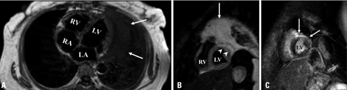
161 there was a non-ischemic sub-epicardial to mid wall late gado-
linium enhancement involving the anterior wall with extension to a small portion of the anterior septum/anterolateral walls (Fig. 2, Supplementary movie 2). Those findings are in keep- ing with infiltration of the myocardial wall by the mediastinal lymphoma rather than an external compression.
She was treated with 3 cycles of combination chemotherapy (R-ESHAP; rituximab plus etoposide, cytarabine, cisplatinum, and methylprednisolone). Follow-up contrast-enhanced CT showed marginal improvement of the size and extent of the mediastinal mass. Unfortunately, the patient died after com- pletion of chemotherapy and her mode of death was unwit- nessed.
Diffuse large B-cell lymphoma is an uncommon category of primary mediastinal B-cell lymphoma that originates in the thymus. It is more prevalent in women and young adults. It usually presents with systemic symptoms, shortness of breath, chest discomfort, and palpable lymph nodes. It rarely mani- fests as an intracardiac mass and likely remains silent and of- ten diagnosed on autopsy. With ventricular invasion, patients usually manifest with ventricular dysfunction which carries a dismal prognosis as noted in our patient.1-3)
The incorporation of multi-modality imaging is very im- portant in the diagnosis and management of cardiac/extracar- diac masses. Discrimination between cardiac infiltration by lymphoma and primary cardiac tumors is complicated. CMR and contrast enhanced CT are the most valuable tools in de-
marcating direct invasion by nearby mediastinal masses from primary cardiac tumors.4)5) In our case, CMR was very useful in depicting a direct infiltration of the myocardium by the mediastinal lymphoma rather than intracavitary involvement or merely an external compression by the mass.
Supplementary movie legends
Movie 1. Transthoracic echocardiogram parasternal short axis view showing a large mass encircling the anterior and lat- eral left ventricular walls causing akinesis.
Movie 2. Cardiac magnetic resonance short axis cine image revealing a low signal intensity mass encircling the anterior and lateral walls of the left ventricle causing hypokinesis.
References
1. Savage KJ. Primary mediastinal large B-cell lymphoma. Oncologist 2006;11:488-95.
2. O’Mahony D, Peikarz RL, Bandettini WP, Arai AE, Wilson WH, Bates SE. Cardiac involvement with lymphoma: a review of the literature.
Clin Lymphoma Myeloma 2008;8:249-52.
3. Yang CC, Tsai HW, Lai ST, Wu HC, Lo CY, Chang Y. Mediastinal diffuse large B-cell lymphoma invading the left atrium mimicking coronary artery disease with a mural thrombus. J Chin Med Assoc 2012;75:606-9.
4. Goldman M, Matthews R, Meng H, Bilfinger T, Kort S. Evaluation of cardiac involvement with mediastinal lymphoma: the role of innovative integrated cardiovascular imaging. Echocardiography 2012;29:E189-92.
5. Bley TA, Zeiser R, Ghanem NA, Hackanson B, Brink I, Langer M.
High grade cardiac lymphoma vitality monitoring by gadolinium-enhanced magnetic resonance imaging (MRI). In Vivo 2005;19:689-93.
Fig. 2. A: Axial T1-weighted imaging showing the mass inseparable from the left ventricular wall (arrows). B: Short-axis T2-weighted imaging showing myocardial edema in the anterior and lateral walls of the left ventricle (arrowheads) with high signal intensity of the mass (arrow). C: Short-axis late gadolinium enhancement imaging showing non-ischemic sub-epicardial to mid wall late enhancement involving the anterior wall with extension to a small portion of anterior septum/anterolateral walls (arrows). LA: left atrium, LV: left ventricle, RA: right atrium, RV: right ventricle.
A B C