ISSN: 2233-601X (Print) ISSN: 2093-6516 (Online)
− 419 −
Received: August 23, 2018, Revised: October 3, 2018, Accepted: October 16, 2018, Published online: December 5, 2018
Corresponding author: Harish Pal, Department of General and Minimal Access Surgery, Max Super Speciality Hospital, W-3, Vaishali Sector 1, Delhi-NCR, 201012, India
(Tel) 91-0120-4173000 (Fax) 91-1204173010 (E-mail) [email protected]
© The Korean Society for Thoracic and Cardiovascular Surgery. 2018. All right reserved.
This is an open access article distributed under the terms of the Creative Commons Attribution Non-Commercial License (http://creativecommons.org/
licenses/by-nc/4.0) which permits unrestricted non-commercial use, distribution, and reproduction in any medium, provided the original work is properly cited.
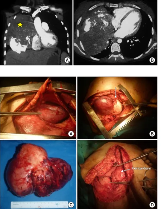
An Aggressive Large Epithelioid Hemangioendothelioma of the Anterior Mediastinum in a Young Woman
Roman Dutta 1 , Harish Pal 2 , Garima Garg 3 , Sambit Mohanty 3
1