A Survey on Current Trends of Breast Imaging Practices in Korea
한국의 유방 영상 실무의 현황에 관한 조사
Bo La Yun, MD1 , Sun Mi Kim, MD1* , Mijung Jang, MD1, Nariya Cho, MD2, Boo-Kyung Han, MD3
1Department of Radiology, Seoul National University Bundang Hospital, Seoul National University College of Medicine, Seongnam, Korea
2Department of Radiology, Seoul National University Hospital, Seoul National University College of Medicine, Seoul, Korea
3Department of Radiology, Samsung Medical Center, Sungkyunkwan University School of Medicine, Seoul, Korea
Purpose This study aimed to analyze the current practices of breast imaging among Korean ra- diologists.
Materials and Methods We invited members of the Korean Society of Breast Imaging (KSBI) working as breast radiologists to participate in a survey on the current practices in breast imag- ing, and investigated how quality assurance (QA), medical audits, and imaging-histologic dis- cordance were conducted.
Results The survey involved 312 members of the KSBI, and 116 (37%) responded to the 21 questions in the questionnaire. Most of the respondents were women (87%), aged below 50 years (82.7%), and working at university or tertiary hospitals (68.1%), who had varying levels of experience in breast imaging. Of the 116 respondents, 114 (96.6%) followed the Breast Imaging Reporting and Data System (BI-RADS) to interpret breast imaging. Out of 116, 72 (62.1%) inter- preted 20 or more mammograms per day, 84 (72.4%) performed 10–30 breast ultrasound scans per day, and 65 (56%) interpreted fewer than 5 breast MRI scans per day. Among the respon- dents, 82% performed mammography QA, 76.9% evaluated imaging-histologic correlations, and only 38.9% performed medical audits.
Conclusion The institutions and working patterns of breast radiologists were diverse. Although many respondents did not conduct medical audits, most of them followed BI-RADS when inter- preting breast imaging, performing QA, and evaluating imaging-histologic correlations.
Index terms Breast; Quality Control; Medical Audits; Radiologist
INTRODUCTION
Breast cancers are increasing rapidly worldwide, with a 20% increased incidence in
Received May 23, 2018 Revised July 21, 2018 Accepted December 18, 2018
*Corresponding author Sun Mi Kim, MD Department of Radiology, Seoul National University Bundang Hospital, Seoul National University College of Medicine, 82 Gumi-ro 173beon-gil, Bundang-gu, Seongnam 13620, Korea.
Tel 82-31-787-7617 Fax 82-31-787-4070 E-mail [email protected] This is an Open Access article distributed under the terms of the Creative Commons Attribu- tion Non-Commercial License (https://creativecommons.org/
licenses/by-nc/4.0) which permits unrestricted non-commercial use, distribution, and reproduc- tion in any medium, provided the original work is properly cited.
ORCID iDs Sun Mi Kim https://
orcid.org/0000-0003-0899-3580 Bo La Yun
https://
orcid.org/0000-0002-5457-7847
2012 compared to that in 2008. Moreover, in Korea, breast cancer incidence in 2000 was 5906 and has increased steadily, and reached 18381 in 2014 (1, 2). Therefore, breast cancer is the second most common cancer among women in Korea, following thyroid cancer. According to the Ministry of Health and Welfare’s National Cancer Registration Project, breast cancer accounted for 19.3% of all cancers in female patients in 2014 (3). This increase in breast can- cer incidence in Korea is probably caused by the effects of Westernized diets including high amounts of fat and calories, and a rise in total estrogen exposure due to decreased frequency of pregnancy and breastfeeding, as well as increased age at childbirth, early menarche, late menopause, and obesity. With public interest in women’s health rising, breast cancer screen- ing has become a part of the government-sponsored cancer-screening program. As a result, the frequency of early detection of breast cancer has increased. The proportion of patients with early breast cancer (stage 0 or stage 1) from 32.6% in 2000 to 60.6% in 2015 (4). As the number of screening programs has increased, the number of breast imaging examinations has also increased. Although the number of breast examinations has risen, there have been no reports of actual practices in breast imaging in Korea, for example, the daily workload, standardization of imaging interpretation, performance of quality assurance (QA), medical auditing, and so on. The purpose of our study was to report the current practices in breast imaging among Korean radiologists.
MATERIALS AND METHODS
From October 2017 to December 2017, we invited 312 members of the Korean Society of Breast Imaging (KSBI) working as breast radiologists to participate in a survey by e-mail on the current practices in breast imaging. An e-mail message from the investigators with a link to a web survey was sent to members of the KSBI with known e-mail addresses via a list server.
Follow-up emails were sent to non-respondents. Follow-up telephone calls were not made.
The survey instrument was a 21-question online questionnaire implemented through the Sur- veyMonkey online survey tool (SurveyMonkey, Portland, OR, USA). The questions were de- signed by two investigators and revised several times before the administration of the instru- ment to ensure question clarity and utility. This survey consisted of 21 questions divided into four categories (Appendix 1): questions about the baseline characteristics of the respondents, daily practices in breast imaging, performing medical audits and QA, and interest in a dedi- cated breast imaging reading system incorporating QA and medical auditing.
RESULTS
BASELINE CHARACTERISTICS OF THE RESPONDENTS
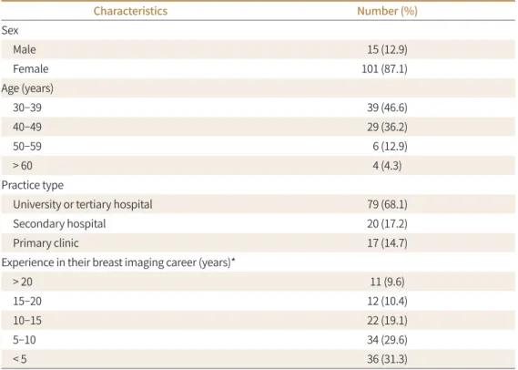
One hundred and sixteen individual responses to the survey were received. Most respon- dents were below 50 years of age (82.7%) and women (87%). The types of hospitals that the breast radiologists who participated in the survey worked at were as follows: 79 (68.1%), uni- versity or tertiary hospitals; 20 (17.2%), secondary hospitals; and 17 (14.7%), primary clinics.
Four respondents whom were classified as working in primary clinics mentioned that they are worked at screening centers. The respondents’ experience during their breast imaging
career was over 20 years in 11, 10–20 years in 34, 5–10 years in 34, and 5 years or less in 36.
One respondent skipped this question. Table 1 shows the detailed baseline characteristics of the respondents.
DAILY WORKLOAD
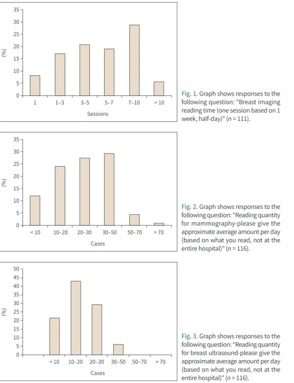
Regarding dedicated working time in breast imaging, most radiologists commonly worked 7–10 sessions per week (32, 28.8%) followed by 3–5 sessions (23, 20.1%) (Fig. 1). One-hundred- fourteen of 116 (96.6%) respondents followed the Breast Imaging Reporting and Data System (BI-RADS). The physicians in their 50s and 30s working at the primary institution did not use BI-RADS because they used another system more familiar to them. The daily numbers of mammograms read were 30–50 for 34 radiologists (29.3%), 20–30 for 32 (27.6%), 10–20 for 28 (24.1%), fewer than 10 for 16 (12.1%), and over 70 for one (0.9%) (Fig. 2). The daily numbers of breast ultrasound examinations performed were 10–20 for 50 radiologists (43.1%), 20–30 for 32 (29.3%), fewer than 10 for 25 (21.6%), and 30–50 for seven (6%) (Fig. 3). The daily numbers of breast MRI scans read were less than five for 65 radiologists (56%), 5–10 for 16 (13.8%), 10–15 for three (2.6%), 20–30 for three (2.6%), 15–20 for one (0.9%), and zero for 28 (24.1%) (Fig. 4).
The average reading durations for mammography were 1–2 min for 50 radiologists (44.6%), 30 sec to 1 min for 35 (31.3%), 2–3 min for 14 (12.5%), less than 30 sec for 7 (6.3%), 3–4 min for three (2.7%), 4–5 min for three (2.7%). Four radiologists skipped the question. The average duration required to perform and interpret ultrasound scans were 5–10 min for 50 (45.5%), 10–15 min for 28 (25.5%), less than 5 min for five (23.6%), and 15–20 min for six (5.5%); six ra- diologists skipped the question. The average reading durations for MRI were 10–15 min for 33
Table 1. Characteristics of Korean Breast Imaging Survey Responders and Hospitals
Characteristics Number (%)
Sex
Male 15 (12.9)
Female 101 (87.1)
Age (years)
30–39 39 (46.6)
40–49 29 (36.2)
50–59 6 (12.9)
> 60 4 (4.3)
Practice type
University or tertiary hospital 79 (68.1)
Secondary hospital 20 (17.2)
Primary clinic 17 (14.7)
Experience in their breast imaging career (years)*
> 20 11 (9.6)
15–20 12 (10.4)
10–15 22 (19.1)
5–10 34 (29.6)
< 5 36 (31.3)
*One respondent skipped this question.
35 30 25 20 15 10 5 0
(%)
< 10 10–20 20–30 30–50 50–70 > 70 Cases
35 30 25 20 15 10 5 0
(%)
1 1–3 3–5 5–7 7–10 > 10 Sessions
50 45 40 35 30 25 20 15 10 5 0
(%)
< 10 10–20 20–30 30–50 50–70 > 70 Cases
Fig. 1. Graph shows responses to the following question: “Breast imaging reading time (one session based on 1 week, half-day)” (n = 111).
Fig. 2. Graph shows responses to the following question: “Reading quantity for mammography-please give the approximate average amount per day (based on what you read, not at the entire hospital)” (n = 116).
Fig. 3. Graph shows responses to the following question: “Reading quantity for breast ultrasound-please give the approximate average amount per day (based on what you read, not at the entire hospital)” (n = 116).
radiologists (39.3%), 5–10 min for 29 (34.5%), 15–20 min for 12 (14.3%), less than 5 min for four (4.8%), while 32 did not perform MRI interpretation.
MEDICAL AUDITING AND QA
Most respondents (91, 82%) performed QA for mammography, and 20 (18%) did not; five respondents skipped the question. The intervals for performing QA were every 3 months for 57 radiologists (63.3%), 3–6 months for 14 (15.6%), less than 3 months for 13 (14.4%), 1 year for four (4.4%), 6–12 months for two (2.2%), and one respondent skipped the question. The aver-
age durations for performing QA were less than 10 min for 29 radiologists (34.9%), 10–20 min for 27 (32.5%), 20–30 min for 19 (22.9%), 45–60 min for five (6%), 30–45 min for three (3.6%), and eight skipped the question.
In contrast, most respondents (66, 61.1%) did not perform a medical audit. Forty-two (38.9%) performed medical auditing and nine skipped the question.
Most respondents (83, 76.9%) performed imaging-histologic correlation and discussed the findings with the clinician in cases of discordance; 25 (23.2%) reported not performing imag- ing-histologic correlation and eight skipped the question. The intervals for performing imag- ing-histologic correlation were 1–2 weeks for 37 radiologists (41.1%), less than 1 week for 30 (33.3%), 2–3 weeks for eight (8.9%), 1 month for six (6.7%), 1–3 months for two (2.2%), 3–6 months for one, over 6 months for 1, no regular interval for five, and 26 skipped the question.
INTEREST REGARDING A DEDICATED BREAST IMAGING READING SYSTEM INCORPORATING QA AND MEDICAL AUDITING
Most respondents (108, 97.3%) were willing to try out a BI-RADS-based interpretation system that can complete automated reports and manage data that have been read, and three (2.7%) were unwilling; five skipped the question.
Most respondents (109, 97.3%) were willing to use a reading system that displays candi- dates for clinical image QA, and is capable of medical auditing and three (2.7%) were unwill- ing; four skipped the question.
DISCUSSION
According to the statistical data provided by the Health Insurance Review and Assessment Service in Korea, breast MRI frequency increased twofold from 27072 cases in 2013 to 40286 cases in 2016, and breast ultrasound frequency increased more than tenfold from 12004 cases in 2013 to 126132 cases in 2016 (5). Despite a dramatic increase in the frequency of breast can- cer imaging owing to the increased breast cancer prevalence and public and media attention toward breast cancer, there has been little consideration of medical auditing, or other issues related to breast imaging interpretation.
From this survey, most of them (28.8%) worked 7–10 sessions per week. The median daily Fig. 4. Graph shows responses to the
following question: “Reading quanti- ty for breast MRI-please give the ap- proximate average amount per day (based on what you read, not at the entire hospital)” (n = 116).
60 50 40 30 20 10 0
(%)
No MRI < 5 5–10 10–15 15–20 20–30 > 30 Cases
workload was 30 routine four-view mammography procedures, 20 whole-breast ultrasound procedures, and five breast MRI procedures. An exact comparison of this workload to those in other countries is not possible because our performance and medical insurance systems are different. However, when the data were converted to work relative value units (RVU) from the USA, then 45.55 RUV would be the 50th percentile according to the Association of Admin- istrators in Academic Radiology (AAARAD) survey conducted in the fiscal year 2016 (6, 7).
When converting individual responses to RUV, the mean was 50.8 RUV (range, 7.6–123.4; 114 responses available). The mean RUV at a university hospital or tertiary hospital (58.1, n = 79) and mean RUV at a primary or secondary hospital (33.1, n = 35) were statistically significantly different (p < 0.001, independent t-test). In reference to the AAARAD survey from the fiscal year 2016, 39 RUV/day was the 25th percentile, 47 was the 50th percentile, 59 was the 75th percentile, and 75 was the 90th percentile. The survey responses showed that the lower 36%
corresponded to the 25th percentile, 14% to the 50th percentile, 33.3% to the 75th percentile, and 15.8% to the 90th percentile (6, 7). These comparisons did not include biopsies with higher RUV than the routine image interpretation; thus, the true RUV for Korean breast radi- ologists are greater than that shown in this survey’s results. Moreover, biopsy is more com- monly performed at university and tertiary hospitals, and the difference in RUV values be- tween hospital types is more significant.
Among the respondents, 18% did not perform QA. This is because there was another col- league responsible for QA. The most common interval was 3 months because the clinical im- aging evaluation and phantom imaging evaluation performed by physicians were done in this time interval. Over 80% of respondents were conduction QA because of the legal require- ment for the mammography QA; however, medical auditing was performed by fewer than 40%. Fortunately, 76.9% of respondents performing imaging-histologic correlation discussed findings with clinicians in discordant biopsy cases. The false-negative rate of breast imaging, which is the main problem in breast cancer screening, is known to be 10–30%, and it is known that the density of the breast parenchyma (breast density), the quality of mammography, and the quality of the reader affect mammography accuracy (8). In May 2011, the Ministry of Health and Welfare and National Cancer Center conducted a “Cancer Conquest Forum,”
which revealed that only 0.6% of patients with cancer were found to have breast cancer (9).
As a measure to supplement the low breast cancer detection rate in national breast cancer screening, continuous education is being provided for mammography screening, but no medical auditing has been conducted for the readings. In the United States in the early 1980s, the Quality Assurance Certification Program for mammography institutes was initiated un- der the leadership of the Society of Radiation Oncology and with the Food and Drug Admin- istration’s approval. In 1992, the mammography quality standard law was enacted and the mammography facility, radiation dose, worker regulations, regular image quality evalua- tions, and certification systems were implemented (10). The content of the breast readings should be qualified by examinations only for those who have been educated for a certain pe- riod of time or more; this stipulates rigorous requirements for equipment, standardizes read- ing methods, daily, weekly, 3 months, 6 months, designates one person who is responsible for image quality management for each institution, designates one radiologist who is respon- sible for image quality management activities, and conducts medical audits of the reading re-
sults (10). Medical auditing provides an ideal target for mammography screening results, in- cludes the accuracies of surveillance by the radiologist and the medical audit, and notifies the patient of the results. As a result, in the United States, improvements in quality control com- pared to that in the 1960s increased the incidence of breast cancer detected by mammogra- phy compared with findings by promotion, and especially the discovery of small breast can- cers less than 1 cm was increased (11, 12). In Korea, regulations were revised and newly established on the safety management of diagnostic radiological apparatus by the Ordinance of Health and Welfare on January 13, 2001, and include matters concerning X-ray field exam- inations, the compression band size test, the average wire dose test, the phantom image eval- uation test, and so on.
Image quality control for mammography has been implemented in Korea since 2004, but the inspection standard is not strict compared to that in the USA. Furthermore, there is no regulation of medical auditing. During the last 20 years, there has been greatly increased breast imaging according to the numbers; however, presently, improvement of quality is warranted.
This study has several limitations. First, only 37.2% of candidates responded. Further, this study may demonstrate selection bias, because only those who responded voluntarily were involved. In addition, many respondents worked in university or tertiary hospitals, and thus the results are not reflective of the practices in primary clinics. In addition, since most re- spondents were working in referral hospitals, most of the examinations were probably per- formed for diagnostic purposes; however, the proportion of diagnostic and screening exami- nations have not been investigated. Second, the survey was distributed only to KSBI members. The practices of other doctors including breast surgeons in large, private breast clinics were not reflected, and hence it is difficult to represent all the current breast imaging practices. Third, in the comparison of breast radiologist workloads, we could not reflect differ- ences in the number of patients or breast radiologists in each hospital and did not include tasks such as biopsy, outpatient consultations, or conferences.
Despite these limitations, this study could be important in providing information on the current practices of breast imaging in Korea including how imaging is performed and inter- preted, and the performance of QA, medical auditing, and imaging-histologic discordance.
The employing institutions and working patterns of breast radiologists were diverse. Most re- spondents followed BI-RADS when interpreting breast imaging and performed QA and imag- ing-histologic correlation, although many were not conducting medical audits.
Conflicts of Interest
The authors have no potential conflicts of interest to disclose.
Acknowledgments
This research was supported by the Basic Science Research Program through the National Research Foundation of Korea (NRF) funded by the Ministry of Education (No. 2017R1D1A1B03033975).
REFERENCES
1. Cancer statistics. Incidence of breat cancer. Available at. http://www.cancer.go.kr/. Published 2014. Ac- cessed Aug 14, 2017
2. Korean Statistical Information Service. Incidence of breat cancer. Available at. https://kosis.kr/. Published 2016. Accessed Aug 14, 2017
3. Jung KW, Won YJ, Oh CM, Kong HJ, Lee DH, Lee KH. Cancer statistics in Korea: incidence, mortality, surviv- al, and prevalence in 2014. Cancer Res Treat 2017;49:292-305
4. Korean Breast Cancer Society. Korean Breast Cancer Society breast cancer facts and figures 2017. Available at. http://www.kbcs.or.kr/sub02/sub04.html/. Accessed Dec 26, 2017
5. Healthcare Bigdata Hub. Breast MRI frequency 2017. Available at. http://opendata.hira.or.kr/. Accessed Jul 2, 2017
6. Centers for Medicare & Medicaid Services. Physician fee schedule CMS-1654-F. Available at. https://www.
cms.gov/Medicare/Medicare-Fee-for-Service-Payment/PhysicianFeeSched/PFS-Federal-Regulation-Notic- es-Items/CMS-1654-F.html?DLPage=1&DLEntries=10&DLSort=2&DLSortDir=descending. Accessed Nov 15, 2016
7. AAARAD Survey FY16. Association of Administrators in Academic Radiology. Available at. https://wp.
aaarad.org/wp/works/journal/. Accessed Dec 26, 2017
8. Kolb TM, Lichy J, Newhouse JH. Comparison of the performance of screening mammography, physical ex- amination, and breast US and evaluation of factors that influence them: an analysis of 27,825 patient eval- uations. Radiology 2002;225:165-175
9. Park EC. Evaluation of organized cancer screening program in Korea. Available at. http://www.ndsl.kr/
ndsl/search/detail/report/reportSearchResultDetail.do?cn=TRKO201100004109. Published 2010. Accessed December 26, 2017
10. McLelland R, Hendrick RE, Zinninger MD, Wilcox PA. The American College of Radiology mammography accreditation program. AJR Am J Roentgenol 1991;157:473-479
11. Linver MN, Paster SB, Rosenberg RD, Key CR, Stidley CA, King WV. Improvement in mammography inter- pretation skills in a community radiology practice after dedicated teaching courses: 2-year medical audit of 38,633 cases. Radiology 1992;184:39-43
12. Sickles EA, Ominsky SH, Sollitto RA, Galvin HB, Monticciolo DL. Medical audit of a rapid-throughput mam- mography screening practice: methodology and results of 27114 examinations. Radiology 1990;175:323- 327
한국의 유방 영상 실무의 현황에 관한 조사
윤보라1 · 김선미1* · 장미정1 · 조나리야2 · 한부경3
목적 본 연구는 한국 영상의학과 의사의 유방 영상 실무의 현행을 분석하고자 하였다.
대상과 방법 유방 영상 검사 시행 및 판독현황에 대한 조사를 위하여 유방 영상 판독 전문의 로 일하는 한국 유방영상학회 회원을 대상으로 하여 어떻게 품질관리(quality assurance; 이 하 QA), 의료 감사 및 영상-병리 학적 불일치를 시행하는지 조사하였다.
결과 설문에 한국 유방영상학회 회원 312명이 참여했으며, 설문지의 21개 질문에 116명(37%) 이 응답하였다. 응답자의 대부분은 여성(87%), 50세 미만(82.7%), 대학 또는 3차 병원 근무자 (68.1%)로 다양한 수준의 유방 영상 판독 경험을 보였다. 유방 영상을 판독할 때 유방 영상 판 독 데이터 체계(Breast Imaging Reporting and Data System; 이하 BI-RADS)를 따른 응답자 는 116명 중 114명(96.6%)이었다. 72명(62.1%)이 유방 촬영술을 하루에 20건 이상 해석하였 다. 84명(72.4%)이 하루에 10~30회의 유방 초음파 검사를 실시하였다. 65명(56%)이 하루에 5 회 미만의 유방 자기공명영상(MRI) 판독을 하였다. 대부분 응답자(82%)는 유방 촬영의 품질 관리를 수행하고 있으며, 76.9%에서 영상-병리 상관 관계를 확인했지만 의료 감사를 수행한 비율은 38.9%에 불과하였다.
결론 유방 영상 전문의들이 일하고 있는 기관과 근무 패턴은 다양하였다. 대부분의 응답자는 유방 영상을 판독할 때 BI-RADS를 따랐고 대부분 QA와 영상-병리 상관 관계 분석을 시행하 였으나. 의료 감사는 수행하지 않았다.
1서울대학교 의과대학 분당서울대학교병원 영상의학과,
2서울대학교 의과대학 서울대학교병원 영상의학과,
3성균관대학교 의과대학 삼성서울병원 영상의학과
APPENDIX
APPENDIX 1. SURVEY FORM FOR BREAST IMAGING PRACTICE
All responses to this survey are confidential. If you have any questions about the contents, please contact us at the following address:
1. Sex: Male/Female 2. Age group: 30s/40s/50s/60s
3. What kind of organization do you work for?
Primary clinic/secondary hospital/tertiary hospital/university hospital/other (explanation) 4. Breast imaging career:
Less than 1 year/1–3 years/3–5 years/5–10 years/10–15 years/15–20 years/more than 20 years
5. Breast imaging reading time: (one session based on 1 week, half-day)
Less than one session/1–3 sessions/3–5 sessions/5–7 sessions/7–10 sessions/over 10 ses- sions/(others, please specify)
6. Do you follow the BI-RADS for reading?
① No, I do not follow it (proceed to question 7)
② Yes, I do follow it (proceed to number 8)
7. If you do not follow the BI-RADS for reading, please indicate the reason(s) from the following items:
① I did not learn the BI-RADS
② Clinicians do not like the BI-RADS and want to use an older method of image reading
③ I am using my own system that is comfortable for me
④ Other (explanation): ____________________________________
8. Reading quantity: Please provide an approximate average amount per day (based on what you read, not at the entire hospital).
Mammography: Less than 10 cases/10–20 cases/20–30 cases/30–50 cases/50–70 cases/more than 70 cases/(others)
Breast ultrasound: Less than 10 cases/10–20 cases/20–30 cases/30–50 cases/50–70 cases/
more than 70 cases/(others)
Breast MRI: No MR (0)/Less than 5 cases/5–10 cases/10–15 cases/15–20 cases/20–30 cases/
more than 30 cases/(others)
9. Average reading time: Please provide the approximate average time required.
Mammography (based on the 4 routine views): 30 seconds only/30 seconds–1 minute/1–2
minutes/2–3 minutes/3–4 minutes/4–5 minutes/5 minutes or more/(others)
Breast ultrasound: less than 5 minutes/5–10 minutes/10–15 minutes/15–20 minutes/20–30 minutes/more than 30 minutes/(others)
Breast MRI: No MR (0)/Less than 5 minutes/5–10 minutes/10–15 minutes/15–20 min- utes/20–30 minutes
10. Do you implement image quality evaluation (quality control) for mammography?
① Yes, I do (Proceed to question 11)
② No, I do not (Proceed to question 12)
11. How often and how long does it take to perform clinical image quality assessment (quality control)?
Interval: Less than 1 month/1–2 months/2–3 months/quarterly (every 3 months)/3–6 months/6 months or more/(others)
Time required: Less than 10 minutes/10–20 minutes/20–30 minutes/30–45 minutes/45 minutes–1 hour/1–2 hours/more than 2 hours/(others)
12. Do you perform a medical audit?
① Yes, I do
② No, I do not
13. Do histologic correlations with the histologic examination lead to discordant results? Do you contact the clinical department?
① Yes, I do (Proceed to question 14)
② No, I do not (Proceed to question 15)
14. How often do you perform imaging-histologic correlations?
Less than 1 week/1–2 weeks/2–3 weeks/every month/1–3 months/3–6 months/6 months or more/(others)
15. If you have an inspection system based on BI-RADS that can complete an automated report and manage reading data, would you be willing to try it?
0 (no intention)/1 2 3 (normal)/4 5 (very much)
16. If you have a diagnostic system that displays clinical image quality assessment of the can- didate and is capable of a medical audit, would you be willing to try it?
0 (no intention)/1 2 3 (normal)/4 5 (very much)
![Notice of Gardening Service for the Embassy of the Republic of Korea [Extension of Closing Date]](data:image/gif;base64,R0lGODlhAQABAIAAAP///wAAACH5BAEAAAAALAAAAAABAAEAAAICRAEAOw==)