Clinicopathologic Similarities of the Main and Minor Lesions of Synchronous Multiple Early Gastric Cancer
The detection rate of early gastric cancer (EGC) is increasing due to improvements in diagnostic methods, but synchronous multiple EGC (SMEGC) remains a major problem.
Therefore, we investigated the characteristics of and the correlation between the main and minor lesions of SMEGC. We retrospectively reviewed the medical records of patients with EGC between April 2008 and May 2013. The main lesion was defined as the one with the greatest invasion depth. If lesions had the same invasion depth, the tumor diameter was used to define the main lesion. Of 963 patients who had treatment for EGC, 37 patients with SMEGC were analyzed. The main and minor lesions showed a significant positive correlation of size (r = 0.533, P = 0.001). The main and minor lesions of SMEGC showed the same vertical and horizontal locations at 70.3% and 64.9%, respectively (P = 0.002 and P = 0.002). Macroscopic types were identical in 67.6% (P < 0.001), and 32.4% had identical macroscopic type and location. The main and minor lesions had identical characteristics of invasion depth, presence of lymphovascular invasion (LVI), and differentiation in 78.4%, 83.8%, and 83.8%, respectively. Differentiation, LVI, and invasion depth (microscopic characteristics) were simultaneously the same in 62.2%. The location, macroscopic type, and 3 microscopic characteristics were matched in 27%. The main and minor lesions of SMEGC have similar clinicopathologic characteristics. Therefore, the possibility of SMEGC should not be neglected in cases of EGC, considering an
understanding of the characteristics and association of lesions.
Keywords: Stomach; Endoscopy; Gastrectomy; Neoplasms, Multiple Primary, Synchronous
Jung Ho Kim,1,2,3* Seok Hoo Jeong,1* Jina Yeo,1 Woon Kee Lee,4
Dong Hae Chung,5 Kyoung Oh Kim,1,2 Jun-Won Chung,1,2 Yoon Jae Kim,1,2,3 Kwang An Kwon,1,2,3
and Dong Kyun Park1,2
1Department of Internal Medicine, Gachon University Gil Medical Center, Incheon, Korea;
2Depatment of Internal Medicine, School of Medicine, Gachon University, Incheon, Korea;
3Gachon Medical Research Institute, Gachon University Gil Medical Center, Incheon, Korea;
4Department of Surgery, Gachon University Gil Medical Center, Gachon University School of Medicine, Incheon, Korea; 5Department of Pathology, Gachon University Gil Medical Center, Gachon University School of Medicine, Incheon, Korea
* Jung Ho Kim and Seok Hoo Jeong have contributed equally to this work.
Received: 1 November 2015 Accepted: 11 March 2016 Address for Correspondence:
Dong Kyun Park, MD
Department of Internal Medicine, Gachon University Gil Medical Center, 21 Namdong-daero 774-beon-gil, Namdong-gu, Incheon 21565, Korea
E-mail: pdk66@gilhospital.com
Funding: This research was supported by the Gachon University Gil Medical Center (Grant number: 2014-05-02 and 2014-19- 02) and, this work was supported by the 2016 grant from the Incheon Medical Association, Korea.
http://dx.doi.org/10.3346/jkms.2016.31.6.873 • J Korean Med Sci 2016; 31: 873-878
INTRODUCTION
The detection rate of early gastric cancer (EGC) is increasing due to improvements in diagnostic methods. EGC is defined as a lesion confined to the gastric mucosa or submucosa, without risk of regional lymph node metastasis (1). While advanced gastric cancer (AGC) has a 5-year survival rate of around 30%- 60%, EGC has a good prognosis with a survival rate greater than 90% (2). Therefore, missing the presence of a synchronous can- cer is more important for patients with EGC than AGC.
Two or more malignant cancer lesions in the stomach are of- ten diagnosed during upper gastrointestinal endoscopy and are known as synchronous multiple gastric cancer (SMGC). Re- cently, the incidence of multiple gastric cancer is increasing due to development of endoscopic technology and the increas-
ing use of a more detailed pathologic examination. SMGC is re- ported more commonly in association with EGC than with AGC, and SMGC associated with EGC is reported to account for 5%- 15% of all gastric cancer cases (3-6). Minimally invasive resec- tion procedures such as endoscopic resection and laparoscopic surgery have been widely used in patients with EGC because of the desire to increase the quality of life (7,8). Therefore, SMGC is a very important issue to the clinician because there is a pos- sibility of missing the presence of a cancer in the remnant stom- ach after minimal resection.
In particular, small lesions of SMGC can easily be overlooked.
Patients with an overlooked cancer miss their chance for early diagnosis and treatment, and the gastric cancer is then allowed to progress to advanced cancer. For this reason, it is important to clarify the association between main and minor lesions in
synchronous multiple early gastric cancer (SMEGC) that could lead to missing lesions or recurrence. However, no studies have investigated the characteristics and association of the main and minor lesions of SMEGC. Therefore, the aim of this study was to elucidate the clinicopathologic characteristics of and the asso- ciation between the main and minor lesions of SMEGC.
MATERIALS AND METHODS Study population
We retrospectively reviewed the medical records of patients who were diagnosed with EGC and were treated by endoscopic resection or surgical resection between April 2008 and May 2013 at Gachon University Gil Medical Center, Incheon, Korea. This study targeted patients who underwent initial treatment of EGC, and focused on main and minor lesions that were discovered during the first treatment of EGC. Informed written consent with adequate explanation was obtained from each patient be- fore the treatment, and each treatment had been undertaken with curative intent.
Clinicopathologic characteristics
The location, macroscopic type, and the histological findings of the gastric cancers were categorized according to the Japanese Gastric Cancer Association criteria (9). In this system, the longi- tudinal location of the stomach is anatomically divided into three portions, the upper third (UT), middle third (MT), and lower third (LT). The cross-sectional circumference of the stomach is divided into four equal parts: anterior wall (AW), lesser curva- ture (LC), posterior wall (PW), and greater curvature (GC). The macroscopic type of tumor was divided into three types: (i) ele- vated (I, IIa and IIa + IIb); (ii) flat (IIb); (iii) depressed (IIc, IIc + III and III). The maximum diameter of the cancer lesion was measured as the tumor size.
The histological types were grouped as either differentiated types (papillary adenocarcinoma, well and moderately differ- entiated tubular adenocarcinomas) or undifferentiated types (poorly differentiated tubular adenocarcinoma, signet-ring cell carcinoma, and mucinous adenocarcinoma).
Definition
While solitary EGC was defined as a single malignant focus in a patient with EGC, SMEGC was defined as having two or more malignant foci. SMGC was defined according to Moertel’s crite- ria as follows (10): 1) each lesion is a pathologically proven ma- lignancy; 2) each lesion is clearly separated from the others by microscopically normal gastric wall; and 3) the possibility that one of the lesions represents a metastatic tumor or a local ex- tension must be ruled out.
The main and minor lesions of gastric cancer were also de- fined, in accordance with Moertel’s criteria, as follows: 1) if the
depth of invasion of two or more lesions is equal, the one with the greatest diameter is regarded as the main lesion (also known as major lesions), with other lesions regarded as minor lesions (also known as accessory lesions) (10); and 2) if the depths of invasion are different in two or more lesions, the one with the greatest depth of invasion is regarded as the main lesion, with other lesions regarded as minor lesions. When there are more than three EGC lesions, the secondary main lesion is regarded as the minor lesion.
Procedure
The type of surgery (subtotal or total gastrectomy) and the ex- tent of lymphadenectomy were determined by the attending surgeon depending on the location of the cancer and the intra- operative conditions of the patient. Curative R0 resection and D2 lymph node dissection according to the Japanese Gastric Cancer Association were performed when possible. As previ- ously reported, endoscopic submucosal dissection was per- formed accordance to standard methods (11,12). All of the pa- tients underwent computed tomography with/without endo- scopic ultrasonography before treatment.
Statistical analysis
Statistical analysis was performed using SPSS 12.0 software (IBM SPSS Statistics, IBM Corporation, Armonk, NY, USA) for MS Windows®. Categorical variables are presented as absolute numbers or percentages, whereas continuous variables are presented as means ± standard deviation (SD). Continuous data were analyzed using the independent t-test, and other cat- egorical data were analyzed using the χ2 or Fisher’s exact test.
We used a linear by linear association and correlation analysis to determine the interrelationship between main and minor le- sions in patients with SMEGC. P values < 0.05 were considered statistically significant.
Ethics statement
This study was approved by the institutional review board of the Gachon University Gil Medical Center (IRB No. GAIRB2015-234).
Informed consent was waived by the board.
RESULTS
Patient characteristics
A total of 963 Korean patients who underwent endoscopic or surgical resection were diagnosed with EGC. Among these pa- tients, 37/963 (3.8%) patients diagnosed with SMEGC were in- cluded in this study. The mean age of the SMEGC patients was 64.3 ± 9.6 years, and 33 (89.2%) patients were men. Among the 37 patients, 20 received surgery as their first treatment. Thirty- five patients had no lymph node metastasis, and two had lymph node metastasis. Twenty-eight patients had two lesions and
nine patients had more than three lesions.
Correlation between main and minor lesions of patients with SMEGC
In patients with SMEGC, the mean size of the main and minor lesions was 28.0 ± 23.0 mm and 12.3 ± 7.5 mm, respectively.
The main and minor lesion size showed a significant moderate positive correlation (r = 0.533, P = 0.001), as the size of the mi- nor lesion increased when the main lesion increased in Fig. 1.
The comparison of characteristics between the main and mi- nor lesions of SMEGC is summarized in Table 1. The main and minor lesions of SMEGC were mostly located at LT of the stom- ach, 64.9% and 46.0%, respectively. When the main lesion was at UT of the stomach, 83.3% of the minor lesions were also at UT, and when the main lesion was at MT of the stomach, 85.7%
of the minor lesions were also at MT. Similarly, when the main lesion was at LT, 62.5% of the minor lesions were also at LT. When
the main lesion was at AW, GC, PW, or LC of the stomach, 37.5%, 66.7%, 57.1%, and 84.6% of the minor lesions were found at the same location, respectively. The main and minor lesions of SM- EGC showed the same vertical and horizontal locations in 70.3%
and 64.9% of cases (P = 0.002 and P = 0.002), respectively.
As for macroscopic types, 50% of the minor lesions were the elevated type when the main lesion was elevated, 58.3% of the minor lesions were the flat type when the main lesion was flat, and 82.4% of the minor lesions were the depressed type when the main lesion was depressed. The association of macroscopic type was statistically significant at 67.6% for both the main and minor lesions of SMEGC to share the same type (P < 0.001).
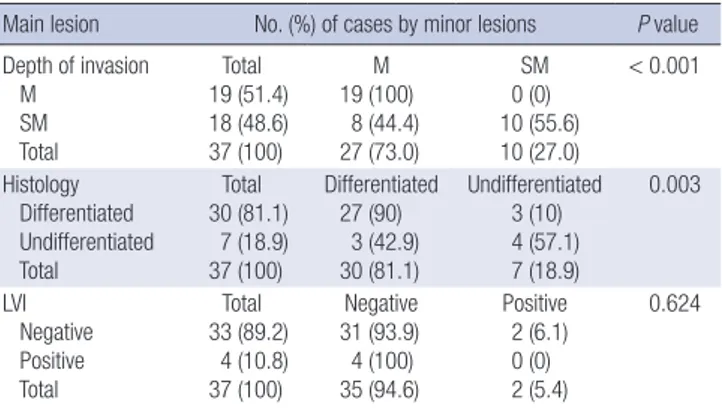
The other comparisons of characteristics between the main and minor lesions of SMEGC are summarized in Table 2. When the main lesion was a differentiated type, 90% of the minor le- sions were differentiated, an association that was statistically significant (P = 0.003). When the main lesion did not have lym- phovascular invasion (LVI), 93.9% of the minor lesions did not have LVI, and although 6.1% of the minor lesions had LVI, this did not reach statistical significance. Close to half (48.6%) of the main lesions were submucosal (SM) cancer, and when the main
Table 1. Comparison of characteristics between main and minor lesions of SMEGC
Main lesions No. (%) of cases by minor lesions P value
Vertical location UT MT LT Total
Total 6 (16.2) 7 (18.9) 24 (64.9) 37 (100)
UT 5 (83.3) 0 (0) 3 (12.5) 8 (21.6)
MT 0 (0) 6 (85.7) 6 (25.0) 12 (32.4)
LT 1 (16.7) 1 (14.3) 15 (62.5) 17 (46.0)
0.002
Horizontal location AW
GC PW LC Total
Total 8 (21.6) 9 (24.3) 7 (19.0) 13 (35.1) 37 (100)
AW 3 (37.5) 1 (11.1) 1 (14.3) 1 (7.7) 6 (16.2)
GC 0 (0) 6 (66.7) 1 (14.3) 1 (7.7) 8 (21.6)
PW 3 (37.5) 2 (22.2) 4 (57.1) 0 (0) 9 (24.3)
LC 2 (25.0) 0 (0) 1 (14.3) 11 (84.6) 14 (37.8)
0.002
Macroscopic type Elevated Flat Depressed Total
Total 8 (21.6) 12 (32.4) 17 (46.0) 37 (100)
Elevated 4 (50) 2 (16.7) 0 (0) 6 (16.2)
Flat 4 (50) 7 (58.3) 3 (17.6) 14 (37.8)
Depressed 0 (0) 3 (25) 14 (82.4) 17 (46.0)
< 0.001
UT, upper third; MT, mid third; LT, low third; AW, anterior wall; GC, great curvature; PW, posterior wall; LC, lesser curvature.
Table 2. Comparisons of additional characteristics of main and minor lesions in SMEGC Main lesion No. (%) of cases by minor lesions P value Depth of invasion
M SM Total
Total 19 (51.4) 18 (48.6) 37 (100)
M 19 (100)
8 (44.4) 27 (73.0)
SM 0 (0) 10 (55.6) 10 (27.0)
< 0.001
Histology Differentiated Undifferentiated Total
Total 30 (81.1)
7 (18.9) 37 (100)
Differentiated 27 (90)
3 (42.9) 30 (81.1)
Undifferentiated 3 (10) 4 (57.1) 7 (18.9)
0.003
LVI Negative Positive Total
Total 33 (89.2)
4 (10.8) 37 (100)
Negative 31 (93.9) 4 (100) 35 (94.6)
Positive 2 (6.1) 0 (0) 2 (5.4)
0.624
M, mucosa; SM, submucosa; LVI, lymphovascular invasion.
Fig. 1. Correlation of tumor size in main and minor lesions of SMEGC. The line indi- cates a moderate linear relationship of tumor size between the main and minor lesions of SMEGC (r = 0.533, P = 0.001).
SMEGC, synchronous multiple early gastric cancer.
Tumor size of minor lesions (mm)
Tumor size of main lesions (mm)
0 20 40 60 80 100 120
40 35 30 25 20 15 10 5 0
lesion was SM cancer, 55.6% of the minor lesions were also SM cancer (P < 0.001).
The matching characteristics of the main and minor lesions The main and minor lesions were mainly differentiated types (both 81.1%). The main and minor lesions were the same dif- ferentiated types in 83.8% of cases. Twenty-six out of 37 (70.3%) showed both identical differentiation and LVI. Three microscop- ic characteristics, differentiation, LVI, and invasion of depth, were the same in both main and minor lesions in 62.2% of cas- es. Both the three microscopic characteristics and the type were matched in 43.2%, and 27% of cases had all six factors matched in Fig. 2.
DISCUSSION
SMEGC detected in the gastric mucosa after endoscopic resec- tion or minimally invasive surgery is a major problem. Atten- tion to the possibility is always required, because if not detected during initial treatment, early treatment opportunities may be lost because SMEGC will only be detected at an advanced stage.
Therefore, we investigated characteristics within the lesions of SMEGC, in order to increase the possibility of finding anoth- er lesion and decrease the number of missed gastric cancers. In particular, this study focused on EGC to compare the character- istics of the initial carcinogenic process. SMEGC studies focus- ing mainly on the associations between main and minor lesions, such as this study, are rare. In addition, this study used a strict definition of SMEGC, and unlike other studies (7,13,14), target- ed SMEGC that were concurrently treated with multiple EGC during initial treatment.
This study was conducted on 37 (3.8%) of 963 EGC patients.
The prevalence of the disease was lower than has been reported
in previous studies, 5%-15% (3-6). The reason for the low preva- lence in this study is thought to be due to several factors. First, the definition of SMEGC differs among the previous studies, and in some studies, it is defined to include any second lesion that has occurred within a year (7,13,14). However, in this study it was strictly defined to be limited to multiple EGC that were detected during initial treatment. Secondly, the difference in prevalence may be due to the difference in race and region. Sim- ilar to this study, other studies held in South Korea have report- ed SMEGC prevalence of 3%-8% (15,16).
In this study, the main lesion was significantly larger than the minor lesion, and there is an association in that as the main le- sion becomes larger so does the minor lesion. Macroscopic types were identical type in 25/37 (67.6%), and the vertical and hori- zontal location in 17/37 (50%) were simultaneously identical.
Finally, 12/37 (32.4%) showed both identical macroscopic type and location at the same time. This result shows that in over 1/3 of SMEGCs, the gross appearance of both main and minor le- sions is similar and they have similar locations. These results correspond to previous studies of gastric cancer (5,14). They support the “collision tumor phenomenon” theory where the main and minor lesions occur adjacent to each other. However, it must be taken into account that regardless of the characteris- tics of the main lesion, the proportion of the minor lesions be- ing located at UT and MT, and the occurrence of flat types are high (17).
For the microscopic findings, the similarity of invasion depth, presence of LVI, and the level of differentiation between the main and minor lesions were shown to be high: 29/37 (78.4%), 31/37 (83.8%), and 31/37 (83.8%), respectively. In 23/37 (62.2%), these three factors were identical. These results support the “field car- cinogenesis” hypothesis, where the entire gastric mucosa has an identical carcinogenic background. According to this hypo- thesis, the risk factors and precancerous lesions such as Helico- bacter pylori infection, atrophic gastritis, and intestinal meta- plasia occur mainly in the distal stomach. The main and minor lesions of this study all occurred in the distal stomach as well.
The strength of this study lies in its elucidation of the ratio of identical characteristics present in the macroscopic and micro- scopic findings of main and minor lesions in SMEGC having statistical significance. In the case of multiple gastric cancers occurring within the same individual, the genetic and environ- mental backgrounds as well as the level of exposure to carcino- gens is considered to be uniform across the entire mucosa of the stomach.
According to the results of this study, the identical character- istics between the main and minor lesions showed that micro- scopic findings were more similar than macroscopic findings that included location and gross appearance. Even if carcino- genesis begins, environmental factors that could be changed by continuous stimuli such as Helicobacter pylori, intestinal meta- Fig. 2. The matching characteristics between the main and minor lesions of SMEGC.
The main and minor lesions had the same differentiation in 83.8% of cases. Three microscopic characteristics, including differentiation, LVI, and depth of invasion, were the same in 62.2% of the main and minor lesions. Both the three microscopic char- acteristics and the type were matched in 43.2%. All six factors matched in 27.0%, including the three microscopic characteristics, macroscopic type, and vertical and horizontal location.
SMEGC, synchronous multiple early gastric cancer; LVI, lymphovascular invasion; VL, vertical location; HL, horizontal location.
Matching properties
Differentiation 31/37 (83.8%)
26/37 (70.3%) 23/37 (62.2%)
16/37 (43.2%) 13/37 (35.1%) 10/37 (27.0%) Differentiation + LVI
Differentiation + LVI + Depth Differentiation + LVI + Depth + Type Differentiation + LVI + Depth + Type + VL Differentiation + LVI + Depth + Type + VL + HL
Number of patients (%)
plasia, and life style, can affect the outcome of macroscopic ap- pearance.
In molecular biology, it is known that gastric cancer has a high rate of genetic diversity, which is the base of its molecular phenotype (18,19). These molecular phenotypes primarily con- tribute to histologic features and determine clinical diversity.
Many mechanisms other than environmental factors are inter- connected. As further study is required on the effects of molec- ular phenotypes on morphological clinical characteristics, our results could contribute to encourage a focus on molecular and biologic analysis of SMEGC.
Because the development of endoscopic techniques is incre- asing the diagnostic rate of EGC, minimal invasive treatment is becoming more important. In addition, because it is known that the concurrence rate of SMGC in EGC is higher than that of AGC, the characteristics of minor lesions are clinically impor- tant (14). Therefore, it is critical to accurately detect SMEGC ini- tially. Once an endoscopist has detected an EGC, a more me- ticulous endoscopic examination should be considered to iden- tify a second EGC lesion. If the matching properties between the main and minor lesions are not overlooked when EGC is first detected or during follow-up, the possibility of missing can- cers will be reduced.
The predisposing conditions of SMGC were reported in sev- eral previous studies: male, elderly people and intestinal types of gastric cancer were more likely to be susceptible (7). Most multiple gastric cancers are found in the MT and LT rather than the UT. SMGC is more likely to occur in association with well to moderately differentiated tumors than poorly differentiated ones (4,14,20). Therefore, based on our results, clinicians should pay careful attention to finding other lesions in patients with these characteristics.
In conclusion, the main and minor lesions of SMEGC share similar clinicopathologic characteristics. If synchronous EGC is overlooked, the risk of recurrence will increase and prognosis will be poor. Therefore, when EGC is detected, the possibility of SMEGC should not be neglected, taking into account our un- derstanding of the characteristics of the main and minor lesions.
DISCLOSURE
The authors have no potential conflicts of interest to disclose.
AUTHOR CONTRIBUTION
Conception and coordination of the study: Park DK, Kim JH.
Acquisition of data: Jeong SH, Kim JH, Chung JW. Data review:
Lee WK, Chung DH, Kim KO, Kim YJ. Statistical analysis: Kwon KA. Manuscript preparation: Kim JH, Jeong SH. Revision: Yeo J.
Manuscript approval: all authors.
ORCID
Jung Ho Kim http://orcid.org/0000-0002-6944-473X Seok Hoo Jeong http://orcid.org/0000-0001-9197-1184 Jina Yeo http://orcid.org/0000-0002-7923-8729 Woon Kee Lee http://orcid.org/0000-0001-9494-4026 Dong Hae Chung http://orcid.org/0000-0002-4538-0989 Kyoung Oh Kim http://orcid.org/0000-0002-5365-2550 Jun-Won Chung http://orcid.org/0000-0002-0869-7661 Yoon Jae Kim http://orcid.org/0000-0001-8477-6823 Kwang An Kwon http://orcid.org/0000-0002-2947-2111 Dong Kyun Park http://orcid.org/0000-0002-2862-6641 REFERENCES
1. Nasu J, Doi T, Endo H, Nishina T, Hirasaki S, Hyodo I. Characteristics of metachronous multiple early gastric cancers after endoscopic mucosal resection. Endoscopy 2005; 37: 990-3.
2. Maehara Y, Kakeji Y, Oda S, Baba H, Sugimachi K. Tumor growth patterns and biological characteristics of early gastric carcinoma. Oncology 2001;
61: 102-12.
3. Kato M, Nishida T, Yamamoto K, Hayashi S, Kitamura S, Yabuta T, Yoshio T, Nakamura T, Komori M, Kawai N, et al. Scheduled endoscopic surveil- lance controls secondary cancer after curative endoscopic resection for early gastric cancer: a multicentre retrospective cohort study by Osaka University ESD study group. Gut 2013; 62: 1425-32.
4. Otsuji E, Kuriu Y, Ichikawa D, Okamoto K, Hagiwara A, Yamagishi H. Clini- copathologic characteristics and prognosis of synchronous multifocal gas- tric carcinomas. Am J Surg 2005; 189: 116-9.
5. Nakajima T, Oda I, Gotoda T, Hamanaka H, Eguchi T, Yokoi C, Saito D. Meta- chronous gastric cancers after endoscopic resection: how effective is an- nual endoscopic surveillance? Gastric Cancer 2006; 9: 93-8.
6. Peng J, Wang Y. Epidemiology, pathology and clinical management of multiple gastric cancers: a mini-review. Surg Oncol 2010; 19: e110-4.
7. Nitta T, Egashira Y, Akutagawa H, Edagawa G, Kurisu Y, Nomura E, Tan- igawa N, Shibayama Y. Study of clinicopathological factors associated with the occurrence of synchronous multiple gastric carcinomas. Gastric Can- cer 2009; 12: 23-30.
8. Bang CS, Baik GH, Shin IS, Kim JB, Suk KT, Yoon JH, Kim YS, Kim DJ. He- licobacter pylori eradication for prevention of metachronous recurrence after endoscopic resection of early gastric cancer. J Korean Med Sci 2015;
30: 749-56.
9. Japanese Gastric Cancer Association. Japanese classification of gastric carcinoma: 3rd English edition. Gastric Cancer 2011; 14: 101-12.
10. Moertel CG, Bargen JA, Soule EH. Multiple gastric cancers; review of the literature and study of 42 cases. Gastroenterology 1957; 32: 1095-103.
11. Kim JH, Kim YJ, An J, Lee JJ, Cho JH, Kim KO, Chung JW, Kwon KA, Park DK, Kim JH. Endoscopic features suggesting gastric cancer in biopsy-prov- en gastric adenoma with high-grade neoplasia. World J Gastroenterol 2014; 20: 12233-40.
12. Kang KJ, Kim KM, Min BH, Lee JH, Kim JJ. Endoscopic submucosal dis- section of early gastric cancer. Gut Liver 2011; 5: 418-26.
13. Seo JH, Park JC, Kim YJ, Shin SK, Lee YC, Lee SK. Undifferentiated histol-
ogy after endoscopic resection may predict synchronous and metachro- nous occurrence of early gastric cancer. Digestion 2010; 81: 35-42.
14. Isobe T, Hashimoto K, Kizaki J, Murakami N, Aoyagi K, Koufuji K, Akagi Y, Shirouzu K. Characteristics and prognosis of synchronous multiple early gastric cancer. World J Gastroenterol 2013; 19: 7154-9.
15. Choi J, Kim SG, Im JP, Kang SJ, Lee HJ, Yang HK, Kim JS, Kim WH, Jung HC, Song IS. Lymph node metastasis in multiple synchronous early gas- tric cancer. Gastrointest Endosc 2011; 74: 276-84.
16. Kim HM, Kim HK, Lee SK, Cho JH, Pak KH, Hyung WJ, Noh SH, Kim CB, Lee YC, Song SY, et al. Multifocality in early gastric cancer does not in- crease the risk of lymph node metastasis in a single-center study. Ann Surg Oncol 2012; 19: 1251-6.
17. Kitamura K, Yamaguchi T, Okamoto K, Otsuji E, Taniguchi H, Hagiwara A, Sawai K, Takahashi T. Clinicopathologic features of synchronous multifo-
cal early gastric cancers. Anticancer Res 1997; 17: 643-6.
18. Tan IB, Ivanova T, Lim KH, Ong CW, Deng N, Lee J, Tan SH, Wu J, Lee MH, Ooi CH, et al. Intrinsic subtypes of gastric cancer, based on gene expres- sion pattern, predict survival and respond differently to chemotherapy.
Gastroenterology 2011; 141: 476-85, 485.e1-11.
19. Ryu JW, Kim HJ, Lee YS, Myong NH, Hwang CH, Lee GS, Yom HC. The proteomics approach to find biomarkers in gastric cancer. J Korean Med Sci 2003; 18: 505-9.
20. Ribeiro U Jr, Jorge UM, Safatle-Ribeiro AV, Yagi OK, Scapulatempo C, Per- ez RO, Corbett CE, Alves VA, Zilberstein B, Gama-Rodrigues J. Clinico- pathologic and immunohistochemistry characterization of synchronous multiple primary gastric adenocarcinoma. J Gastrointest Surg 2007; 11:
233-9.