http://dx.doi.org/10.5090/kjtcs.2013.46.1.33 ISSN: 2233-601X (Print) ISSN: 2093-6516 (Online)
Department of Thoracic and Cardiovascular Surgery, Gachon University Gil Medical Center, Gachon University
†This manuscript was presented at the 42th Academic Meeting of the Korean Thoracic and Cardiovascular Society.
Received: June 29, 2012, Revised: August 22, 2012, Accepted: August 24, 2012
Corresponding author: Chul Hyun Park, Department of Thoracic and Cardiovascular Surgery, Gachon University Gil Medical Center, Gachon University, 21 Namdong-daero 774beon-gil, Namdong-gu, Incheon 405-760, Korea
(Tel) 82-32-460-8426 (Fax) 82-32-460-3668 (E-mail) [email protected]
C
The Korean Society for Thoracic and Cardiovascular Surgery. 2013. All right reserved.
CC
This is an open access article distributed under the terms of the Creative Commons Attribution Non-Commercial License (http://creative- commons.org/licenses/by-nc/3.0) which permits unrestricted non-commercial use, distribution, and reproduction in any medium, provided the original work is properly cited.
Early and Mid-term Changes of the Distal Aorta after Total Arch Replacement for Acute Type A Aortic Dissection
Chang Hu Choi, M.D., Chul Hyun Park, M.D., Yang Bin Jeon, M.D., So Young Lee, M.D., Jae Ik Lee, M.D., Kook Yang Park, M.D.
Background: Total arch replacement (TAR) is being more widely performed due to recent advances in operative techniques and cerebral protective strategies. In this study, the authors reviewed the relationship between TAR and early- and mid-term changes of the false lumen after TAR in acute type A aortic dissection. Materials and Methods: Twenty-six patients (aged, 54.7±13.3 years) who underwent TAR for acute type A dissection between June 2004 and February 2012 were reviewed. The relationship between the percentage change in the aortic diam- eter and the false lumen patency status was assessed by examining the early and late postoperative computed to- mography imaging studies. Results: There were two in-hospital mortalities, one late death, and three follow-up loses. The mean follow-up duration for the final 21 patients studied was 54±19.0 months (range, 20 to 82 months). The incidence of false lumen thrombosis within 2 weeks of surgery in the proximal, middle, and distal thoracic aorta, and the suprarenal and infrarenal abdominal aorta were 67%, 38%, 38%, 48%, and 33%, re- spectively, and 57%, 67%, 52%, 33%, and 33% for those examined at a mean of 49±18 months after surgery, respectively. The false lumen regressed in 11 patients (42.3%). The aortic diameters were larger in the patients with a patent false lumen than those with a thrombosed false lumen at all levels of the descending aorta (p
<0.05). Conclusion: TAR and a more complete primary tear-resection can be accomplished with a relatively low-risk of morbidity and mortality. Enlargement of the distal aorta significantly correlated with the false lumen pa- tency status.
Key words: 1. Aorta
2. Aortic dissection 3. Aortic arch
INTRODUCTION
The course of an untreated acute type A aortic dissection has the potential to rapidly progress into a catastrophic state.
The known factors that contribute to increasing the mortality rate include the development of pericardial tamponade from aortic rupture, acute myocardial ischemia or infarction due to
involvement of the coronary arteries, extension of dissection into the branch vessels leading to compromised distal organ perfusion, aortic rupture into the pleural space, and aortic valve involvement resulting in acute onset heart failure.
Despite the improvements in surgical techniques and perioper-
ative care, the mortality rate associated with acute type A
aortic dissection remains dismally high (range, 15% to 30%)
Table 1. Preoperatively clinical characteristics (n=26)
Characteristic Value
Sex (male/female) Age (yr) Marfan syndrome Hypertension Diabetes mellitus Malperfusion Brain Coronary Limb
Hemopericardium Cardiac tamponade Aortic rupture
Aortic insufficiency (grade≥3) Intimal tear
Ascending aorta Aortic arch Descending aorta
Ascending & descending aorta Patent false lumen
18/8 54.7±13.3 (28–76)
2 15 2 6 (23.1)
3 2 1 15 (57.7) 4 (15.3) 1 (3.8) 4 (15.3)
4 (15.3) 9 (34.6) 12 (46.2) 1 (3.8) 25 (100)
Values are presented as mean±standard deviation (range) or number (%).
[1-3]. Although the risk of immediate mortality in the acute phase is reduced by performing ascending aortic and hemi- arch repair, these operations are unable to prevent the need for distal reoperation in the future. Total arch replacement (TAR) allows for a more complete resection of the primary tear, but it is associated with a higher surgical risk [4,5].
Recent enhancements in surgical techniques and cerebral pro- tection methods have reduced the immediate postoperative mortality risks of surgery. Nevertheless, the long-term prog- nosis after acute type A dissection surgery remains dis- appointing as patency of the descending aortic false lumen re- mains little affected by the surgical outcome of the primary proximal repair [6-8].
In the present study, we evaluated the false lumen changes in the descending aorta after TAR and studied the relation- ship between the aortic diameter and the false lumen patency status in these patients.
MATERIALS AND METHODS 1) Patients
Fifty-three patients underwent emergency or urgent surgery for acute type A aortic dissection between June 2004 and February 2012. TAR was performed in 26 of these patients (49%). The mean follow-up duration was 54±19.0 months (range, 20 to 82 months). The ages ranged from 28 to 76 years (mean 54.7±13.3 years), and there were 18 men and 8 women. The preoperative characteristics of aortic dissection included Marfan syndrome in 2 patients (7.7%), brain malper- fusion in 3 (11.5%), coronary malperfusion in 2 (7.7%), limb malperfusion in 1 (3.8%), hemopericardium in 15 (57.7%), cardiac tamponade in 4 (15.3%), aortic rupture in 1 (3.8%), and severe aortic insufficiency in 4 (15.3%). The primary in- timal tear was located in the ascending aorta in 4 patients (15.3%), in the aortic arch in 9 (34.6%), in the descending aorta in 12 (46.2%), and in the ascending and descending aorta in 1 (3.8%) (Table 1). TAR was performed for the fol- lowing conditions: 1) an intimal tear in the arch (excluding the minor curvature), 2) a tear in the descending aorta, 3) massive arch dissection, 4) Marfan syndrome, 5) an aneur- ysmally dilated arch, and 6) an atheromatous arch. Three pa- tients were lost during follow-up (one had travelled overseas
and 2 patients were lost for unknown reasons), and there were 2 in-hospital deaths. Thus, 21 patients were included in the present analyses. The patency status of the distal aortic false lumen during follow-up was assessed by comparing the early (within 2 weeks of surgery) and last computed tomog- raphy (CT) images (mean 49±18 months after surgery). The present study was approved by our institutional review board with a waiver for requirement of patient consent.
2) Surgical procedures
All of the operations were performed using open distal
anastomosis with hypothermic circulatory arrest, as pre-
viously described in the literature [9]. The cerebral protection
method has evolved over time with antegrade selective cere-
bral perfusion (SCP) being preferred to retrograde cerebral
perfusion (RCP). RCP was performed in the first 3 patients
and then we converted to SCP for the succeeding 23
patients. The aortic procedures were performed through a
median sternotomy under hypothermic circulatory arrest at a
target temperature of 25
oC. The SCP, which was performed
through an arterial cannula inserted into a right axillary ar-
tery side graft, was maintained at a flow rate of 10 mL/kg/min and adjusted to a right radial artery pressure of around 50 to 70 mmHg.
The proximal ascending aorta was first excised, after which the dissected layers of the aortic root were reinforced with Teflon felt. The aortic valve was resuspended for supra- commissural aorta replacement in the majority of the cases after the distal proximal aorta was cross clamped. The aortic arch was then opened. SCP was delivered through the right axillary artery by clamping the innominate artery and se- lectively inserting an auto-inflatable balloon cannula into the left common carotid artery and/or the left subclavian artery.
The remaining portions of the ascending aorta and the aortic arch were then resected. The stump of the descending aorta was reinforced with Teflon felt as described for the ascending aorta. A segment of a Dacron graft, which served as an ele- phant trunk, was inserted into the descending aorta, when in- dicated, after which the distal end of a commercially avail- able quadrifurcated arch graft was anastomosed to the stump of the descending aorta. Systemic perfusion was performed through the perfusion side arm of the quadrifurcated arch graft. The proximal portion of the arch graft was anasto- mosed to the previously repaired ascending aortic stump after systemic perfusion. The neck vessels were separately anasto- mosed to the quadrifurcated arch graft in series during sys- temic re-warming. During the procedure, the cerebral oxygen saturation was maintained at a constant level. The con- comitant procedures included aortic valve resuspension in 18 patients, composite root replacement in 4, coronary artery by- pass to the right coronary artery in 2, an atrial septal defect in 1, and the elephant trunk technique in 6 patients.
3) Data collection and statistical analysis
We analyzed the early and midterm results through a retro- spective review of the medical records. Thoracoabdominal CT scanning was carried out to assess the size changes in the distal dissecting aneurysm and the distal false lumen status during follow-up. A CT scan was performed within 2 weeks of surgery (early) and at the last follow-up (mean 49±18 months after surgery). The false lumen patency was evaluated in the horizontal plane at five predefined levels, which div- ided the descending thoracic aorta into proximal, mid, and
distal thirds, and the infra-diaphragmatic abdominal aorta into suprarenal and infrarenal portions. The maximal aortic diame- ters were also measured at the corresponding segments of the distal aorta. The false lumen was described as ‘patent’ if the aortic false lumen at each respective level was patent or par- tially thrombosed, and described as being ‘thrombotic’ if the false lumen was thrombotic without any dye enhancement or being completely obliterated (Fig. 1). In order to evaluate the aortic diameter in relationship to the false lumen patency sta- tus, the aortic diameters at all segments were measured and expressed as percentages of their initial sizes. The Wilcoxon signed rank test and the Mann-Whitney test were used for the analysis, which was performed using statistical software IBM SPSS ver. 19.0 (IBM Co., Armonk, NY, USA).
RESULTS
Several complications arose during or after surgery (bleeding in 5, pericardial effusion in 1, mediastinitis in 1, and right axillary arterial injury in 1). Two patients (7.7%) died during hospitalization, both caused by multi-organ mal- perfusion and gastrointestinal bleeding. The intimal tears in these patients were located near the left common carotid ar- tery and just below the left subclavian artery. No post- operative neurological complications occurred in any of the surviving patients. Late descending aortic repairs were per- formed in 3 patients at a mean of 64.5±20.5 months after surgery. During the mean follow-up period of 54±19 months (range, 20 to 82 months), there was one late death due to an aortoesophageal fistula at 59 months. This male patient had an enlarged thoracic aorta with a diameter of 5.0 cm and a thrombosed false lumen. On the CT images, a completely thrombosed or obliterated false lumen was observed during the early period in the proximal, middle, and distal thoracic descending aorta and in the suprarenal and infrarenal abdomi- nal aorta in 14 (67%), 8 (38%), 8 (38%), 10 (48%), and 7 patients (33%), respectively, and in 12 (57%), 13 (67%), 11 (52%), 8 (33%), and 8 patients (33%), respectively in the late period (Fig. 2). These findings showed no significant differ- ences between the early and late CT images (p>0.05).
However, in three patients (14%), the thrombosed false lumen
in the early period had converted to a partially thrombosed or
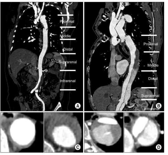
Fig. 1. The conditions of false lu- men were evaluated in the horizontal plane at five levels of the proximal, middle and distal 1/3 descending thoracic aorta and suprarenal and in- frarenal abdominal aorta. (A) False lumen was obliterated in thoracic and suprarenal abdominal aorta. (B) False lumen was partially throm- bosed or patent below the middle thoracic aorta. False lumens are rep- resented as obliterated and throm- bosed in (C), but patent in (D).
Aortic diameters were measured at the level of maximal aortic dilatation.
a patent false lumen in the late period, whereas the patent thoracic aortic false lumen in 11 patients had regressed (42.3%) by the time the late CT images were obtained.
The mean of the maximal aortic diameters at the five seg- ments where the false lumen was thrombotic or obliterated in the early stage were 3.9±0.3 cm, 3.7±0.7 cm, 3.2±0.5 cm, 3.1±0.6 cm, and 2.4±0.6 cm, respectively, and were 3.7±0.6 cm, 3.8±1.0 cm, 3.1±0.4 cm, 2.9±0.5 cm, and 2.5±0.5 cm in the late stage. On the other hand, the mean of the maximal aortic diameters with patent false lumens in the early stage were 3.8±0.3 cm, 3.4±0.5 cm, 3.2±0.2 cm, 3.1±0.4 cm, and 2.6±0.3 cm, respectively, and were 4.5±0.9 cm, 4.3±1.0 cm, 3.8±0.5 cm, 3.7±0.7 cm, and 3.2±0.4 cm in the late stage (Fig. 3). The percentage change in the descending aortic di- ameter from the early to late follow-up period was greater in the patients with a patent false lumen than in those with a thrombosed false lumen at all levels of the aorta (p<0.05) (Table 2).
DISCUSSION
The primary goal of acute type A aortic dissection is en- abling immediate survival, that is, all other considerations in- cluding those regarding the quality of the long-term outcome are of secondary importance [10]. From this perspective, lim- iting the surgery to ascending aortic or hemiarch replacement is compatible with an optimal surgical practice. Furthermore, recent advances in diagnostic methods and surgery have im- proved outcomes and reduced the overall mortality to less than 10% in centers have expertise in aortic dissection [5,11].
In the present study, the mortality rate associated with
TAR was 7.7% (2/26 patients). Despite the limited experience
with this procedure and the relatively small volume of sur-
geries performed at our institution, the mortality outcome was
less related to the operative factors and more related to the
preoperative patient risk factors. This is consistent with the
tendency for surgeons to be generally less aggressive on pa-
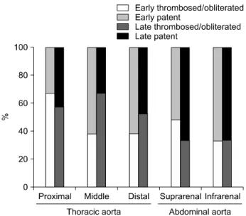
Fig. 2. Statuses of descending aortas after total arch replacement as determine by early and late follow-up computed tomography.
Left columns describe the early state and right columns describe the late state. The lower portion of each column depicts throm- bosed or obliterated false lumen and the upper portion depicts the patent false lumen.
tients with greater instability. According to the International Registry of Acute Aortic Dissection stratification, unstable pa- tients with identifiable risk factors had nearly twice the mor- tality risk of stable patients (30.0% vs. 15.5%, respectively) [3]. At our institution, there have been no proximal reopera- tions in patients that have been operated on for complicated root pathologies such as in Marfan syndrome with severely dilated sinuses or valvular regurgitation requiring root re- placement surgery with a composite valved graft. Therefore, the preoperative risk factors were more important in affecting the outcome than the surgical complexity.
Whether or not complete resection of the entry site in the acute state is always necessary is controversial. Some studies have shown no influence by the completeness of the entry site resection on the false lumen thrombosis [6,12], whereas others have shown the false lumen persisting after proximal surgery in up to 50% to 80 % of the cases depending on the completeness of the primary tear resection [5,13]. Kazui et al.
[4] reported a hospital mortality rate of 4% and a freedom from reoperation rate of 75% at 10 years after TAR. Based on previous studies that have reported an increased correla- tion between failing to resect the intimal tear and an in-
creased incidence of late reoperation, Halstead et al. [10]
speculated that a decreased incidence of distal reoperation might be attributable to the completeness of the intimal tear resection at the time of the acute dissection. Furthermore, having a patent false lumen, which may be due to a persis- tent intimal tear, has been observed to significantly increase the aortic growth rates even in those patients whose aortic di- ameters were in fact actually relatively small immediately af- ter surgery [10]. Although the primary objective of the initial operation in these patients is immediate survival, we aimed to achieve not only the immediate survival but also an optimal long-term outcome. We believe that the surgical principle of completely resecting the intimal tear during the initial oper- ation whenever possible is conducive to achieving this objective.
Chronic enlargement of the residual aortic arch and the de- scending aorta after primary surgery is one of the major causes of poor patient satisfaction during follow-up. Park et al. [7] reported aneurysmal dilatation occurring in nearly half of all the patients after the initial surgery, with distal reopera- tion being required in 15.6% of them at 33.6 months. It was also observed that aortic dilatation occurred more frequently in thoracic aortas with a patent or enlarged false lumen, a large aortic diameter, in patients with Marfan syndrome, younger patients, or males [7].
In the present study, false lumen changes in the thoracic
descending aortas were checked serially by computed tomog-
raphy from the early to the late follow-up period. The rate of
complete thrombosis at the level of the descending thoracic
aorta was 38% to 67% in the early and 52% to 57% in the
late period. In 11 patients (42.3%), the patent thoracic aortic
false lumen on the early CT imaging studies had regressed to
total obliteration in the late follow-up images. On the other
hand, the abdominal aorta did not grow to clinically sig-
nificant levels in most of the patients despite the persistence
of a patent false lumen. The false lumen in the two Marfan
syndrome patients was patent. In three other patients, the
false lumen was thrombosed in the early period but became
partially thrombosed or patent in the late period. Therefore,
as the false lumen patency status may change with time and
thereby affect the aortic growth rate, the attending physician
should be mindful of the pattern of the descending thoracic
Fig. 3. Percentage changes of diameters in the descending aorta. Percentage changes in aortic diameter when the false lu- men remained patent as compared with a thrombosed false lu- men was greater at all levels of the descending aorta during the follow-up period (p<0.05). (A) Proximal thoracic aorta. (B) Suprarenal abdominal aorta. (C) Middle thoracic aorta. (D) Infra- renal abdominal aorta. (E) Distal thoracic aorta.
aortic false lumen status in these patients.
Previous studies have reported thrombotic closure or ob- literation of the descending aortic false lumen occurring more frequently in the early stage in patients undergoing TAR and modified elephant trunk procedure [5,14]. Since 2008, TAR with the elephant trunk technique has been performed at our
institution in 6 patients with a true lumen that was large
enough to accept the elephant trunk graft. Over a mean fol-
low-up of 27.7±10.5 months (range, 16 to 44 months) the
false lumen had regressed in 4 of the patients, and the mean
of the maximum diameter in the thoracic aorta was 3.22±0.29
cm. The false lumen was patent below the middle thoracic
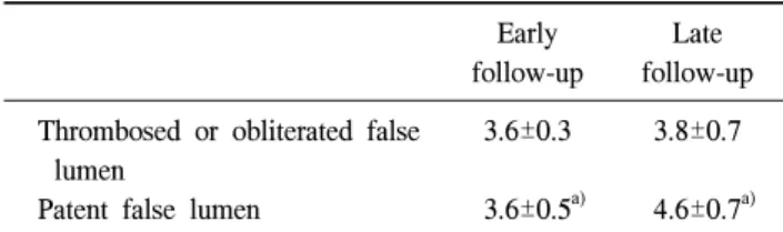
Table 2. The diameter of aorta at the level where the maximal dilatation occurred in the thoracic aorta
Early follow-up
Late follow-up Thrombosed or obliterated false
lumen
Patent false lumen
3.6±0.3
3.6±0.5
a)3.8±0.7
4.6±0.7
a)Follow-up 54±19.0 months (range, 20 to 82 months).
a)