Small cell carcinomas are malignancies derived from neuroendocrine cells. Neuroendocrine cells are distrib- uted throughout the gastrointestinal tract, pancreas, lung, thyroid, adrenal gland, and many other organs (1).
The gastrointestinal tract has the largest population of neuroendocrine cells, but neuroendocrine malignancies are rare in the colon or rectum (2), accounting for less than 1 percent of all colorectal cancer (3). A PubMed search revealed about twenty case reports for small cell carcinoma of the rectum in the English literature.
Like its pulmonary counterpart, extrapulmonary small cell carcinoma is aggressive, with rapid local pro- gression and early regional and distant spread.
Therefore, the prognosis of patients with this neoplasm is generally poor, especially in extrapulmonary small
cell carcinoma involving the gastrointestinal tract (4, 5).
This report presents computed tomographic (CT) im- ages, a laparoscopic biopsy, and an excisional biopsy of a case of small cell carcinoma of the rectum.
Case Report
A 57-year-old male patient was evaluated for com- plaints of bowel habit changes for the past three months, intermittent hematochezia, and tenesmus. The patient had no history of trauma or any previous surgery. A physical examination revealed the presence of a firm mass in the midrectum. The level of carci- noembryonic antigen (CEA) was normal and the other laboratory findings also were within normal ranges.
Chest radiography showed no pathological findings.
An abdominal CT scan performed on a 64-channel mul- ti-slice CT scanner revealed an approximately 7-cm seg- mented bulky mass with irregular margins encircling the upper rectum, with perirectal fat infiltration and massive perirectal/internal iliac lymph node enlarge-
J Korean Soc Radiol 2009;60:37-40
─ 37 ─
CT Finding of Small Cell Carcinoma of the Rectum:
A Case Report
1Suk Ki Jang, M.D.,In Oak Ahn, M.D., So Ya Paik, M.D.2, Su Min Kang, M.D., Jin Young Yoo, M.D., Dae Bong Kim, M.D.
1Department of Radiology, Bundang Jesaeng General Hospital
2Department of Pathology, Bundang Jesaeng General Hospital Received April 29, 2008 ; Accepted October 15, 2008
Address reprint requests to : In Oak Ahn, M.D., Department of Radiology, Bundang Jesaeng General Hospital, 255-2, Seohyun-dong, Bundang-gu, Sungnam-si, Gyungki-do, 463-774, Korea.
Tel. 82-31-779-0051 Fax. 82-31-779-0062 E-mail: [email protected]
Extrapulmonary small cell carcinoma is a rare neoplasm. It is a highly aggressive, malignant tumor with rapid local progression and early metastasis. We report a case of small cell carcinoma arising in the rectum that presented as a bowel habit change in a 57-year-old man. Physical examination revealed a firm, fixed mass in the rectum.
Computed tomography of the mass revealed a circumferential bulky mass with an ir- regular margin with perirectal fat infiltration and enlarged regional lymph nodes.
Histologic examination of the resected mass was consistent with small cell carcinoma of the rectum.
Index words :Rectal neoplasms Carcinoma, small cell
Tomography, X-ray computed
ment (Fig. 1A, B). Colonoscopy revealed a fungating mass with friable mucosa in the rectal wall about 5 cm from the anal verge. Colonoscopic biopsies were consis- tent with small cell carcinoma of the rectum.
A low anterior resection was performed for complete excision of the mass. Gross examination of the resected
specimen showed a huge ulcerofungating mass measur- ing 11.5 cm×6 cm. Macroscopically, a cross-section re- vealed a gray-white solid, firm, neoplastic tissue extend- ing to the perirectal fat tissue. Histologic examination re- vealed the tumor to be composed of sheets of small, un- differentiated cells. A high-power view showed typical
Suk Ki Jang, et al: CT Finding of Small Cell Carcinoma of the Rectum
─ 38 ─
A B
C
D
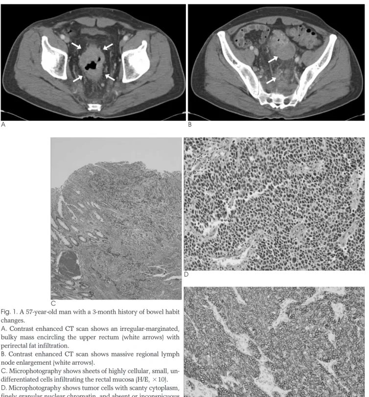
Fig. 1. A 57-year-old man with a 3-month history of bowel habit changes.
A. Contrast enhanced CT scan shows an irregular-marginated, bulky mass encircling the upper rectum (white arrows) with perirectal fat infiltration.
B. Contrast enhanced CT scan shows massive regional lymph node enlargement (white arrows).
C. Microphotography shows sheets of highly cellular, small, un- differentiated cells infiltrating the rectal mucosa (H/E, ×10).
D. Microphotography shows tumor cells with scanty cytoplasm, finely granular nuclear chromatin, and absent or inconspicuous nucleoli (H/E, ×100).
E. Immunostaining shows diffuse and intense membrane stain- ing in the small cell carcinoma cells (CD56, ×100). E
�
�
�
�
�
�
cytologic features of small cell carcinoma: scanty cyto- plasm, finely granular nuclear chromatin, and absent or inconspicuous nucleoli (Fig. 1C, D). Immunohistoche- mical staining revealed these tumor cells to be diffusely positive for the neuroendocrine marker, CD56 (Fig. 1E).
A chest CT scan was performed 1 month and 6 months after surgery, and a follow-up abdominal CT scan was performed 6 months after the operation. One year has elapsed since the surgery and the patient is do- ing relatively well, with no evident tumor recurrence or distant metastasis.
Discussion
According to the recent World Health Organization (WHO) classification scheme (2000), (neuro) endocrine tumors are divided into well-differentiated endocrine tu- mors, well-differentiated endocrine carcinomas, and poorly differentiated endocrine carcinomas. The most poorly differentiated endocrine carcinomas are small cell carcinomas (6).
Colorectal small cell carcinoma is a relatively rare tu- mor, with an overall incidence of less than 1 percent among all colorectal cancers (1, 3). The most common location for this type of cancer is the rectum, followed by the cecum and sigmoid colon (1).
Extrapulmonary small cell carcinoma is a distinct dis- ease entity with unique biological behavior and progno- sis (4, 5). They frequently show neuroendocrine differ- entiation by immunohistochemical staining for neuron- specific enolase, chromogranins, and synaptophysin (7).
Neuroendocrine cancers of the gastrointestinal tract manifest a highly aggressive behavior (1, 2), even more than their adenocarcinoma counterpart of the same stage (1, 3). Liver and lymph-node involvement are found early in 70 to 80 percent of patients, and neither size, location, nor number of primary lesions have prog- nostic value in these tumors (1, 8).
The diagnosis of extrapulmonary small cell carcinoma of the colon should be made only when metastasis from a lung tumor has been excluded (9). Chest CT may be better than chest radiography for excluding primary pul- monary small cell carcinoma and for accurate tumor staging in patients with suspected extrapulmonary small cell carcinoma. Although an initial chest CT scan was not performed here, chest radiographs were normal at presentation and there was not evidence of a pulmonary lesion on follow-up chest CT. This evidence seem suffi- cient for diagnosing extrapulmonary small cell carcino-
ma.
Few reports on small cell carcinoma of the rectum have included radiological findings. Kim et al (9) first re- ported radiological findings of a poorly enhancing, bulky exophytic mass encircling the colon on CT, but only a pathological specimen was obtained from colono- scopic biopsy. Here, small cell carcinoma of the rectum appeared on the CT scan image as a circumferential bulky rectal mass with irregular margins, perirectal fat infiltration, and enlarged regional lymph nodes, which matched with gross specimens.
Although small cell carcinoma of the rectum tends to present with irregular margins, bulky mass, and large lymphadenopathy, these CT findings show considerable overlap with adenocarcinoma or lymphoma. Even en- dorectal ultrasound findings of small cell carcinoma of the rectum resemble adenocarcinoma (10).
In summary, we present the CT findings of a very rare case of extrapulmonary small cell carcinoma of the rec- tum. The lesion appeared on the contrast-enhanced CT scan as an irregular-marginated, bulky mass with mas- sive perirectal/internal iliac lymph node enlargement.
This tumor is a rare type of rectal cancer.
References
1. Cebrian J, Larach SW, Ferrara A, Williamson PR, Trevisani MF, Lujan HJ, et al. Small-cell carcinoma of the rectum. Report of two cases. Dis Colon Rectum 1999;42:274-277
2. Corman ML. Colon & Rectal Surgery. 3rd ed. Philadelphia: J.B.
Lippincott com, 1993:748
3. Yaziji H, Broghamer WL. Primary small cell undifferentiated car- cinoma of the rectum associated with ulcerative colitis. South Med J 1996;89:921-924
4. Remick SC, Hafez GR, Carbone PP. Extrapulmonary small cell carcinoma: a review of the literature with emphasis on therapy and outcome. Medicine 1987;66:457-471
5. Galanis E, Frytak S, Lloyd RV. Extrapulmonary small cell carcino- ma. Cancer 1997;79:1729-1736
6. Chang S, Choi D, Lee SJ, Lee WJ, Park MH, Kim SW, et al.
Neuroendocrine neoplasms of the gastrointestinal tract: classifica- tion, pathologic basis, and imaging features. Radiographics 2007;27:1667-1679
7. Matsui K, Kitagawa M, Miwa A, Kuroda Y, Tsuji M. Small cell car- cinoma of the stomach: a clinicopathologic study of 17 cases. Am J Gastroenterol 1991;86:1167-1175
8. Sterling RK. Ectopic ACTH syndrome associated with anorectal carcinoma. Report of a case and review of the literature. Dig Dis Sci 1993;38:955-959
9. Kim HC, Park SI, Park SJ, Shin HC, Oh MH, Kim HH, et al. Small cell carcinoma of the colon: barium study and CT findings. Br J Radiol 2005;78:255-256
10. Murphy JM, Daly P, Wilson GF. Endorectal ultrasound in a small cell carcinoma of the rectum; staging and assessment of response to chemotherapy. Clin Radiol 1999;54:696-698
J Korean Soc Radiol 2009;60:37-40
─ 39 ─
Suk Ki Jang, et al: CT Finding of Small Cell Carcinoma of the Rectum
─ 40 ─
대한영상의학회지 2009;60:37-40
직장 소세포암의 CT 소견: 증례 보고1
1분당제생병원 영상의학과
2분당제생병원 진단병리과
장석기∙안인옥∙백소야2∙강수민∙유진영∙김대봉
폐 외 소세포암은 드문 종양이며 빠른 국소 진행과 조기 전이를 보이는 매우 공격적인 악성 종양이다. 저자들은 배 변 습관 변화를 주소로 내원한 57세 남자 환자의 직장에 발생한 소세포암 1예를 경험하였기에 컴퓨터단층촬영 및 조 직 병리소견을 함께 보고하고자 한다. 이학적 검사에서 직장에 단단하고 고정된 종괴가 만져졌다. 컴퓨터단층촬영에 서 이 종괴는 불규칙한 경계의 조영증강을 보이는 환형의 거대 종괴였으며 직장주위 지방 침윤과 커진 국소 림프절 이 있었다. 최종 병리 진단은 직장의 소세포암이었다.