ABSTRACT
Despite the advent of the drug-eluting stents (DES) and improved stent design, in-stent restenosis (ISR) remains a challenging problem. The currently available options for treatment of ISR include angioplasty alone, repeat stenting with DES or drug-coated balloons. Several recent studies have compared the available options for treating ISR in an attempt to identify the preferred therapeutic strategy. In this review, we will discuss the currently available therapeutic strategies for the management of patients with ISR and the evidence supporting their use.
Keywords: Restenosis; Angioplasty; Balloon; Coronary; Drug-eluting stent
INTRODUCTION
Although the use of coronary stents brought about a dramatic improvement in patients' clinical and procedural outcomes, the long-term outcome of stent implantation remains significantly constrained by the risk of developing in-stent restenosis (ISR) over time.
1)ISR which is currently defined as a >50% stenosis of a previously stented segment, occurs in as many as 30% of all patients receiving bare metal stents (BMS).
2)Despite the advent of the drug-eluting stents (DES) and improved stent design, the rates of ISR in patients treated with DES are as high as 10%.
3)Specifically, the widespread adoption of DES for small arteries, long lesions, complex coronary lesions, diabetes, and a history of bypass surgery has in fact been the trigger for significant numbers of patients re-presenting with DES restenosis in contemporary clinical practice.
4)5)The treatment of patients with ISR continues to remain a challenge, and currently available options include angioplasty alone, repeat stenting with DES or drug-coated balloons (DCB).
6)Recent meta-analyses have performed comparisons of these available options in an attempt to identify the preferred therapeutic modality. Therefore, this review will discuss the currently available therapeutic strategies for the management of patients with ISR and the evidence supporting their use.
Review Article
Received: Apr 2, 2018 Accepted: Apr 10, 2018 Correspondence to Eun-Seok Shin, MD, PhD
Division of Cardiology, Department of Internal Medicine, Ulsan University Hospital, University of Ulsan College of Medicine, 877, Bangeojinsunhwando-ro, Dong-gu, Ulsan 44033, Korea.
E-mail: [email protected] Copyright © 2018. The Korean Society of Cardiology
This is an Open Access article distributed under the terms of the Creative Commons Attribution Non-Commercial License (https://
creativecommons.org/licenses/by-nc/4.0) which permits unrestricted noncommercial use, distribution, and reproduction in any medium, provided the original work is properly cited.
ORCID iDs Ae-Young Her
https://orcid.org/0000-0002-9990-6843 Eun-Seok Shin
https://orcid.org/0000-0002-9169-6968 Conflict of Interest
The authors have no financial conflicts of interest.
Author Contributions
Conceptualization: Shin ES; Supervision: Shin ES; Writing - original draft: Her AY; Writing - review & editing: Shin ES.
Ae-Young Her , MD, PhD
1, and Eun-Seok Shin , MD, PhD
21
Division of Cardiology, Department of Internal Medicine, Kangwon National University School of Medicine, Chuncheon, Korea
2
Division of Cardiology, Department of Internal Medicine, Ulsan University Hospital, University of Ulsan College of Medicine, Ulsan, Korea
Current Management of In-Stent
Restenosis
DEFINITION AND CLASSIFICATION OF ISR
ISR is defined as the gradual re-narrowing of a stented coronary artery lesion due to arterial damage with subsequent neointimal tissue proliferation.
7)The angiographic definition of ISR remains a binary event defined as a stenosis within the stented segment or its edge (5-mm segments adjacent to the stent) of >50% of the vessel diameter as determined by coronary angiography.
2)3)The clinical definition of ISR requires the presence of >50% diameter in-stent stenosis and one of the following: clinical symptoms of recurrent angina, objective signs of ischemia (electrocardiography changes), positive coronary hemodynamic assessment with fractional flow reserve (FFR) <0.80, intravascular ultrasound (IVUS) minimum cross- sectional area <4 mm
2(6 mm
2for left main), or restenosis with ≥70% reduction in lumen diameter even in the absence of clinical symptoms or signs.
7)The Mehran system is a morphological classification created for BMS-ISR lesions (pattern I:
focal, pattern II: diffuse, pattern III: proliferative, and pattern IV: occlusive) which can help to predict the need for repeat revascularization (19%, 35%, 50%, and 98%, respectively).
2)This classification scheme has also been shown to have prognostic value in DES-ISR.
8)Additionally, the American College of Cardiology/American Heart Association classification has been validated in patients with ISR: lesions B2 and C are more frequently associated with suboptimal acute results; a higher restenosis rate; and poorer long-term clinical outcomes.
9)CHARACTERIZAION AND PRESENTATION OF ISR
Mounting evidence strongly suggests that there are significant differences between ISR in BMS and DES,
10)11)with the main disparities, time of presentation, morphological patterns, underlying substrate, and the response to interventions. The time course of neointimal accumulation differs considerably between DES and BMS, which is a manifestation of the delayed arterial healing that appears to characterize the vascular response to DES implantation.
12-14)Moreover, compared with BMS-ISR, DES-ISR tends to be focal, particularly at the stent edge or in areas of stent fracture. Lack of diffuse neointimal hyperplasia in DES may be due to the high overall suppression of neointimal growth by DES unless there is mechanical stent failure.
10)15)In addition, focal neoatherosclerosis occurs not only more frequently, but also significantly earlier in DES-ISR compared with BMS-ISR.
16)Assessing the underlying etiology for ISR is critical for guiding and optimizing repeat
interventions to prevent repeated ISR. The most well recognized and preventable cause
for ISR is stent under-expansion, and this is considered a major factor triggering ISR after
either BMS or DES implantation.
17)This problem may be due to stent under-sizing, low
deployment pressures, or extensive vessel calcification leading to stent under-deployment
or under-expansion.
18)Stent misplacement or stents not fully covering the underlying lesion
are other important risk factors for ISR. Geographic miss leaves a characteristic “candy-
wrapper” angiographic appearance at the edge of a stented segment and is thought to lead
to ISR because of stent-related edge dissection, poor endothelialization and subsequent
proliferation of the atherosclerotic plaque burden.
19)Stent fractures may also trigger focal ISR
as they cause similar problems to edge disease except within the stent. Finally, the adoption
of DES has led to the recognition of drug resistance and local hypersensitivity reactions as
another possible cause of ISR.
7)Intracoronary imaging, which can be performed with either IVUS or optical coherence tomography (OCT), plays an important role in evaluating the potential mechanism of
ISR.
17)20)IVUS can detect the presence of neointimal hyperplasia within the stent, stent under-
expansion, stent fracture or edge restenosis, and the borders of the external elastic lamina for vessel sizing enabling optimization of stent expansion.
17)However, due to its superior axial resolution (15 µm), OCT provides better detailed images of the vessel-lumen interface, the neointimal tissue, and the strut distribution.
11)18)It has enabled more detailed evaluation of the ISR etiology and has highlighted the morphologic differences between BMS- and DES-ISR. OCT in BMS-ISR typically shows a homogeneous high-signal tissue band, which is characteristic of neointimal hyperplasia rich in smooth muscle cells.
11)In contrast, DES- ISR is typically characterized by a focal, heterogeneous and layered intrastent tissue band, which represents hypocellular neointima with high proteoglycan or fibrin content which likely occurs in the setting of neoatherosclerosis. Specific findings that are also suggestive of neoatherosclerosis include neointimal rupture, thin-cap fibroatheroma, lipid pools, macrophage accumulation, and evidence of non-occlusive thrombosis.
20)Although ISR had traditionally been thought to represent a relatively benign clinical entity with predominantly stable clinical presentation, more recent studies suggest that a significant number of patients with ISR present with acute coronary syndrome.
21)22)This acute clinical presentation is likely to be related to the neoatherosclerotic process described for DES, which is more likely to follow the typical atherosclerotic cascade of coronary occlusion secondary to neoatherosclerotic plaque rupture and thrombus formation.
23)It is possible that late stent thrombosis is just a step in the continuum of the neoatherosclerotic process seen in DES-ISR. Conversely, the natural history of asymptomatic patients with angiographic restenosis is favorable.
24)Therefore, treatment of asymptomatic patients (oculostenotic reflex) should be avoided whenever possible.
25-27)Similar to de novo lesions, the functional significance of ISR should be assessed using a pressure wire. Prospective studies have validated the use of FFR for clinical decision making in ISR, and have found that deferring revascularization in patients with an FFR of >0.75 is safe and appropriate.
28)29)TREATMENT OF ISR
Medical/surgical treatment
There is little evidence to support medical treatments for ISR. Although abciximab was considered to be of particular value in patients with ISR in early studies, larger trials failed to confirm any clinical benefit.
30)31)Similarly, oral sirolimus was initially considered to be of potential value in these patients, however, the lack of long-term efficacy and the higher incidence of adverse drug effects have shown it to be a poor option.
32)33)Finally, coronary surgery may be considered in patients with recalcitrant ISR, particularly in those with a diffuse ISR pattern or associated significant disease in other major vessels.
15)Plain old balloon angioplasty (POBA)
POBA is one of the earliest treatments that has been used in patients with ISR.
34)The
procedure is technically straightforward and is consistently associated with satisfactory
acute results and a very low incidence of complications.
35)The immediate angiographic
improvement following POBA results from both axial and longitudinal tissue extrusion
as well as further stent expansion.
36)37)Results are particularly favorable in patients with a
focal pattern of ISR and when stent/native artery size mismatch has been identified with
intravascular imaging.
2)High-pressure balloon dilatation and the use of a non-compliant balloon is often necessary to obtain optimal results.
23)In general, a balloon to artery ratio of 1.1 to 1 is recommended for sizing when treating ISR.
35)One of the limitation of POBA is that sub-acute tissue re-intrusion back to the lumen tends to occur within minutes of the last balloon inflation.
38)This explains the “early lumen loss” phenomenon detected in POBA studies in ISR, a finding also associated with subsequent recurrent restenosis. Additionally, edge-related complications should be carefully avoided during aggressive balloon dilations.
Balloon slippage outside the stent (“water-melon seeding” phenomenon), which occurs more often in severe and diffuse narrowing when balloons are oversized, can lead to edge dissections and suboptimal outcomes.
39)Progressive balloon upsizing as well as the use of short low profile balloons can help avoid this phenomenon and edge-related complications.
40)Cutting and scoring balloon therapy
The cutting balloon is an attractive and simple technique for treatment of ISR. Theoretically, the device deeply incises neointimal tissue and may favor its subsequent extrusion. The lateral blades of the device anchor the balloon within the target lesion, preventing balloon slippage-related complications. Initial observational data suggested that cutting balloons may have superior efficacy compared to POBA, a finding which was associated with a lower rate of target lesion revascularization (TLR) (12.5% vs. 40%) at follow-up.
41)42)However, in the largest randomized trial (Restenosis Cutting Balloon Evaluation Trial [RESCUT]), cutting balloon angioplasty comparing POBA failed to show an improvement in angiographic restenosis or in the rate of clinical events at late follow-up.
43)Cutting balloon angioplasty was associated however with the need to use fewer balloons, less additional stenting, and a lower rate of balloon slippage (6.5% vs. 25%).
Scoring balloons are based on the same principle as cutting balloons but are especially attractive in patients with ISR due to their superior flexibility and deliverability.
35)The
Intracoronary Stenting and Angiographic Results: Optimizing Treatment of Drug-Eluting Stent In-Stent Restenosis 4 (ISAR-DESIRE IV trial) assessed the use of scoring balloons prior to DCB treatment of DES-ISR.
44)The results showed superior angiographic outcomes at 6 to 8 months in the scoring balloon arm, but failed to show any significant difference in clinical outcomes.
Debulking techniques
Debulking techniques such as directional/rotational atherectomy and excimer laser are a novel treatment for ISR through their physical removal of neointimal tissue or neoatherosclerotic plaque. It was believed that after the initial removal of excess stenotic tissue by the debulking device, just a low-pressure balloon post-dilation is required to avoid additional vessel wall injury. Early observational studies suggested that the use of laser or rotational atherectomy, followed by a POBA post-dilation, was superior to conventional POBA alone in ISR.
45)Directional atherectomy was also assessed in early studies, but this was soon abandoned because it was not well suited for small or distal vessels, which are common locations for ISR.
46)The excimer laser showed good results in some cases but eventually proved to have poorer ablation capability compared with rotational atherectomy.
45)The value of rotational atherectomy in patients with BMS-ISR was evaluated in 2 randomized
trials. In Randomized Trial of Rotational Atherectomy Versus Balloon Angioplasty for Diffuse
In-Stent Restenosis (ROSTER), rotational atherectomy reduced the amount of residual tissue
within the stent and the rate of TLR at follow-up, compared with POBA alone.
47)On the other
hand, in the Angioplasty Versus Rotational Atherectomy for Treatment of Diffuse In-Stent
Restenosis Trial (ARTIST), which compared rotational atherectomy with POBA alone, lower restenosis rates, an improved safety profile and superior clinical outcomes were seen in the POBA group.
48)Recently, the value of debulking techniques in patients with DES-ISR has been re-evaluated with the latest study showing greater acute luminal gain after percutaneous coronary intervention with excimer laser atherectomy.
49)Reassuringly, even though excimer laser atherectomy was used for DES-ISR in significantly more complex lesions, the long- term clinical outcomes were favorable. Therefore, although debulking techniques are not considered to be a routine treatment of ISR, they can be considered as a pre-treatment option for undilatable ISR lesions, especially those as a result of severely under-expanded stents or calcified intrastent neoatherosclerosis.
50)51)Vascular brachytherapy
Brachytherapy was one of the most promising treatment options for patients with neointimal hyperplasia related to BMS-ISR. It involved temporary intracoronary deposition of a
radioactive isotope within the diseased segment, which led to significantly reduced clinical and angiographic restenosis rates. Randomized clinical trials in patients with ISR showed it to be more effective in preventing ISR progression and improving clinical outcomes than either POBA or debulking procedures with laser or atherectomy.
52)53)However, the advent of DES signaled the end of brachytherapy. The 2 large randomized clinical trials which compared the efficacy of brachytherapy versus DES in patients with BMS-ISR were Sirolimus- Eluting Stents versus Vascular Brachytherapy for In-Stent Restenosis Within Bare-Metal Stents (SISR) and Paclitaxel-Eluting Stents versus Vascular Brachytherapy for In-Stent Restenosis Within Bare-Metal Stents (TAXUS V ISR). Both showed that DES were superior in decreasing restenosis rates and the need for revascularization as compared to brachytherapy at long-term follow-up.
54)55)Disappointingly whilst observational studies of DES-ISR suggested a role for brachytherapy,
56)no randomized trials comparing it to DES or DCB therapy have ever been conducted. Finally, the complexity of the procedure, as well as issues with radioprotection/radiation dosing, led to the virtual abandonment of this strategy.
Repeat stenting with BMS
Early studies suggested that the problem of early tissue loss, which was seen with POBA, was virtually eliminated with the use of BMS, which gave credence to the possible superiority of stenting over POBA in the treatment of ISR.
38)In BMS-ISR, IVUS studies also demonstrated that repeat stenting was the best strategy to obtain a larger acute lumen gain and better immediately results post procedure.
57)In the Restenosis Intra-stent Balloon Angioplasty Versus Elective Stenting (RIBS I) trial, patients with BMS-ISR, were randomized to receive either POBA or repeat BMS implantation, with acute angiographic results being significantly better after BMS placement due to a larger acute gain.
58)However, at 6-month follow-up, significant late lumen loss in the BMS group resulted in the final angiographic appearance being similar in both groups. To date, large randomized trials assessing the value of BMS in patients with DES-ISR are lacking.
Repeat stenting with DES
In de novo lesions, DES produce a profound inhibition of neointimal proliferation.
59)60)Therefore, the use of DES has become an attractive option in the treatment of neointimal
hyperplasia in BMS-ISR. The Intracoronary Stenting or Angioplasty for Restenosis
Reduction-Drug-Eluting Stents for In-Stent Restenosis (ISAR-DESIRE) trial was the first
randomized study assessing the value of DES in patients with BMS-ISR.
61)The rate of
recurrent restenosis was significantly lower with sirolimus- (14.3%) and paclitaxel-DES
(21.7%) compared with POBA alone (44.6%). Similar results were also shown in a subsequent meta-analysis comparing these 2 DES for BMS-ISR.
62)In the Restenosis Intrastent: Balloon Angioplasty Versus Elective Sirolimus-Eluting Stenting (RIBS II) trial, which compared sirolimus-DES versus POBA in patients with BMS-ISR, patients with sirolimus-DES had a significantly lower restenosis rate (11%) and superior long-term clinical outcomes.
63)In addition, IVUS imaging confirmed the dramatic reduction of neointimal proliferation seen after the use of sirolimus-DES. The 4-year long-term follow-up study demonstrated a sustained clinical benefit from DES placement without any significant increase in major adverse cardiac events (MACE).
64)Unfortunately, the treatment of DES-ISR is more challenging, and overall, the outcomes in patients requiring treatment for DES-ISR are worse compared with patients with BMS-
ISR.
7)65)Early observational studies suggested that DES provided superior results compared
with other strategies such as POBA or cutting balloon angioplasty.
66)Investigators have proposed that DES-ISR that results from a mechanical complication (such as stent under- sizing, edge dissection or stent fracture) can be successfully overcome by placing another DES. However, debate regarding whether to use a DES eluting the same or a similar type of drug (homo-DES approach) versus a switch to a different type of drug (hetero-DES approach) has continued.
67)The benefits of a switch approach are based on the hypothesis that it might overcome drug resistance or polymer-related problems. In Intracoronary Stenting and Angiographic Results: Drug Eluting Stents for In-Stent Restenosis 2 (ISAR-DESIRE 2) trial of sirolimus-DES-ISR, the hetero-DES strategy using a paclitaxel-DES failed to reduce restenosis or target vessel revascularization rates compared to repeat stenting with sirolimus- DES.
65)The Restenosis Intra-Stent: Balloon Angioplasty vs Drug-Eluting Stent (RIBS III) trial also compared the DES-switch approach to same-stent implantation. Although there was no significant difference between the hetero-DES and homo-DES approach, the study suggested that the use of second-generation DES was superior to first-generation DES, and intravascular imaging for treatment guidance had improved angiographic and clinical outcomes.
68)Despite these benefits of repeat stenting with DES in the management of DES-ISR, current data suggests that 10–20% of these patients will go on to develop recurrent ISR.
61)64)Bioresorbable vascular scaffolds (BVS)
BVS have also been proposed as treatment for patients with ISR. The main advantages are that the device eventually disappears from the vessel wall, avoiding the presence of multiple stent layers, and prevents early lumen loss associated with tissue retraction seen in balloon angioplasty.
69)Some small studies have established that BVS placement in the treatment of ISR is safe and feasible.
70)71)In 65 patients with ISR treated with BVS, clinical outcomes at 1 year revealed a TLR rate of about 12%, and all of these patients avoided having a permanent second layer of stent struts.
71)Nevertheless, since no randomized trial evaluating the effectiveness of BVS in management of ISR has yet been performed, the routine use of this strategy cannot be recommended.
DCB
The development of DCB enabled deliver of anti-proliferative drug to the area of ISR without
leaving behind an additional layer of stent strut. Although the value of DCB in de novo lesions
remains controversial, the use of DCB has been to proven to be very effective in patients
with both BMS-ISR and DES-ISR (Table 1). The initial study of BMS-ISR demonstrated that
DCB were superior to POBA alone.
72)The 6-month angiographic results were significantly
improved in the DCB group (late loss: 0.33 mm vs. 0.74 mm, p<0.002). The subsequent
larger randomized study compared paclitaxel-DES placement to paclitaxel-coated balloon application in BMS-ISR. At 6-month follow-up, DCB significantly reduced the primary endpoint of the study (angiographic late loss: 0.17 mm vs. 0.38 mm, p=0.03), although minimal lumen diameter and diameter stenosis were similar in both arms.
73)Recently, the Restenosis Intra-stent: Drug-eluting Balloon vs. Everolimus-eluting Stent (RIBS V) trial conducted a randomized comparison of DCB with second-generation everolimus-DES in patients with BMS-ISR.
74)This study showed better late angiographic findings in the DES arm (minimal lumen diameter: 2.01 mm vs. 2.36 mm, p<0.001), but showed similar rates of restenosis and clinical outcomes. Therefore, the overall non-inferior outcomes with DCB treatment as compared with DES placement in several studies seem to support the use of DCB for treatment of BMS-ISR, especially in situations where additional stent layers are undesirable or bleeding events.
The value of DCB in patients with DES-ISR has also been assessed. An initial small randomized study demonstrated that in patients with DES-ISR, DCB provided superior clinical and angiographic results compared with POBA alone (late lumen loss: 0.18 mm vs. 0.72 mm, p=0.001).
22)The efficacy of DCB in patients with DES-ISR was subsequently confirmed in a multicenter, randomized trial including patients with any type of DES-ISR (late loss: 0.43 mm vs. 1.03 mm in POBA arm, p<0.001).
75)Moreover, another controlled study suggested that DCB are equivalent to paclitaxel-DES in patients with DES-ISR (late lumen loss: 0.46 mm vs. 0.55 mm, non-inferiority p<0.001) at 9 months follow-up.
76)Recently, the larger Intracoronary Stenting and Angiographic Results: Drug Eluting Stents for In-Stent Restenosis (ISAR-DESIRE 3) randomized trial investigated the efficacy of DCB versus paclitaxel-DES versus conventional POBA alone in patients with limus DES-ISR.
40)The results demonstrated that DCB were non-inferior to paclitaxel-DES (diameter stenosis: 38.0% vs.
37.4%, non-inferiority p=0.007) and that both DCB and paclitaxel-DES were superior to POBA alone. In summary, the data from the meta-analyses of available randomized clinical trials suggest that DCB are superior to POBA alone and similar to first-generation DES in patients with BMS- or DES-ISR.
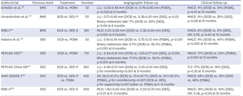
77)Table 1. Randomized clinical trials of DCB on treatment of ISR
Author/trial Previous stent Treatment Number Angiographic follow-up Clinical follow-up
Scheller et al.
72)BMS DCB vs. POBA 52 LLL: 0.03±0.48 mm (DCB) vs. 0.74±0.86 mm (POBA),
p=0.002 at 6 months MACE: 4% (DCB) vs. 31% (POBA),
p=0.01 at 12 months
Unverdorben et al.
73)BMS DCB vs. DES-P 131 LLL: 0.17±0.42 mm (DCB) vs. 0.38±0.61 mm (DES), p=0.03 MACE: 9% (DCB) vs. 22% (DES), p=0.08 at 12 months
Binary restenosis rate: 7% (DCB) vs. 20% (DES), p=0.06 at 6 months
RIBS V
74)BMS DCB vs. DES-E 189 MLD: 2.01±0.60 mm (DCB) vs. 2.36±0.60 mm (DES),
p<0.001 at 9 months MACE: 8% (DCB) vs. 6% (DES),
HR: 0.76, p=0.60 at 12 months Habara et al.
22)DES DCB vs. POBA 50 LLL: 0.18±0.45 mm (DCB) vs. 0.72±0.55 mm (POBA), p=0.001 MACE: 4% (DCB) vs. 40% (POBA),
p=0.005 at 12 months Binary restenosis rate: 8.7% (DCB) vs. 62.5% (POBA),
p<0.001 at 6 months
PEPCAD-DES
75)DES DCB vs. POBA 110 LLL: 0.43±0.61 mm (DCB) vs. 1.03±0.77 mm (DES), p<0.001, MACE: 17% (DCB) vs. 50% (POBA), p<0.001 at 12 months
Binary restenosis rate: 17.2% (DCB) vs. 58.1% (POBA), p<0.001 at 6 months
PEPCAD China ISR
76)DES DCB vs. DES-P 220 LLL: 0.46±0.51 mm (DCB) vs. 0.55±0.61 mm (DES),
p for noninferiority<0.001 at 9 months TLF: 17% (DCB) vs. 16% (DES), p=0.52 at 12 months
ISAR-DESIRE 3
40)DES DCB vs. DES-P
vs. POBA 402 DS: 38.0±21.5% (DCB) vs. 37.4±21.7% (DES) vs. 54.1±25.0%
(POBA), p for noninferiority=0.007 (DCB vs. DES), p for superiority<0.001 (other vs. POBA) at 6–8 months
MACE: 24% (DCB) vs. 19% (DES) vs. 46% (POBA)
RIBS IV
78)DES DCB vs. DES-E 309 MLD: 1.80±0.60 mm (DCB) vs. 2.03±0.70 mm (DES),
p<0.01 at 6–9 months MACE: 18% (DCB) vs. 10% (DES),
HR: 0.58, p=0.04 at 12 months
BMS = bare-metal stents; DCB = drug-coated balloons; DES = drug-eluting stents; DES-E = everolimus drug-eluting stents; DES-P = paclitaxel drug-eluting
stents; DS = diameter stenosis; HR = hazard ratio; ISR = in-stent restenosis; MACE = major adverse cardiac events; LLL = late lumen loss; MLD = minimal lumen
diameter; POBA = plain old balloon angioplasty; TLF = target lesion failure.
Finally, whether DCB proves comparable to repeat stenting with second-generation DES in patients with DES-ISR remains controversial. In the recently published Restenosis Intra-stent of Drug-eluting Stents: Paclitaxel-Eluting Balloon vs Everolimus-Eluting Stent (RIBS-IV) trial which compared second-generation everolimus-eluting DES to DCB for treatment of DES-ISR, both angiographic and clinical outcomes favored second-generation DES over DCB (minimal lumen diameter: 2.03 mm vs. 1.80 mm, p<0.01; MACE: 10% vs. 18%, p=0.04) at 6- to 9-month follow-up.
78)In addition, whether the efficacy of DCB can be further improved by optimal lesion preparation with scoring/cutting balloon remains unknown and the ongoing ISAR-DESIRE 4 randomized trial will address this issue.
Comparison of all treatment modalities
Two recent large meta-analyses were conducted to clarify which strategy is the best treatment modality for ISR. Siontis et al.
79)included 27 trials with a total of 5,923 patients at 6 months to 1 year follow-up. The primary outcome of this analysis was percent diameter stenosis at follow- up, and the secondary endpoint included binary restenosis, rates of TLR, myocardial infarction or death. All modalities included POBA alone, debulking techniques, brachytherapy, BMS, DES, and DCB. Repeat stenting with everolimus-DES was found to be statistically superior to all other modalities for both the primary outcome as well as for binary restenosis rates and TLR.
DCB appeared to be the second most preferable treatment but did not achieve a significant difference over sirolimus or paclitaxel-eluting stents. Giacoppo et al.
80)included 24 trials with a total of 4,880 patients, and the primary outcomes were TLR rates and angiographic late lumen loss. Both DCB and DES were superior to other treatment modalities based on the predefined clinical outcomes. Angiographic outcomes favored DCB or DES over all other modalities, however late lumen loss appeared to be slightly lower in the DCB arm compared with DES.
CONCLUSION
Although the development of DES has reduced the incidence of ISR, treatment of ISR remains a challenging clinical problem. Current clinical data suggest that among various available therapeutic modalities, second-generation DES and DCB provide the best clinical and angiographic results in patients with ISR. Implantation of more than 2 metal stents in repeated ISR lesions is likely to have a detrimental effect on long-term outcomes, even though newer DES may improve the treatment of ISR lesions. Further studies are required to clarify the role of these current therapeutic modalities which may help improve clinical outcomes in those with ISR.
REFERENCES
1. Stone GW, Ellis SG, Cannon L, et al. Comparison of a polymer-based paclitaxel-eluting stent with a bare metal stent in patients with complex coronary artery disease: a randomized controlled trial. JAMA 2005;294:1215-23.
PUBMED | CROSSREF
2. Mehran R, Dangas G, Abizaid AS, et al. Angiographic patterns of in-stent restenosis: classification and implications for long-term outcome. Circulation 1999;100:1872-8.
PUBMED | CROSSREF
3. Kuntz RE, Baim DS. Defining coronary restenosis. Newer clinical and angiographic paradigms. Circulation 1993;88:1310-23.
PUBMED | CROSSREF
4. Win HK, Caldera AE, Maresh K, et al. Clinical outcomes and stent thrombosis following off-label use of drug-eluting stents. JAMA 2007;297:2001-9.
PUBMED | CROSSREF
5. Aoyama T, Ishii H, Toriyama T, et al. Sirolimus-eluting stents vs bare metal stents for coronary intervention in Japanese patients with renal failure on hemodialysis. Circ J 2008;72:56-60.
6. Goel SS, Dilip Gajulapalli R, Athappan G, et al. Management of drug eluting stent in-stent restenosis: a systematic review and meta-analysis. Catheter Cardiovasc Interv 2016;87:1080-91.
7. Dangas GD, Claessen BE, Caixeta A, Sanidas EA, Mintz GS, Mehran R. In-stent restenosis in the drug- eluting stent era. J Am Coll Cardiol 2010;56:1897-907.
PUBMED | CROSSREF
8. Solinas E, Dangas G, Kirtane AJ, et al. Angiographic patterns of drug-eluting stent restenosis and one-year outcomes after treatment with repeated percutaneous coronary intervention. Am J Cardiol 2008;102:311-5.
PUBMED | CROSSREF
9. Alfonso F, Cequier A, Angel J, et al. Value of the American College of Cardiology/American Heart Association angiographic classification of coronary lesion morphology in patients with in-stent restenosis. Insights from the Restenosis Intra-stent Balloon angioplasty versus elective Stenting (RIBS) randomized trial. Am Heart J 2006;151:681.e1-681.e9.
10. Alfonso F. Treatment of drug-eluting stent restenosis the new pilgrimage: quo vadis? J Am Coll Cardiol 2010;55:2717-20.
PUBMED | CROSSREF
11. Byrne RA, Joner M, Tada T, Kastrati A. Restenosis in bare metal and drug-eluting stents: distinct mechanistic insights from histopathology and optical intravascular imaging. Minerva Cardioangiol 2012;60:473-89.
PUBMED
12. Kimura T, Yokoi H, Nakagawa Y, et al. Three-year follow-up after implantation of metallic coronary-artery stents. N Engl J Med 1996;334:561-6.
PUBMED | CROSSREF
13. Byrne RA, Iijima R, Mehilli J, et al. Durability of antirestenotic efficacy in drug-eluting stents with and without permanent polymer. JACC Cardiovasc Interv 2009;2:291-9.
PUBMED | CROSSREF
14. Collet CA, Costa JR, Abizaid A, et al. Assessing the temporal course of neointimal hyperplasia formation after different generations of drug-eluting stents. JACC Cardiovasc Interv 2011;4:1067-74.
PUBMED | CROSSREF
15. Alfonso F, Pérez-Vizcayno MJ, Cruz A, et al. Treatment of patients with in-stent restenosis. EuroIntervention 2009;5 Suppl D:D70-8.
16. Nakazawa G, Otsuka F, Nakano M, et al. The pathology of neoatherosclerosis in human coronary implants bare-metal and drug-eluting stents. J Am Coll Cardiol 2011;57:1314-22.
PUBMED | CROSSREF
17. Fujii K, Mintz GS, Kobayashi Y, et al. Contribution of stent underexpansion to recurrence after sirolimus- eluting stent implantation for in-stent restenosis. Circulation 2004;109:1085-8.
PUBMED | CROSSREF
18. Alfonso F, Sandoval J, Cardenas A, Medina M, Cuevas C, Gonzalo N. Optical coherence tomography: from research to clinical application. Minerva Med 2012;103:441-64.
PUBMED
19. Angiolillo DJ, Sabatá M, Alfonso F, Macaya C. "Candy wrapper" effect after drug-eluting stent implantation: déjà vu or stumbling over the same stone again? Catheter Cardiovasc Interv 2004;61:387-91.
20. Kang SJ, Mintz GS, Akasaka T, et al. Optical coherence tomographic analysis of in-stent neoatherosclerosis after drug-eluting stent implantation. Circulation 2011;123:2954-63.
PUBMED | CROSSREF
21. Cassese S, Byrne RA, Tada T, et al. Incidence and predictors of restenosis after coronary stenting in 10 004 patients with surveillance angiography. Heart 2014;100:153-9.
PUBMED | CROSSREF
22. Habara S, Mitsudo K, Kadota K, et al. Effectiveness of paclitaxel-eluting balloon catheter in patients with sirolimus-eluting stent restenosis. JACC Cardiovasc Interv 2011;4:149-54.
PUBMED | CROSSREF
23. Nicolais C, Lakhter V, Virk HU, et al. Therapeutic options for in-stent restenosis. Curr Cardiol Rep 2018;20:7.
PUBMED | CROSSREF
24. Hernández RA, Macaya C, Iñiguez A, et al. Midterm outcome of patients with asymptomatic restenosis after coronary balloon angioplasty. J Am Coll Cardiol 1992;19:1402-9.
PUBMED | CROSSREF
25. Pinto DS, Stone GW, Ellis SG, et al. Impact of routine angiographic follow-up on the clinical benefits of paclitaxel-eluting stents: results from the TAXUS-IV trial. J Am Coll Cardiol 2006;48:32-6.
PUBMED | CROSSREF
26. Kim YH, Her AY, Rha SW, et al. Routine angiographic follow-up versus clinical follow-up after percutaneous coronary intervention in acute myocardial infarction. Yonsei Med J 2017;58:720-30.
PUBMED | CROSSREF
27. Kim YH, Her AY, Rha SW, et al. Routine angiographic follow-up versus clinical follow-up in patients with multivessel coronary artery diseases following percutaneous coronary intervention with drug-eluting stents: a nested case-control study within a Korean population. Coron Artery Dis 2017;28:307-14.
PUBMED | CROSSREF
28. Lopez-Palop R, Pinar E, Lozano I, Saura D, Picó F, Valdés M. Utility of the fractional flow reserve in the evaluation of angiographically moderate in-stent restenosis. Eur Heart J 2004;25:2040-7.
PUBMED | CROSSREF
29. Nam CW, Rha SW, Koo BK, et al. Usefulness of coronary pressure measurement for functional evaluation of drug-eluting stent restenosis. Am J Cardiol 2011;107:1783-6.
PUBMED | CROSSREF
30. Mukherjee D, Reginelli JP, Moliterno DJ, et al. Unexpected mortality reduction with abciximab for in-stent restenosis. J Invasive Cardiol 2000;12:540-4.
PUBMED
31. Moustapha A, Assali AR, Sdringola S, et al. Abciximab administration and clinical outcomes after percutaneous intervention for in-stent restenosis. Catheter Cardiovasc Interv 2002;56:184-7.
32. Hausleiter J, Kastrati A, Mehilli J, et al. Randomized, double-blind, placebo-controlled trial of oral sirolimus for restenosis prevention in patients with in-stent restenosis: the Oral Sirolimus to Inhibit Recurrent In-stent Stenosis (OSIRIS) trial. Circulation 2004;110:790-5.
PUBMED | CROSSREF
33. Kufner S, Hausleiter J, Ndrepepa G, et al. Long-term risk of adverse outcomes and new malignancies in patients treated with oral sirolimus for prevention of restenosis. JACC Cardiovasc Interv 2009;2:1142-8.
PUBMED | CROSSREF
34. Macander PJ, Roubin GS, Agrawal SK, Cannon AD, Dean LS, Baxley WA. Balloon angioplasty for treatment of in-stent restenosis: feasibility, safety, and efficacy. Cathet Cardiovasc Diagn 1994;32:125-31.
PUBMED | CROSSREF
35. Alfonso F, Byrne RA, Rivero F, Kastrati A. Current treatment of in-stent restenosis. J Am Coll Cardiol 2014;63:2659-73.
PUBMED | CROSSREF
36. Alfonso F, Pérez-Vizcayno MJ, Hernández R, et al. Long-term outcome and determinants of event-free survival in patients treated with balloon angioplasty for in-stent restenosis. Am J Cardiol 1999;83:1268-70, A9.
37. Mehran R, Mintz GS, Popma JJ, et al. Mechanisms and results of balloon angioplasty for the treatment of in-stent restenosis. Am J Cardiol 1996;78:618-22.
PUBMED | CROSSREF
38. Alfonso F, Garcia P, Fleites H, et al. Repeat stenting for the prevention of the early lumen loss
phenomenon in patients with in-stent restenosis. Angiographic and intravascular ultrasound findings of a randomized study. Am Heart J 2005;149:e1-8.
PUBMED | CROSSREF
39. Alfonso F, Pérez-Vizcayno MJ, Gómez-Recio M, et al. Implications of the “watermelon seeding”
phenomenon during coronary interventions for in-stent restenosis. Catheter Cardiovasc Interv 2005;66:521-7.
PUBMED | CROSSREF
40. Byrne RA, Neumann FJ, Mehilli J, et al. Paclitaxel-eluting balloons, paclitaxel-eluting stents, and balloon angioplasty in patients with restenosis after implantation of a drug-eluting stent (ISAR-DESIRE 3): a randomised, open-label trial. Lancet 2013;381:461-7.
PUBMED | CROSSREF
41. Montorsi P, Galli S, Fabbiocchi F, Trabattoni D, Ravagnani PM, Bartorelli AL. Randomized trial of conventional balloon angioplasty versus cutting balloon for in-stent restenosis. Acute and 24-hour angiographic and intravascular ultrasound changes and long-term follow-up. Ital Heart J 2004;5:271-9.
PUBMED
42. Adamian M, Colombo A, Briguori C, et al. Cutting balloon angioplasty for the treatment of in-stent restenosis: a matched comparison with rotational atherectomy, additional stent implantation and balloon angioplasty. J Am Coll Cardiol 2001;38:672-9.
PUBMED | CROSSREF
43. Albiero R, Silber S, Di Mario C, et al. Cutting balloon versus conventional balloon angioplasty for the treatment of in-stent restenosis: results of the restenosis cutting balloon evaluation trial (RESCUT). J Am Coll Cardiol 2004;43:943-9.
PUBMED | CROSSREF
44. Kufner S, Joner M, Schneider S, et al. Neointimal modification with scoring balloon and efficacy of drug-coated balloon therapy in patients with restenosis in drug-eluting coronary stents: a randomized controlled trial. JACC Cardiovasc Interv 2017;10:1332-40.
PUBMED | CROSSREF
45. Mehran R, Dangas G, Mintz GS, et al. Treatment of in-stent restenosis with excimer laser coronary angioplasty versus rotational atherectomy: comparative mechanisms and results. Circulation 2000;101:2484-9.
PUBMED | CROSSREF
46. Sanchez PL, Rodriguez-Alemparte M, Colon-Hernandez PJ, et al. Directional coronary atherectomy vs. rotational atherectomy for the treatment of in-stent restenosis of native coronary arteries. Catheter Cardiovasc Interv 2003;58:155-61.
47. Sharma SK, Kini A, Mehran R, Lansky A, Kobayashi Y, Marmur JD. Randomized trial of rotational atherectomy versus balloon angioplasty for diffuse in-stent restenosis (ROSTER). Am Heart J 2004;147:16-22.
PUBMED | CROSSREF
48. vom Dahl J, Dietz U, Haager PK, et al. Rotational atherectomy does not reduce recurrent in-stent restenosis: results of the angioplasty versus rotational atherectomy for treatment of diffuse in-stent restenosis trial (ARTIST). Circulation 2002;105:583-8.
PUBMED | CROSSREF
49. Ichimoto E, Kadohira T, Nakayama T, De Gregorio J. Long-term clinical outcomes after treatment with excimer laser coronary atherectomy for in-stent restenosis of drug-eluting stent. Int Heart J 2018;59:14-20.
PUBMED | CROSSREF
50. Kobayashi Y, Teirstein P, Linnemeier T, Stone G, Leon M, Moses J. Rotational atherectomy (stentablation) in a lesion with stent underexpansion due to heavily calcified plaque. Catheter Cardiovasc Interv 2001;52:208-11.
51. Alfonso F, Sandoval J, Nolte C. Calcified in-stent restenosis: a rare cause of dilation failure requiring rotational atherectomy. Circ Cardiovasc Interv 2012;5:e1-2.
PUBMED | CROSSREF
52. Leon MB, Teirstein PS, Moses JW, et al. Localized intracoronary gamma-radiation therapy to inhibit the recurrence of restenosis after stenting. N Engl J Med 2001;344:250-6.
PUBMED | CROSSREF
53. Waksman R, White RL, Chan RC, et al. Intracoronary gamma-radiation therapy after angioplasty inhibits recurrence in patients with in-stent restenosis. Circulation 2000;101:2165-71.
PUBMED | CROSSREF
54. Holmes DR Jr, Teirstein P, Satler L, et al. Sirolimus-eluting stents vs vascular brachytherapy for in-stent restenosis within bare-metal stents: the SISR randomized trial. JAMA 2006;295:1264-73.
PUBMED | CROSSREF
55. Stone GW, Ellis SG, O'Shaughnessy CD, et al. Paclitaxel-eluting stents vs vascular brachytherapy for in- stent restenosis within bare-metal stents: the TAXUS V ISR randomized trial. JAMA 2006;295:1253-63.
PUBMED | CROSSREF
56. Torguson R, Sabate M, Deible R, et al. Intravascular brachytherapy versus drug-eluting stents for the treatment of patients with drug-eluting stent restenosis. Am J Cardiol 2006;98:1340-4.
PUBMED | CROSSREF
57. Mintz GS, Hoffmann R, Mehran R, et al. In-stent restenosis: the Washington Hospital Center experience.
Am J Cardiol 1998;81:7E-13E.
PUBMED | CROSSREF
58. Alfonso F, Zueco J, Cequier A, et al. A randomized comparison of repeat stenting with balloon angioplasty in patients with in-stent restenosis. J Am Coll Cardiol 2003;42:796-805.
PUBMED | CROSSREF
59. Stefanini GG, Holmes DR Jr. Drug-eluting coronary-artery stents. N Engl J Med 2013;368:254-65.
PUBMED | CROSSREF
60. Stettler C, Wandel S, Allemann S, et al. Outcomes associated with drug-eluting and bare-metal stents: a collaborative network meta-analysis. Lancet 2007;370:937-48.
PUBMED | CROSSREF
61. Kastrati A, Mehilli J, von Beckerath N, et al. Sirolimus-eluting stent or paclitaxel-eluting stent vs balloon angioplasty for prevention of recurrences in patients with coronary in-stent restenosis: a randomized controlled trial. JAMA 2005;293:165-71.
PUBMED | CROSSREF
62. Dibra A, Kastrati A, Alfonso F, et al. Effectiveness of drug-eluting stents in patients with bare-metal in- stent restenosis: meta-analysis of randomized trials. J Am Coll Cardiol 2007;49:616-23.
PUBMED | CROSSREF
63. Alfonso F, Perez-Vizcayno MJ, Hernandez R, et al. A randomized comparison of sirolimus-eluting stent with balloon angioplasty in patients with in-stent restenosis: results of the Restenosis Intrastent: Balloon angioplasty versus elective sirolimus-eluting Stenting (RIBS-II) trial. J Am Coll Cardiol 2006;47:2152-60.
PUBMED | CROSSREF
64. Alfonso F, Perez-Vizcayno MJ, Hernandez R, et al. Long-term clinical benefit of sirolimus-eluting stents in patients with in-stent restenosis results of the RIBS-II (Restenosis Intra-stent: Balloon angioplasty vs.
elective sirolimus-eluting Stenting) study. J Am Coll Cardiol 2008;52:1621-7.
PUBMED | CROSSREF
65. Byrne RA, Cassese S, Windisch T, et al. Differential relative efficacy between drug-eluting stents in patients with bare metal and drug-eluting stent restenosis; evidence in support of drug resistance:
insights from the ISAR-DESIRE and ISAR-DESIRE 2 trials. EuroIntervention 2013;9:797-802.
PUBMED | CROSSREF
66. Latib A, Mussardo M, Ielasi A, et al. Long-term outcomes after the percutaneous treatment of drug- eluting stent restenosis. JACC Cardiovasc Interv 2011;4:155-64.
PUBMED | CROSSREF
67. Kastrati A, Byrne R. New roads, new ruts: lessons from drug-eluting stent restenosis. JACC Cardiovasc Interv 2011;4:165-7.
PUBMED | CROSSREF
68. Alfonso F, Perez-Vizcayno MJ, Dutary J, et al. Implantation of a drug-eluting stent with a different drug (switch strategy) in patients with drug-eluting stent restenosis. Results from a prospective multicenter study (RIBS III [Restenosis Intra-stent: Balloon angioplasty versus drug-eluting Stent]). JACC Cardiovasc Interv 2012;5:728-37.
PUBMED | CROSSREF
69. Alfonso F, Garcia J, Perez-Vizcayno MJ, et al. New stent implantation for recurrences after stenting for in-stent restenosis: implications of a third metal layer in human coronary arteries. J Am Coll Cardiol 2009;54:1036-8.
PUBMED | CROSSREF
70. Rivero F, Bastante T, Cuesta J, Benedicto A, Restrepo JA, Alfonso F. Treatment of in-stent restenosis with bioresorbable vascular scaffolds: optical coherence tomography insights. Can J Cardiol 2015;31:255-9.
PUBMED | CROSSREF
71. Jamshidi P, Nyffenegger T, Sabti Z, et al. A novel approach to treat in-stent restenosis: 6-and 12-month results using the everolimus-eluting bioresorbable vascular scaffold. EuroIntervention 2016;11:1479-86.
PUBMED | CROSSREF
72. Scheller B, Hehrlein C, Bocksch W, et al. Treatment of coronary in-stent restenosis with a paclitaxel- coated balloon catheter. N Engl J Med 2006;355:2113-24.
PUBMED | CROSSREF
73. Unverdorben M, Vallbracht C, Cremers B, et al. Paclitaxel-coated balloon catheter versus paclitaxel-coated stent for the treatment of coronary in-stent restenosis. Circulation 2009;119:2986-94.
PUBMED | CROSSREF
74. Alfonso F, Perez-Vizcayno MJ, Cardenas A, et al. A randomized comparison of drug-eluting balloon versus everolimus-eluting stent in patients with bare-metal stent-in-stent restenosis: the RIBS V Clinical Trial (Restenosis Intra-stent of Bare Metal Stents: paclitaxel-eluting balloon vs. everolimus-eluting stent). J Am Coll Cardiol 2014;63:1378-86.
PUBMED | CROSSREF
75. Rittger H, Brachmann J, Sinha AM, et al. A randomized, multicenter, single-blinded trial comparing paclitaxel-coated balloon angioplasty with plain balloon angioplasty in drug-eluting stent restenosis: the PEPCAD-DES study. J Am Coll Cardiol 2012;59:1377-82.
PUBMED | CROSSREF
76. Xu B, Gao R, Wang J, et al. A prospective, multicenter, randomized trial of paclitaxel-coated balloon versus paclitaxel-eluting stent for the treatment of drug-eluting stent in-stent restenosis: results from the PEPCAD China ISR trial. JACC Cardiovasc Interv 2014;7:204-11.
PUBMED | CROSSREF
77. Indermuehle A, Bahl R, Lansky AJ, et al. Drug-eluting balloon angioplasty for in-stent restenosis: a systematic review and meta-analysis of randomised controlled trials. Heart 2013;99:327-33.
PUBMED | CROSSREF
78. Alfonso F, Pérez-Vizcayno MJ, Cárdenas A, et al. A prospective randomized trial of drug-eluting balloons versus everolimus-eluting stents in patients with in-stent restenosis of drug-eluting stents: the RIBS IV randomized clinical trial. J Am Coll Cardiol 2015;66:23-33.
PUBMED | CROSSREF
79. Siontis GC, Stefanini GG, Mavridis D, et al. Percutaneous coronary interventional strategies for treatment of in-stent restenosis: a network meta-analysis. Lancet 2015;386:655-64.
PUBMED | CROSSREF
80. Giacoppo D, Gargiulo G, Aruta P, Capranzano P, Tamburino C, Capodanno D. Treatment strategies for coronary in-stent restenosis: systematic review and hierarchical Bayesian network meta-analysis of 24 randomised trials and 4880 patients. BMJ 2015;351:h5392.
PUBMED | CROSSREF