97
Korean Circulation J 2007;37:97-102 ⓒ2007, The Korean Society of Circulation
Predictive Factors for Restenosis after Drug-Eluting Stent Implantation
Cheol Whan Lee, MD and Seung-Jung Park, MD
Department of Medicine, Asan Medical Center, University of Ulsan, Seoul, Korea ABSTRACT
Background and Objectives:Despite the dramatic reduction in restenosis conferred by drug-eluting stents (DES), restenosis remains a significant problem for real-world patients. Restenosis is a complex phenomenon, and a variety of stent-, drug-, patient- and lesion-related factors have been studied as the determinants of restenosis after DES implantation. Methods and Results:The stent delivery system, the polymer and the drug are integral components of DES, and these are the device-specific factors that can affect restenosis. While the sirolimus- eluting Cypher stent appears to provide better outcomes than the paclitaxel-eluting Taxus stent in high-risk patient groups with complex lesions, such differences between the two DES are not apparent in the low-risk groups.
Diabetic patients are generally prone to restenosis after percutaneous coronary intervention, but there are conflicting findings regarding the impact of diabetes mellitus on restenosis after DES implantation. The post- intervention final lumen area continues to be the most important determinant of restenosis after DES implanta- tion, indicating that a greater stented area contributes to a decreased rate of restenosis even in the DES era. Non- uniform strut distribution and stent fracture also contribute to the development of restenosis after DES im- plantation. In addition, the risk of restenosis increases linearly according to lesion length, and a “full metal jacket” approach in small vessels is related to a high risk of DES failure. Conclusion:Small vessel disease, diffuse disease and the type of DES are important predictors of restenosis after DES implantation. However, predicting restenosis remains difficult, and this indicates the need for further studies in order to ultimately identify those patients who are at high risk for DES failure. (Korean Circulation J 2007;37:97-102)
KEY WORDS:Stents;Risk factors;Coronary restenosis.
Introduction
Restenosis has become the major therapeutic chal- lenge for interventional cardiology since the introduc- tion of percutaneous coronary intervention.1-5) Drug- eluting stents (DES) have dramatically reduced the risk of restenosis, and they have made a significant impact on the practice of interventional cardiology. Initial studies have shown that these devices reduce the target vessel revascularization rates to ~5% in simple lesions, and this rate approaches the target vessel revascula- rization rate of bypass surgery.1-5) However, restenosis remains a major clinical issue in real-world patients due to the complexity of many lesions. In addition, DES may increase the risk of late stent thrombosis due to delayed endothelialization, which results in the
need for prolonged dual antiplatelet therapy.6) Further advances in stent technology may be required before a
‘true’ cure for coronary artery disease is established.
The present review discusses the clinical, lesional and procedural predictors of angiographic restenosis after DES implantation, and this may help guide the practical use of DES.
Balloon Angioplasty
Restenosis is a major limitation of balloon angioplasty.
A number of clinical and anatomic factors had been reported to be predictive of restenosis after percutaneous coronary balloon angioplasty.7-10) Diabetic patients have a much higher risk of restenosis than do non-diabetic patients, with the reported restenosis rates ranging from 50-70%. Lesion- and procedure-related factors, including ostial disease, diffuse disease, a heavy plaque burden and a small post-intervention lumen diameter, have also been reported to be predictive factors. However, the predictive power of these latter factors is very limited.
Atherosclerotic plaques can be compressed or stretch-
Received:February 22, 2007 Accepted:February 26, 2007
Correspondence:Seung-Jung Park, MD, Division of Cardiology, Asan Medical Center, University of Ulsan, 388-1 Pungnap-dong, Songpa-gu, Seoul 138-736, Korea
Tel: 82-2-486-5918, Fax: 82-2-3010-3150 E-mail: [email protected]
ed; this results in severe laceration during revasculariza- tion, and restenosis can result from such injury. Con- strictive arterial remodeling rather than neointimal hyperplasia is a major mechanism of restenosis after balloon angioplasty.11)
Bare-metal Stents
Coronary stenting prevents arterial remodeling by placing mechanical scaffolding in the vessel. While the use of bare-metal stents has led to improved acute and long-term outcomes, this success has been limited by the development of restenosis.12)13) Several reports have evaluated the impact of baseline and procedural char- acteristics on the risk of subsequent restenosis after bare metal stent implantation, and a number of high- risk parameters such as diabetes, lesion length and vessel size have been consistently identified in most studies (Table 1).14-16) Intravascular ultrasound variables, includ- ing the in-stent area, the extent of the preexisting plaque, stenting of total occlusions and a history of diabetes mellitus, as well as implantation of a long stent, have all been shown to predict in-stent restenosis.17) In addition, the thickness of the stent strut has also been found to play an important role in the development of restenosis, with thin-strut stents reported to cause less neointima proliferation.18) Of these variables, the rela- tionship between the final lumen size and restenosis has been well validated, leading to the principle of
“bigger is better”.19) However, the final lumen size pre- dicted the occurrence of restenosis in only 30% of pa- tients, and the disadvantage of a larger lumen is exag- gerated neointimal hyperplasia. In contrast to restenosis after balloon angioplasty, in-stent restenosis is exclusively caused by intimal hyperplasia because stents prevent the remodeling process.20) Thus, the factors that affect neointima formation primarily influence in-stent res- tenosis, an observation which has led to the development of DES.
Drug-eluting Stents
DES has revolutionized the treatment of coronary atherosclerosis by dramatically reducing restenosis.1-5) After their initial success in stable patients with simple, de novo lesions, the use of DES has been extended to
high-risk patients and complex lesions. The clinical impact of DES for treating small vessel disease, diffuse disease, multivessel disease or for patients with diabetes awaits further evidence from multicenter trials. While Food and Drug Administration(FDA) approval of DES applied to only a narrow patient population, so-called
“off-label” use now accounts for at least 60% of DES implantion.6) Such use has lacked thorough analysis, and it may be associated with a higher risk of stent thrombosis, death or myocardial infarction compared to on-label use. In addition, the risk of restenosis in- creases with the wide, unrestricted use of DES. In a real- world population, the restenosis rate is not negligible, and identification of the patients at high risk for res- tenosis is still required to better guide therapy. Restenosis is a complex phenomenon that may be associated with a variety of stent-, drug-, patient- and lesion-related factors.
Stent-related factors
The stent delivery system is composed of polymer and the drug, and these are integral components of DES. Drugs should be evenly delivered if a stent ex- pands, and a stent platform with regular strut spacing appears to be optimal for uniform drug delivery. Two drugs, sirolimus and taxol, have been extensively in- vestigated as components of DES, and they have FDA approval for clinical use.2) Paclitaxel inhibits the assembly of tubulin into stable microtubules, which is essential for cellular division and cell migration, and both cellular division and cell migration are involved in the restenosis process. Likewise, sirolimus inhibits smooth muscle cell proliferation, matrix production and inflammation.
Other drugs currently being tested include the rapa- mycin analogs everolimus and ABT-578, and these appear to be very effective in inhibiting restenosis. In addition to the drug type and polymer composition, the optimal dosing and release kinetics for drugs may also affect restenosis and vascular healing.
Two types of DES are currently widely used in clinical practice: the paclitaxel-eluting Taxus stent and the sirolimus-eluting Cypher stent. While both these DESs are durable and effectively prevent restenosis, there is ongoing debate as to the potential superiority of one device over the other.21) Several studies have compared the efficacy of the two DES when they are used as the primary therapy for coronary artery disease, and some conflicting observations have resulted. The REALITY trial22) is the largest prospective, randomized, multi- center comparison study of the two DES. The trial enrolled 1,353 patients with de novo native coronary lesions, and the primary endpoint was the in-lesion binary stenosis rate at 8 months. The trial found that the Cypher stent was associated with less late loss than the Taxus stent(0.09±0.43 vs. 0.31±0.44 mm, respec-
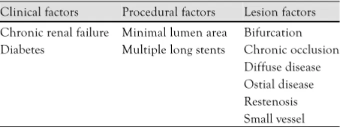
Table 1. Predictors of restenosis after bare-metal stenting Clinical factors Procedural factors Lesion factors Chronic renal failure
Diabetes
Minimal lumen area Multiple long stents
Bifurcation Chronic occlusion Diffuse disease Ostial disease Restenosis Small vessel
tively, p<0.001). There was no difference between the stents in terms of the restenosis rate(9.6% vs. 11.1, respectively, p=0.31) and clinical outcomes(10.7% vs.
11.4, respectively, p=0.73). The SIRTAX trial23) was a randomized, 1,012-patient, single blind comparison of Cypher and Taxus stents. In contrast to the REALITY trial, the SIRTAX study found that the Taxus stent was associated with a higher major adverse cardiac event rate and a higher target lesion revascularization rate. Likewise, the ISAR-DIABETES randomized trial24) that compared Cypher and Taxus stents in 250 diabetic patients with de novo lesions found the restenosis rate associated with the Cypher stent was lower than that associated with the Taxus stent(6.9% vs. 16.5%, respectively, p=
0.03). That study also showed that the two stents did not differ in terms of the target lesion revascularization rates(6.4% vs. 12.2%, respectively, p=0.13). We recently reported the results of the Long-DES II trial,25) which compared the use of Cypher and Taxus stents in 500 patients with long(≥25 mm) native coronary lesions.
The study found that the Cypher stent was associated with a lower in-segment binary restenosis rate than the Taxus stent(3.3% versus 14.6%, respectively, relative risk: 0.23, p<0.001). The in-stent late loss of the lumen diameter was 0.09±0.37 mm in the Cypher group and 0.45±0.55 mm in the Taxus group(p<0.001). There was no significant difference between the stents in terms of the incidence of death or myocardial infarction after 9 months of follow-up. These findings suggest that for long native coronary artery disease, the Cypher stent is superior in terms of restenosis and target vessel failure.
Kandzari et al.26) compared the recently introduced zotarolimus-eluting stent with the sirolimus-eluting stent in terms of the relative clinical efficacy, the angiographic outcomes and safety in 436 patients with de novo native coronary lesions. The study found that the zotarolimus- eluting stent was associated with greater angiographic in-segment late lumen loss(0.34±0.44 mm vs. 0.13±
0.32 mm, respectively, p<0.001) and in-segment binary angiographic restenosis(11.7% vs. 4.3%, respectively, p
=0.04) compared with the sirolimus-eluting stent. The two stents were found to be similar in terms of clinically driven target lesion revascularization(6.3% zotarolimus vs. 3.5% sirolimus, p=0.34) and target vessel failure (12.0% zotarolimus vs. 11.5% sirolimus, p=1.0).
Overall, the current available data indicates that using the Cypher stent results in less late lumen loss than using the Taxus or Endeavor stents.21) These differences may reflect the metal platform design, the polymer and/or the pharmacological agents. The extent of late loss correlates with target lesion revascularization, and its influence on restenosis is related to the baseline risk of restenosis. Therefore, in terms of restenosis, while the Cypher stent may provide better outcomes than the Taxus or Endeavor stents for complex lesions that have
a high risk for restenosis, such a difference may not exist in low risk lesion groups.
Patient-related factors
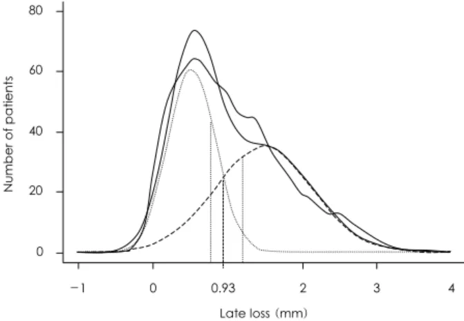
The frequency-distribution curves of the angiographic indices of restenosis after bare-metal stent placement have a bimodal pattern, indicating the existence of two distinct populations with different propensities for restenosis(Fig. 1). Kastrati et al.27) also reported that the risk of a lesion developing restenosis after stent im- plantation was 2.5 times higher if a companion lesion had restenosis, and this was independent of the analyzed patient risk factors.28) These findings suggest that uni- dentified patients factors may exert a critical influence on the development of restenosis after baremetal stent implantation. However, the pattern of angiographic late loss differs between thse lesions treated with DES and and the lesions treated with bare-metal stents. The dis- tribution of late loss for the Cypher stent appeared largely skewed to the right with a Gaussian distribution.29) In addition, we found that late loss was higher in the Taxus stent group than in the Cypher stent group, demon- strating that late lumen loss tended to favor the Cypher
Fig. 1. Late lumen loss at follow-up for 1,062 lesions after bare-metal stents implantation.
80
60
40
20
-1 0
0 0.93 2 3 4 Late loss (mm)
Number of patients
Fig. 2. Frequency distribution of the late loss values for the Cypher and Taxus stents.
-1.2 -0.8 -0.4 0.0 0.4 0.8 1.2 1.6 2.0 2.4 2.8 3.2 Late loss (mm)
250
200
150
100
50
0
Frequency
Sirolimus-eluting stent Paclitaxel-eluting stent
stent over the Taxus stent(Fig. 2).30) However, it remains uncertain whether the likelihood of restenosis for a lesion is greater when a companion lesion has developed restenosis after DES implantation.
There are conflicting results from the previous reports regarding the effect of diabetes on restenosis after DES implantation.31-33) While some authors have concluded that diabetes is an independent predictor of restenosis, others have reported that diabetes per se may not be an independent risk factor for repeat revascularization.
Local variables such as small vessels and diffuse disease may be more important for predicting restenosis than simply diabetes. However, only a relatively small number of diabetic patients, and even fewer insulin-dependent diabetics, have been studied, so further studies are required to ascertain whether diabetes is a predictor of restenosis in the DES era. Overall, the reports in the literature suggest that clinical variables are not strong predictors of which patients will or will not develop restenosis after DES implantation.
Procedure- and lesion-related factors
The post-intervention final lumen area has been doc- umented to be the most powerful predictor of restenosis
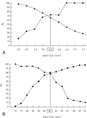
after both balloon angioplasty and bare-metal stent implantation. Several studies have shown that in pa- tients receiving DES implants, the post-intervention final lumen area is the most important determinant of restenosis, suggesting that a greater stent area contributes to a decreased rate of restenosis, even in those patients with implanted DES.31) Intravascular ultrasound studies have revealed that the independent predictors of angio- graphic restenosis after Cypher stent implantation are the post-procedural final minimum stent area and the stented length of the artery. We have shown that the angiographic restenosis rate is highest in lesions with a stent area <5.5 mm2 and a stent length >40 mm(Fig.
3).33) Non-uniform strut distribution also contributes to intimal hyperplasia after Cypher stent implantation in de novo lesions, and this suggests a gap between the stent struts may be associated with a decrease in local drug delivery, which may then contribute to the devel- opment of restenosis.34) In addition, stent fracture is rarely related to very focal intrastent restenosis despite complete abolition of intimal hyperplasia in the re- mainder of the stented segment.35) Overall, the published reports have suggested that residual stenosis is a signi- ficant component of the restenosis problem, and this indicates that achieving a larger lumen area with ade- quate stent expansion remains an important strategy for reducing restenosis, even in the DES era.
While the lesion length and stent length correlate with restenosis, lesion length is an independent pre- dictor of restenosis.36) A small increase in the ratio of the in-stent length to the lesion length has a profound effect in reducing that margin effect. These findings indicate that stent length has less influence on res- tenosis when using DES compared with using bare metal stents,36)37) and this supports the current strategy of complete lesion coverage. However, a full metal jacket approach(stented length >60 mm) in small vessels has been linked with a high risk of DES failure(Fig. 4).38)
Fig. 3. The sensitivity and specificity curves identified the optimal cut- off values of the final minimum stent CSA (A) & stent length (B) that predicted angiographic restenosis after Cypher implantation.
CSA: cross sectional area.
3.5 4.0 4.5 5.0 55.55 6.0 6.5 7.0 7.5 Stent CSA (mm2)
100 90 80 70 60 50 40 30 20 10 0
(%)
10 15 20 25 30 35 5405 45 50 55 60 65 70 Stent CSA (mm2)
100 90 80 70 60 50 40 30 20 10 0
(%)
B A
Fig. 4. Effects of lesion length on the restenosis rate in small coronary arteries (≤2.8 mm).
0 5 10 15 20 25 30 35
<40 40-60 >60
Lesion length (mm)
Restenosis rate
p<0.01
13.0%
29.4%
7.0%
Conclusions
DES have been widely used as a revascularization strategy for a broad variety of clinical and anatomic situations, and restenosis remains a major clinical issue.
Small vessel disease, diffuse disease and long stents have been shown to be predictive variables for restenosis after DES implantation. However, there is a poor cor- relation between these variables and the risk of subse- quent restenosis. Further studies are required in order to identify the factors that can be used to reliably predict whether an individual patient will experience restenosis after DES implantation.
This study was supported by grants from the Cardiovascular Research Foundation, and the Korea Health 21 R&D Project, Ministry of Health & Welfare, Korea(0412-CR02-0704-0001).
REFERENCES
1) Morice MC, Serruys PW, Sousa JE, et al. A randomized com- parison of a sirolimus-eluting stent with a standard stent for coronary revascularization. N Engl J Med 2002;346:1773-80.
2) Moses JW, Leon MB, Popma JJ, et al. Sirolimus-eluting stents versus standard stents in patients with stenosis in a native co- ronary artery. N Engl J Med 2003;349:1315-23.
3) Colombo A, Drzewiecki J, Banning A, et al. Randomized study to assess the effectiveness of slow- and moderate-release polymer- based paclitaxel-eluting stents for coronary artery lesions. Cir- culation 2003;108:788-94.
4) Pache J, Dibra A, Mehilli J, Dirschinger J, Schomig A, Kastrati A. Drug-eluting stents compared with thin-strut bare stents for the reduction of restenosis: a prospective, randomized trial. Eur Heart J 2005;26:1262-8.
5) Ardissino D, Cavallini C, Bramucci E, et al. Sirolimus-eluting vs uncoated stents for prevention of restenosis in small coronary arteries: a randomized trial. JAMA 2004;292:2727-34.
6) Grines CL, Bonow RO, Casey DE Jr, et al. Prevention of pre- mature discontinuation of dual antiplatelet therapy in patients with coronary artery stents: a science advisory from the American Heart Association, American College of Cardiology, Society for Cardiovascular Angiography and Interventions, American College of Surgeons, and American Dental Association, with representa- tion from the American College of Physicians. Circulation 2007;
115:813-8.
7) Bourassa MG , Lesperance J, Eastwood C, et al. Clinical, physio- logic, anatomic and procedural factors predictive of restenosis after percutaneous transluminal coronary angioplasty. J Am Coll Cardiol 1991;18:368-76.
8) van Belle E, Bauters C, Hubert E, et al. Restenosis rates in dia- betic patients: a comparison of coronary stenting and balloon an- gioplasty in native coronary vessels. Circulation 1997;96:1454-60.
9) Peters RJ, Kok WE, di Mario C, et al. Prediction of restenosis after coronary balloon angioplasty: results of PICTURE (Post- intraCoronary Treatment Ultrasound Result Evaluation), a pro- spective multicenter intracoronary ultrasound imaging study.
Circulation 1997;95:2254-61.
10) Hirschfeld JW Jr, Schwartz JS, Jugo R, et al. Restenosis after coronary angioplasty: a multivariate statistical model to relate lesion and procedure variables to restenosis. J Am Coll Cardiol 1991;18:647-56.
11) Mintz GS, Popma JJ, Pichard AD, et al. Arterial remodeling after coronary angioplasty: a serial intravascular ultrasound study.
Circulation 1996;94:35-43.
12) Fischman DL, Leon MB, Baim DS, et al. A randomized com- parison of coronary stent placement and balloon angioplasty in the treatment of coronary artery disease. N Engl J Med 1994;
331:496-501.
13) Carrozza JP Jr, Kuntz RE, Levine MJ, et al. Angiographic and clinical outcome of intracoronary stenting: immediate and long- term results from a large single-center experience. J Am Coll Cardiol 1992;20:328-37.
14) Kastrati A, Schomig A, Elezi S, et al. Predictive factors of restenosis after coronary stent placement. J Am Coll Cardiol 1997;
30:1428-36.
15) Elezi S, Kastrati A, Neumann FJ, Hadamitzky M, Dirschinger J, Schomig A. Vessel size and long-term outcome after coronary stent placement. Circulation 1998;98:1875-80.
16) Ho KKL, Senerchia C, Rodriguez O, et al. Predictors of angio- graphic restenosis after stenting: pooled analysis of 1197 patient with protocol-mandated angiographic follow-up from 5 random- ized stent trials. Circulation 1998;98(Suppl I):I362.
17) Hoffmann R, Mintz GS, Mehran R, et al. Intravascular ultra- sound predictors of angiographic restenosis in lesions treated with Palmaz-Schatz stents. J Am Coll Cardiol 1998;31:43-9.
18) Pache J, Kastrati A, Mehilli J, et al. Intracoronary stenting and angiographic results: strut thickness effect on restenosis outcome (ISAR-STEREO-2) trial. J Am Coll Cardiol 2003;41:1283-8.
19) Kuntz RE, Gibson CM, Nobuyoshi M, Baim DS. Generalized model of restenosis after conventional balloon angioplasty, stent- ing and directional atherectomy. J Am Coll Cardiol 1993;21:15-25.
20) Hoffmann R, Mintz GS, Dussaillant GR, et al. Patterns and me- chanisms of in-stent restenosis: a serial intravascular ultrasound study. Circulation 1996;94:1247-54.
21) Wessely R, Schomig A, Kastrati A. Sirolimus and Paclitaxel on polymer-based drug-eluting stents: similar but different. J Am Coll Cardiol 2006;47:708-14.
22) Morice MC, Colombo A, Meier B, et al. Sirolimus- vs paclitaxel- eluting stents in de novo coronary artery lesions: the REALITY trial: a randomized controlled trial. JAMA 2006;295:895-904.
23) Park MA, Ryu JN, Lim TH, et al. Comparisons of the short-term angiographic outcomes of cypher and taxus stents implanted in the same patient. Korean Circ J 2006;36:600-4.
24) Dibra A, Kastrati A, Mehilli J, et al. Paclitaxel-eluting or siro- limus-eluting stents to prevent restenosis in diabetic patients. N Engl J Med 2005;353:663-70.
25) Kim YH, Park SW, Lee SW, et al. Sirolimus-eluting stent versus paclitaxel-eluting stent for patients with long coronary artery disease. Circulation 2006;114:2148-53.
26) Kandzari DE, Leon MB, Popma JJ, et al. Comparison of zota- rolimus-eluting and sirolimus-eluting stents in patients with native coronary artery disease: a randomized controlled trial. J Am Coll Cardiol 2006;48:2440-7.
27) Kastrati A, Dibra A, Eberle S, et al. Sirolimus-eluting stents vs paclitaxel-eluting stents in patients with coronary artery disease:
meta-analysis of randomized trials. JAMA 2005;294:819-25.
28) Schomig A, Kastrati A, Elezi S, et al. Bimodal distribution of angiographic measures of restenosis six months after coronary stent placement. Circulation 1997;96:3880-7.
29) Kastrati A, Schomig A, Elezi S, Schuhlen H, Wilhelm M, Dirs- chinger J. Interlesion dependence of the risk for restenosis in patients with coronary stent placement in multiple lesions. Cir- culation 1998;97:2396-401.
30) Lemos PA, Mercado N, van Domburg RT, Kuntz RE, O’Neill WW, Serruys PW. Comparison of late luminal loss response
pattern after sirolimus-eluting stent iImplantation or conventional stenting. Circulation 2004;110:3199-205.
31) Lee CW, Park DW, Lee BK, et al. Predictors of restenosis after placement of drug-eluting stents in one or more coronary arteries.
Am J Cardiol 2006;97:506-11.
32) Kastrati A, Dibra A, Mehilli J, et al. Predictive factors of restenosis after coronary implantation of sirolimus- or paclitaxel-eluting stents. Circulation 2006;113:2293-300.
33) Finn AV, Palacios IF, Kastrati A, Gold HK. Drug-eluting stents for diabetes mellitus: a rush to judgment? J Am Coll Cardiol 2005;45:479-83.
34) Hong MK, Mintz GS, Lee CW, et al. Intravascular ultrasound predictors of angiographic restenosis after sirolimus-eluting stent implantation. Eur Heart J 2006;27:1305-10.
35) Takebayashi H, Mintz GS, Carlier SG , et al. Nonuniform strut
distribution correlates with more neointimal hyperplasia after si- rolimus-eluting stent implantation. Circulation 2004;110:3430-4.
36) Sianos G , Hofma S, Ligthart JM, et al. Stent fracture and res- tenosis in the drug-eluting stent era. Catheter Cardiovasc Interv 2004;61:111-6.
37) Yoon HJ, Kim KS, Park HS, et al. Clinical and angiographic factors affect on in-stent restenosis. Korean Circ J 2003;33:
1084-92.
38) Mauri L, O’Malley AJ, Cutlip DE, et al. Effects of stent length and lesion length on coronary restenosis. Am J Cardiol 2004;93:
1340-6.
39) Whang YJ, Kim HS, Kim KM, et al. The efficacy of drug eluting stents on restenosis reduction in small coronary arteries. Korean Circ J 2006;36:450-7.