J. Exp. Biomed. Sci. 15 (2009) 179–185
Repression of PPARγ Activity on Adipogenesis by 17β-estradiol in Differentiated 3T3-L1 Cell
Michung Yoon and Sunhyo Jeong
†Department of Life Sciences, Mokwon University, Taejon 302-729, Korea
In our previous report, we showed that PPARγ does not influence adipogenesis in females with functioning ovaries, indicating that PPARγ activity on adipogenesis is associated with sex-related factors. Among the sex-related factors, estrogen has been recognized as a major factor in inhibiting adiposgenesis in females. Thus, we hypothensized that 17β-estradiol (E) inhibits 3T3-L1 cell adipogenesis by preventing PPARγ activity. E decreased triglyceirde accumulation in differentiated 3T3-L1 cells compared with control group. E also decreased the expression of PPARγ mRNA as well as PPARγ dependent adipocyte-specific genes, such as adipocyte fatty acid binding protein and tumor necrosis factor α.
In addition, E not only decreased luciferase reporter activity by PPARγ, but also transfection of estrogen receptor α (ERα) or ERβ led to decreases in PPARγ reporter gene activation. Moreover, E-activated ERs significantly decreased the luciferase reporter gene activation induced by PPARγ transfection, suggesting that estrogen-activated ERs inhibit PPARγ-dependent transactivation. Accordingly, our results demonstrate that E inhibits the action of PPARγ on adipogenesis through E activated ER, providing evidence that lack of estrogen may potentiate PPARγ action on adipogenesis.
Key Words: 17β-estradiol, PPARγ, ER, Adipogenesis
INTRODUCTION
Adipocytes play a central role in maintaining lipid homeostasis and energy balance in vertebrates by storing triglycerides or releasing free fatty acids in response to changing energy needs. Preadipocytes exits in close pro- ximity to adipocytes and respond to positive energy balance by proliferating and then differentiating into adipocytes (Kirkland et al., 1994). Preadipocyte cell lines are useful models for investigating the adipogenesis process. 3T3-L1 preadipocytes, which can be induced to differentiate into adipocytes in cell culture, is one of the most studied preadipocyte cell line (MacDougald and Lane, 1995;
Gregoire et al., 1998). When mouse 3T3-L1 preadipocytes are treated with inducing mixture that includes insulin,
dexamethasone, and isobutylmetylxanthine (Green et al., 1975; Rubin et al., 1978), they change from an extended fibroblast-like morphology to a round one with cytoplasmic lipid vesicles made of newly biosynthesized triglycerides.
The differentiation of preadipocytes into mature adipo- cytes includes activation of adipogenic gene expression. In recent years, there has been substantial progress in under- standing the transcriptional control of adipogenic gene expression. A number of transcription factors have been identified as important regulators of this process, including the peroxisome proliferators-activated receptor γ (PPARγ) (Zhu et al., 1995; Spiegelman and Filter, 1996).
PPARγ forms an obligate heterodimer with the retinoid X receptor to bind to regulatory elements within the promoters/
enhancers of many adipocyte-specific genes associated with lipid metabolism such as adipocyte fatty acid binding protein (aP2) and lipoprotein lipase (LPL) (Rosen et al., 2000). In the course of differentiation of acipocytes, PPARγ also induce adipogenic genes, including adipsin, tumor necrosis factor α (TNFα) and leptin, which are signaling molecules produced and secreted in adipocytes (Zhang et
*Received: 1September, 2009 / Revised: 18 September, 2009 Accepted: 19 September, 2009
†Corresponding author: Sunhyo Jeong, Department of Life Sciences, Mokwon University, Taejon 302-729, Korea.
Tel: 82-42-829-7589, Fax: 82-42-829-7580 e-mail: [email protected]
al., 1994; Halaas et al., 1995; Kern et al., 1995; Alessi et al., 1997).
Moreover, we showed that PPARγ does not induce adipogenesis in females with functioning ovaries, indicating that PPARγ activity on adipogenesis is influenced by sex- related factors (Yoon and Jeong, 2008). Among the sex- related factors, estrogen has been recognized as a major factor in regulating adipose development and adipogenesis in females. Estrogen favored early osteogenic commitment and inhibited adipogensis of mouse ST2 cells overexpressing either estrogen receptor (ER) α or β, and inhibited adipo- genic commitment of the mesenchymal mouse cell line KS483 (Dang et al., 2002; Okazaki et al., 2002). Estrogen was also reported to stimulate proliferation of human preadipocytes, which remain undifferentiated cells into adipocytes, suggesting that estrogen inhibits adipogenesis (Roncari and Van, 1978). Noticeably, there is a report that estrogen regulates PPARγ activity on adipogenesis in KS483 cells, which concurrently differentiate into osteoblasts and adipocytes (Dang et al., 2002). Accordingly, it is likely that estrogen inhibits adipogenesis of 3T3-L1 cells by preventing PPARγ activity. However, the molecular and cellular mechanisms underlying the metabolic actions of estrogen on adipogensis are poorly understood. Therefore, we sought to investigate the molecular mechanism whereby E inhibits the actions of PPARγ on adipogenesis.
MATERIALS AND METHODS 1. Induction of preadipocyte differentiation
The 3T3-L1 preadipocytes were grown to confluence in DMEM supplemented with 10% fetal bovine serum (FBS),
100 U/ml penicillin, and 100 mg/ml streptomycin at 5%
CO
2and 37℃. Two days postconfluence (day 0), cells were induced to differentiate in differentiation medium [DMEM containing 10% FBS, 0.17 mM isobutylmethylxanthine (IBMX) (Sigma, USA), 0.3 μM dexamethasone (Sigma, USA) and 1 μg/ml insulin (Sigma, USA)] with each com- pounds (troglitazone and 17β-estradiol) for 2 days. Forty- eight hours later (day 2), cells were incubated in DMEM containing only 10% FBS. The medium was replenished at 2-day intervals for additional 4 days. Adipogenesis was determined by staining of lipids with Oil Red O and by the expression of adipocyte-specific markers.
2. Oil Red O staining of differentiated 3T3-L1 Cells were washed twice with PBS, fixed in 10%
formaldehyde for 1 h, washed twice with PBS and stained for 2 h with 4% Oil Red O solution. They were then washed twice with PBS and examined by light microscopy.
3. Analysis of target gene expression
Total RNA of differentiated 3T3-L1 cells was prepared using Trizol reagent (Gibco-BRL, Grand Island, NY) and relative levels of mRNA were assessed by reverse transcription-polymerase chain reaction (RT-PCR). Comple- mentary DNA was synthesized from RNA samples by mixing 2 μg of total RNA and 0.5 μg of the reverse primer in a total volume of 14 μl in water, heating the mixture at 75℃ for 15 min, cooling the mixture immediately on ice for 5 min, and adding 5× M-MLV reaction buffer, 10 mM dNTP mixture (Promega) and 200 units M-MLV RT (Promega) in total volume of 25 μl. Samples were incubated at 42℃ for 60 min. A five μl aliquot of the RT reaction
Table 1. Sequences of oligonucleotide primers and PCR conditions
Genes Size (bp) Primer sequences Annealing (℃) Cycle
Forward: 5'-attctggcccaccaacttcgg-3' PPARγ 340
Reverse: 5'-tggaagcctgatgctttatcccca-3' 58 28
Forward: 5'-caaaatgtgtgatgcctttgtg-3' aP2 417
Reverse: 5'-ctcttcctttggctcatgcc-3' 58 24
Forward: 5'-ctcgagtgacaagcccgtag-3' TNFα 387
Reverse: 5'-ttgacctcagcgctgagcag-3' 58 34
Forward: 5'-tggaatcctgtggcatccatgaaa-3' β-actin 350
Reverse: 5'-taaaacgcagctcagtaacagtcc-3' 58 28
was then used for subsequent PCR amplification with specific primers.
Twenty five μl PCR sample contained 5 μl of the RT reaction, 10× buffer with MgCl
2, 10 mM dNTP, 5 units of Tag polymerase (Solgent, Taejon, Korea) and 10 μM of each primer. Primer sequences and PCR conditions are shown in Table 1. PCR was performed in a PTC-100
TMProgrammable Thermal Controller (MJ Research, Watertown, MA, USA). PCR products were electophoresed on a 1%
agarose gel.
4. Transient transfection Assay
The expression vectors for pSG5-mPPARγ and PPRE
3- tk-luc reporter gene were generously provided by Dr.
Gonzalez (National Cancer Institute, NIH, Bethesda, MD).
Expression vectors for pcDNA-ERα and pcDNA-ERβ
gene constructs were generously provided by Dr. Burow (Tulane University Medical Center, New Orleans, LA).
Murine preadipocyte cell line 3T3-L1 cells were routinely cultured in DMEM containing 10% fetal bovine serum (Gibco-Brl, Grand Island, NY), penicillin G (100 U/ml), streptomycin sulfate (100 μg/ml), amphotericin B (0.25 μg/ml) and 2-mercaptoethanol (50 μM). Cells were seeded in 6-well tissue culture plates (2×10
4cells/well) 24 h prior to transfection. For all transfections, 200 ng/well of each of the appropriate plasmids were used. Transfections were performed using Lipofectamine (Invitrogen, Carlsbad, CA) according to the manufacturer's instructions. After 6 h, the culture medium was changed and the test compound, 17β- estradiol (Sigma) was added. After incubation for 24 h in the presence of the aforementioned chemicals, cells were washed twice with phosphate-buffered saline and assayed
Fig. 1. Inhibition of triglyceride accumu- lation by 17β-estradiol in differentiated 3T3-L1 cells. 3T3-L1 preadipocytes were cultured until they were confluent. After 2 days, they were exposed to dexamethasone, isobutylmethylxanthine, and insulin, with or without E (10 μM). At day 6 post- induction, cells were fixed and stained with Oil red O. Magnification is 10×. E; 17β- estradiol.
A B
Fig. 2. Modulation of PPARγ gene expression by 17β-estradiol in differentiated 3T3-L1 cells. (A) Quantitative analyses of RT-PCR.
3T3-L1 preadipocytes were cultured until they were confluent. After 2 days, they were exposed to dexamethasone, isobutylmethylxanthine, and insulin, with or without E (10 μM). At day 6 postinduction, total RNA was extracted and relative levels of PPARγ and β-actin mRNA were assessed by reverse transcription-polymerase chain reaction (RT-PCR). All values are expressed as mean ± SD of R.D.U. (relative density units) using β-actin as a reference. *Significantly different versus control group, P<0.05. (B) Representative RT-PCR photographs from two of three independent experiments are shown. E; 17β-estradiol.
for luciferase and β-galactosidase activity using commercial kits according to the manufacturer's instructions (Promega, Madison, WI).
5. Statistics
Unless otherwise noted, all values are expressed as mean
± standard deviation (SD). All data were analyzed by ANOVA for statistically significant differences between each group.
RESULTS
1. Triglyceride accumulation in differentiated 3T3-L1 cells
We observed whether E regulates triglyceride accumu- lation in 3T3-L1 cells incubated in differentiation medium for 6 days, using Oil Red O staining for neutral lipids (Fig.
1). Triglycerides were decreased in E treated group compared with vehicle-treated control group, suggesting
that E inhibits triglyceride accumulation in differentiated 3T3-L1 cells.
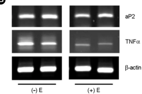
2. Expression of PPARγ dependent adipoctye-specific genes in differentiated 3T3-L1 adipocytes
We determined whether the inhibition of triglyceride accumulation by E in differentiated 3T3-L1 cells was caused by changes in PPARγ and PPARγ-dependent adipocyte- specific gene expression (Fig. 2 and Fig. 3). E decreased PPARγ mRNA levels compared with control group (P<
0.05) (Fig. 2). E also decreased aP2 and TNFα mRNA levels compared with control group (P<0.05) (Fig. 3). These results suggest that E may decrease mRNA levels of PPARγ- dependent adipocyte-specific genes through the inhibition of adipose PPARγ activation, and, thereby, prevented the actions of PPARγ on adiposenesis.
3. PPARγ reporter gene expression
To examine the mechanism by which E inhibited the
A B
Fig. 3. Modulation of adipocyte specific genes expression by 17β-estradiol in differentiated 3T3-L1 cells. (A) Quan- titative analyses of RT-PCR. 3T3-L1 Preadipocytes were cultured until they were confluent. After 2 days, they were exposed to dexamethasone, isobutylmethylxanthine, and insulin, with or without E (10 μM). At day 6 postinduction, total RNA was extracted and relative levels of PPARγ target genes and β-actin mRNA levels were assessed by reverse transcription-polymerase chain reaction (RT-PCR).
All values are expressed as mean ± SD of R.D.U. (relative density units) using β-actin as a reference. *Significantly different versus control group, P<0.05. (B) Representative RT-PCR photographs from two of three independent experiments are shown. E; 17β-estradiol.
actions of PPARγ on adipogensis, 3T3-L1 preadipocytes were transiently transfected with PPARγ, ERα, or ERβ expression constructs and with a luciferase reporter gene construct (PPRE
3-tk-luc) containing three copies of the PPRE from the rat ACOX gene (Figs. 4~6). Cells trans-
fected with PPARγ increased luciferase activity, but E decreased luciferase reporter activity induced by PPARγ transfection (P<0.05) (Fig. 4, lane 4 vs. lane 2). Transfection of ERα or ERβ led to decreases in PPRE luciferase activity (P<0.05), although E did not show additional effects (Fig.
5, lane 2 and 5).
The cells were transiently transfected with PPARγ and ERα or PPARγ and ERβ (Fig. 6). ERα or ERβ transfection decreased PPARγ-induced luciferase activity (lane 3 or 6 vs lane 2) and E-activated ERα or ERβ further reduced the luciferase reporter gene activation by PPARγ transfection compared with ERα- or ERβ-only, respectively (P<0.05) (lane 4 and 5 vs. lane 3, lane 7 and 8 vs. lane 6). These results suggest that E inhibits PPARγ-mediated transactiva- tion through ER activation.
DISCUSSION
The present study demonstrates that E inhibits the actions of PPARγ on adipogenesis in adipocytes. We showed here that E decreased triglyceirde accumulation and the
Fig. 4. Inhibition of PPARγ reporter gene expression by17β-estradiol in the presence of PPARγ. 3T3-L1 Preadipocytes were transiently transfected with expression plasmids for PPARγ and PPRE3-tk-luc reporter. All values are expressed as mean ± SD of three experiments. *Significantly different versus vehicle group (lane 1). P<0.05. **Significantly different versus PPARγ group (lane 2). P<0.05. PPARγ; peroxisome proliferators-activated receptor γ, ER; estrogen receptor, E; 17β-estradiol.
Fig. 6. Inhibition of PPARγ reporter gene expression by 17β- estradiol in the presence of PPARγ and ERα or ERβ. 3T3-L1 preadipocytes were transiently transfected with expression plasmids for PPARγ, PPRE3-tk-luc reporter and ERα or ERβ. #Significantly different versus vehicle group (lane 1). P<0.05. *Significantly different versus PPARγ group (lane 2). P<0.05. **Significantly different versus PPARγ/ERα or ERβ group (lane 3 and lane 6, respectively), P<0.05. PPARγ; peroxisome proliferators-activated receptor γ, ER; estrogen receptor, E; 17β-estradiol.
Fig. 5. Inhibition of PPARγ reporter gene expression by 17β- estradiol in the presence of ERα or ERβ. 3T3-L1 preadipocytes were transiently transfected with expression plasmids for PPRE3-
tk-luc reporter and ERα or ERβ. *Significantly different versus vehicle group (lane 1). P<0.05. PPARγ; peroxisome proliferators- activated receptor γ, ER; estrogen receptor, E; 17β-estradiol.
expression of PPARγ-dependent adipocyte-specific genes, such as aP2 and TNFα, as well as PPARγ gene compared with vehicle-treated control group in differentiated 3T3-L1 cells. Moreover, we suggest that this inhibitory action of E on adipogenesis may be due to the inhibition of PPARγ transactivation.
Hormones are major regulators of adipose tissue and are critical for adipoctye development and function. Estrogen has long been recognized as a major factor in regulating adipose development in females, indicating that it regulates key development events in adipogenesis (Wade et al., 1985).
As for adipogenesis, in vivo and in vitro evidence support the role of estrogen as a negative regulator. ERα knockout mice and aromatase-deficient mice have been reported to have increased adiposity (Heine et al., 2000; Jones et al., 2000). Genistein, which mimics estrogenic action, inhibits adipogenesis in 3T3-L1 cells (Harmon and Harp, 2001).
Similarly, our data showed that E inhibited triglyceride accumulation in differentiated 3T3-L1 cells, suggesting that E is inhibitory regulator on adipogenesis. Furthermore, it was reported that estrogen decreased mRNA levels of aP2 and LPL in KS483 cells, and it is more interesting that estrogen can modulate the adipogenic commitment of the mesenchymal mouse cell line KS483 via transcriptional repression of PPARγ (Dang et al., 2002; Harmon et al., 2002). This reports support our results that E inhibited the expression of PPARγ, and PPARγ-dependent adipogenesis- related adipocyte-specific genes during the differentiation of 3T3-L1 preadipocytes, indicating that E is able to suppress PPARγ actions on adipogenesis in differentiated 3T3-L1 cells.
In the present study, E decreased PPARγ reporter gene activation, as well as ERα or ERβ. Moreover, E decreased PPARγ reporter activity in 3T3-L1 preadipocytes over- expressing ER, suggesting that E inhibits PPARγ trans- activation through the ER activation, accompanied by a blockade of PPARγ dependent adipocyte specific genes and decrease of adipogenesis. The previous reports support our results that E regulated PPARγ transcription in 3T3-L1 cells. PPARγ together with its heteroimeric partner RXRα has been shown to suppress ER-induced target gene expres- sion through competitive binding to an estrogen response
element (ERE) site in the vitellogenin A2 promoter (Keller et al., 1995). ERα and ERβ are also capable of inhibiting ligand-induced PPARγ activation in two different breast cancer cell lines, MDA-MB-231 and MCF-7 breast cancer cells (Wang and Kilgore, 2002). Activation of PPARα or PPARγ by daidzein, phytoestrogens found in soy products, downregulated ERE-luc reporter activity and activation of ERα or ERβ by daidzine downregulated PPARγ tran- scriptional activity (Dang et al., 2004).
Accordingly, it is suggested the possibility that E interferes with PPARγ activity on adipogenesis and that signal cross talk exists between PPARγ and ER. Here we conclude that E may suppress adipocyte formation in 3T3- L1 cells and this event may be mediated by the inhibition of PPARγ actions on adipgenesis through the ER activation.
Acknowledgements
This work was supported by a grant No. KRF-2005-075- C00029 from the Korea Research Foundation and a grant No. 2009-0069150 from the Korea Science and Engineering Foundation.
REFERENCES
Alessi MC, Peiretti F, Morange P, Henry M, nalbone G, Juhan-Vague I. Production of plasminogen activator 1 by human adipose tissue: possible link between viscernal fat accumulation and vascular disease. Diabetes 1997. 46: 860 -867.
Dang ZC, van Bezooijien RL, Karperien M, Papapoulos SE, Lowik CW. Esposure of KS483 cells to estrogen enhances osteogensis and inhibits adipogenesis. J Bone Miner Res.
2002. 17: 394-405.
Dang Z, Löwik CW. The balance between concurrent activation of ERs and PPARs determines daidzein-induced osteogenesis and adipogenesis. J Bone Miner Res. 2004. 19: 853-861.
Green H, Kehinde O. An established preadipose cell line and its differentiation in culture. II. Factors affecting the adipose conversion. Cell 1975. 5: 19-27.
Gregoire FM, Smas CM, Sul HS. Understanding adipocyte differentiation. Physiol Rev. 1998. 78: 783-809.
Halaas JL, Gajiwala KS, Maffei M, Cohen SL, Chait BT, Rabinowitz D, Lallone RL, Burley SK, Friedman JM. Weight-
reducing effects of the plasma protein encoded by the obese gene. Science 1995. 269: 543-546.
Harmon AW, Harp JB. Differential effects of flavonoids on 3T3- L1 adipogenesis and lipolysis. Am J Physiol Cell Physiol.
2001. 280: C807-C813.
Heine PA, Taylor JA, Iwamoto GA, Lubahn DB, Cooke PS.
Increased adipose tissue in male and female estrogen receptor- alpha knockout mice. Proc Natl Acad Sci U S A. 2000. 97:
12729-12734.
Keller H, Givel F, Perroud M, Wahli W. Signaling cross-talk between peroxisome proliferator-activated receptor/retinoid X receptor and estrogen receptor through estrogen response elements. Mol Endocrinol. 1995. 9: 794-804.
Kern PA, Saghizadeh M, Ong JM, Bosch RJ, Deem R, Simsolo RB. The expression of tumor necrosis factor in human adipose tissue. J Clin Invest. 1995. 95: 2111-2119.
Kirkland JL, Hollenberg CH, Kindler S, Gillon WS. Effects of age and anatomic site on preadipocyte number in rat depots.
J Gerontol. 1994. 49: B31-B35.
MacDougald OM, Lane MD. Transcriptional regulation of gene expression during adipocyte differentiation. Annu Rev Biochem. 1995. 64: 345-353.
Jones ME, Thorburn AW, Britt KL, Hewitt KN, Wreford NG, Proietto J, Oz OK, Leury BJ, Robertson KM, Yao S, Simpson ER. Aromatase-deficient (ArKO) mice have a phenotype of increased adiposity. Proc Natl Acad Sci U S A. 2000. 97:
12735-12740.
Okazaki R, Inoue D, Shibata M, Saika M, Kido S, Ooka H, Tomiyama H, Sakamoto Y, Matsumoto T. Estrogen promotes early osteoblast differentiation and inhibits adipocyte differ- entiation in mouse bone marrow stromal cell lines that express estrogen receptor (ER) α or β. Endocrinology 2002.
143: 2349-2356.
Roncari DA, Van RL. Promotion of human adipocyte precursor replication by 17β-estradiol in culture. J Clin Invest. 1978.
62: 503-508.
Rosen ED, Waikey CJ, Puigserver P, Spiegelman BM. Tran- scriptional regulation of adipogenesis. Genes Dev. 2000. 14:
1293-1307.
Rubin CS, Hirsh A, Fung C, Rosen OM. Development of hormone receptors and hormonal responsiveness in vitro.
Insulin receptors and insulin sensitivity in the preadipocyte and adipocyte forms of 3T3-L1 cells. J Biol Chem. 1978.
253: 7570-7578.
Spiegelman BM, Filter JS. Adipogenesis and obesity: rounding out the big picture. Cell 1996. 87: 377-389.
Yoon M, Jeong S. Peroxisome proliferator-acitvated receptor γ is not associated with adipogenesis in female mice. J Exp Biomed Sci. 2008. 14: 139-146.
Wade GN, Gray JM, Bartness TJ. Gonadal influences on adiposity.
Int J Obes. 1985. 9: 83-92.
Wang X, Kilgore MW. Signal cross-talk between estrogen receptor alpha and beta and the peroxisome proliferator-activated receptor gamma1 in MDA-MB-231 and MCF-7 breast cancer cells. Mol Cell Endocrinol. 2002 194: 123-133.
Zhang Y, Proenca R, Maffei M, Barone M, Leopold L, Friedman JM. Positional cloning of the mouse obese gene and its human homologue. Nature 1994. 372: 425-432.
Zhu Y, Qi C, Korenberg JR, Chen XN, Noya D, Rao MS, Reddy JK. Structural organization of mouse peroxisome proliferator- activated receptor gamma (mPPAR gamma) gene: alternative promoter use and different splicing yield two mPPAR gamma isoforms. Proc Natl Acad Sci U S A. 1995. 92: 7921-7925.