Machaerium cuspidatum 메탄올 추출물의 항산화 및 항암활성에 관한 연구
진수정1,2, 오유나1, 박현진1, 권현주1,2, 김병우1,2*
1동의대학교블루바이오소재개발센터
2동의대학교자연생활과학대학생명응용학과
Received: August 4, 2016 / Revised: August 25, 2016 / Accepted: August 29, 2016
서 론
암은전세계적으로사망의주요원인중하나이며, 현대의 학의발달에도불구하고암발생률은점점증가하여 2012년 한해동안전세계에서천4백만명이새롭게암이발병하였으 며 8백만명이암으로사망하였다[41]. 국내의경우, 통계청 자료에따르면 2014년국내 3대사망원인은암, 심장질환, 뇌혈관질환으로전체사망자의 47.7%를차지하며그중암
에의한사망률은 28.6%로가장높았다. 또한국가암등록사
업연례보고서에따르면, 2013년새로발생한암환자수는 약 23만명으로암발생률은 10년전보다약 79.3%가증가하
였다[35]. 반면최근암질환에관한많은연구와치료성과
가나오면서 ’09−’13년암환자의 5년상대생존율은 69.4%로
’01−’05년생존율 53.8% 대비 15.6% 향상되었다[35]. 이러한 긍정적인성과속에서도기존의항암제의정상세포에대한 부작용과내성에관한문제들이지속적으로대두되고있어, 부작용이적으며뛰어난항암활성을보유하는천연물유래 소재의중요성이대두되고있으며, 암세포의증식억제, 자가 사멸유도등의분자적기전을밝히는연구가활발히진행 되고있다[26, 33].
Anti-oxidative and Anti-cancer Activities of Methanol Extract of Machaerium cuspidatum Soojung Jin1,2, You Na Oh1, Hyun-jin Park1, Hyun Ju Kwon1,2, and Byung Woo Kim1,2*
1Blue-Bio Industry Regional Innovation Center, 2Department of Life Science and Biotechnology, College of Natural Sciences and Human Ecology, Dong-Eui University, Busan 47340, Republic of Korea
Machaerium cuspidatum, a canopy liana, is a species of genus legume in the Fabaceae family and contrib- utes to the total species richness in the tropical rain forests. In the present study, we investigated the anti- oxidative and anti-cancer effects of M. cuspidatum and its mode of action. The methanol extract of M.
cuspidatum (MEMC) exhibited anti-oxidative activity with an IC50 value of 1.66 µg/ml, and this was attribut- able to its 2,2-diphenyl-1-picrylhydrazyl (DPPH) radical scavenging capacity. MEMC also exhibited a cyto- toxic effect and induced morphological changes in a dose-dependent manner in several cancer cell lines including human lung adenocarcinoma A549 cells, human hepatocellular carcinoma HepG2 cells, and human colon carcinoma HT29 cells. Moreover, MEMC treatment induced the accumulation of subG1 popu- lation, which is indicative of apoptosis in A549 and HepG2 cells. MEMC-induced apoptosis was confirmed by the increase in Annexin V-positive apoptotic cells and apoptotic bodies using Annexin-V staining and DAPI staining, respectively. Further investigation showed that MEMC-induced apoptosis was associated with the increase in p53 and Bax expression, and the decrease in Bcl-2 expression. In addition, MEMC treatment led to proteolytic activation of caspase-3, 8, and 9 and degradation of poly-ADP ribose poly- merase (PARP). Taken together, these results suggest that MEMC may exert a beneficial anti-cancer effect by inducing apoptosis via both the extrinsic and intrinsic pathways in A549 and HepG2 cells.
Keywords: Anti-cancer, anti-oxidative, apoptosis, A549, HepG2, Machaerium cuspidatum
*Corresponding author
Tel: +82-51-890-2900, Fax: +82-505-182-6951 E-mail: [email protected]
© 2016, The Korean Society for Microbiology and Biotechnology
생체 대사 과정 중에서는 superoxide (1O2−), hydroxy radical (·OH), 과산화수소수(H2O2) 등과같은활성산소종 (Reactive oxygen species)이끊임없이발생하며, 이러한활 성산소는화학적으로높은반응성을지녀강력한산화적스
트레스를유발한다[8]. 대부분의활성산소는생체내항산화
체계에의해제거되나, 방어체계의균형이깨어지는경우세 포구성성분인 DNA, 단백질등을파괴하여암, 노화, 심장
질환등다양한질병을일으킨다[10, 16]. 따라서활성산소를
제거할수있는항산화능을보유하는소재의개발및후보 소재의항산화능보유유무확인은다양한생리활성소재개 발에매우중요하다[7].
Apoptosis는 programmed cell death라고도불리며, 살아 있는세포의 survive/death balance를조절하여세포의항상 성을유지할수있도록한다[17, 21]. Apoptosis 과정에이상 이생겨제대로작동하지못하게되면암, 자가면역질환등
이유발되고, apoptosis가촉진될경우퇴행성질환을야기한
다[5]. 따라서암세포에 apoptosis를유도하여암세포를제 거하는 방법이 암치료를 위한 중요한 전략 중 하나이며,
apoptosis 유도에의한항암활성을보유한물질을발굴하고
그기전을밝히는연구가활발히진행되고있다[13, 23, 28,
40]. Apoptosis는세포의수축, DNA의분절및염색질응축 에따른 apoptotic body를형성하는생화학적특징을보이 며, 사멸수용체를통한외인성경로(extrinsic pathway)와미 토콘드리아를통한내인성경로(intrinsic pathway)의두가
지 경로를 통하여 유도된다[14, 42]. 외인성 경로를 통한
apoptosis는세포외부의다양한신호들이 Fas, 종양괴사인
자수용체 1 (TNF-R1) 등과같은세포표면의사멸수용체
(death receptor)에 결합하여 death inducing signaling complex (DISC)를형성하며유발된다. 이 DISC가다양한신
호전달 시스템을 통하여 caspase-8을 활성화시킴으로써
caspase cascade가시작되고이후단계적으로 caspase-3, -6, -7이활성화된다[3, 11]. 또한내인성경로를통한 apoptosis 의경우, 세포의생화학적변화에따른미토콘드리아의막
전위 변화로 인해 cytochrome c가 세포질로 방출되고
caspase-9, apoptotic protease activating factor (Apaf-1)와 결합하여 apoptosome을형성하게된다. 이후개시 caspase 인 caspase-9가활성화되고실행 caspase인 caspase-3, -6, -7이활성화되어 apoptosis가일어난다[3]. 이때 cytochrome c 방출에는 B-cell lymphoma 2 (Bcl-2) family 단백질 중 pro-apoptotic 분자(Bax, Bak 등)와 anti-apoptotic 분자 (Bcl-2, Bcl-xL 등)의발현변화가중요한역할을하는것으로
알려져있다[1]. 이러한외인성경로또는내인성경로를통
해활성화된실행 caspase는 poly ADP-ribose polymerase (PARP)와같은기질단백질을분해한다. PARP는 DNA 수
복에관여하는효소로 caspase에의해절단되면서정상적인
기능을하지못하게되고 apoptosis를유도하게된다[25].
Machaerium cuspidatum은 Fabaceae 과에속하는캐노 피덩굴식물(canopy liana)로, 열대우림지역에분포하며동 물과사람들에게식량을제공해주는생태학적으로매우중 요한식물이다[4, 32]. Machaerium 속식물들은예로부터설 사, 감기, 아프타성구내염등의치료에사용되었으며, 다양
한 Machaerium 속식물들의생리활성에관한연구가활발
히진행되고있다[20, 24]. 특히최근까지 M. villosum과 M.
floribundum의에탄올추출물의항산화활성, M. aristulatum 과 M. multiflorum의 에탄올 추출물의 항균활성 및 M.
aristulatum 유래화합물의세포사멸효과등에관한연구결
과가보고되었다[2, 9, 31, 38]. 그러나 M. cuspidatum의생 리활성에대해서는아직까지밝혀진바가없으며, 따라서본 연구에서는 M. cuspidatum의 메탄올 추출물(Methanol extract of M. cuspidatum; MEMC)을사용하여 항산화능 및항암활성에관하여분석하였다.
재료 및 방법
시료준비
본실험에사용된 M. cuspidatum 메탄올추출물(MEMC) 은해외생물소재허브센터(한국생명공학연구원)에서구입(분 양번호 FBM117-037) 하여사용하였으며, 95.0% GR급메탄 올을사용하여 15분간 sonication을 10회반복하며 45℃에 서 3일간추출하고여과후농축하여동결건조하였다. 추출 한시료는 100 mg/ml 농도로 dimethyl sulfoxide (DMSO;
Sigma, USA)에용해시켜 4℃에서보관하고세포에처리하
기전에배지에희석하여사용하였다.
DPPH radical 소거 활성 측정
MEMC의 항산화능 보유 유무를 확인하기 위하여 2,2-
diphenyl-1-picrylhydrazyl (DPPH) radical 소거법을이용 한전자공여능을측정하였다. MEMC를농도별(0.51−12.8 µg/
ml)로메탄올에녹여 96-well plate에 160 µl를분주한다음 메탄올에용해된 1.5 × 10⁻⁴ M DPPH 40 µl를혼합하여실 온에서 30분간반응시킨후, microplate reader (Molecular Devices, USA)를이용하여 520 nm에서흡광도를측정하였
다. 시료를 첨가하지 않은 음성 대조군과 비교하여 free
radical 소거활성을백분율로나타내고, 50% 저해농도(IC50) 를 계산하였다. 양성 대조군으로는 대표적인항산화제인 ascorbic acid를사용하였으며, 측정값은 3회반복실험의평 균값으로나타내었다. Free radical 소거능의백분율공식은 다음과같다.
DPPH radical scavenging activity (%)
= {1−(A−B)/C} × 100
A: sample absorbance 520 nm B: color control absorbance 520 nm C: control absorbance 520 nm
세포배양
본실험에사용된인체폐암세포 A549, 인체간암세포
HepG2 및 인체 대장암 세포 HT29는 American Type Culture Collection (ATCC, USA)으로부터구입하여사용하 였으며, 10% fetal bovine serum (FBS) 및 penicillin/
streptomycin이포함된 Dulbecco’s modified Eagle’s medium (DMEM)을사용하여 37℃, 5% CO2조건하에서배양하였다.
WST assay에 의한 세포 성장억제 조사
MEMC가암세포의성장억제에어떠한영향을미치는지 를확인하기위하여 WST (water soluble tetrazolium salt) assay를수행하였다. 먼저 24-well plate에 1−2 × 10⁴ cell의
암세포를분주하여 MEMC를농도별로처리하고 48시간동
안배양한후 WST 시약(Daeillab, Korea)이포함된배지로 교체하였다. 30분간 반응시킨 후 microplate reader로 450 nm에서흡광도를측정하였다. 측정값은 3회반복실험 을하여그에대한평균값으로나타내었으며, 본실험결과를 바탕으로이후적정처리농도를결정하였다.
도립현미경을 이용한 세포의 형태 변화 관찰
MEMC 처리에따른암세포의성장억제및형태변화관
찰을위하여 A549, HepG2 및 HT29를 6-well plate에 1− 2 × 10⁵ cells/well로분주한다음적정농도의 MEMC를처
리하였다. 48시간배양후 MEMC 처리에따른암세포의성
장정도와형태변화를도립현미경을이용하여관찰한다음 Axio Vision program을사용하여사진촬영을하였다.
세포주기 분석
MEMC가 A549와 HepG2의세포주기에미치는영향을알
아보기위하여, 암세포에적정농도의 MEMC를처리한다
음, CycleTESTTM PLUS DNA Reagent Kit (Becton
Dickinson, USA)를사용하여세포주기변화를분석하였다.
각세포는 6-well plate에 1−2 × 10⁵ cells/well의농도로분
주하여부착시킨다음, MEMC를농도별로 48시간처리후
회수하였다. 회수한세포는 PBS로세척한다음, ribonuclease A를실온에서 10분간처리하고 propium iodide (PI) 용액을 첨가하여 4℃에서 10분간염색하였다. 염색된세포는 Flow cytometry (Cell Lab Quanta SC; Beckman Coulter, USA)를
이용하여분석하였다.
세포의 apoptosis 분석
A549 및 HepG2 세포에서 MEMC에 의해 유도되는
apoptosis의정량적 분석을위하여 MuseTM Annexin V &
Dead Cell Kit (Merck Millipore, Germany)를사용하여염 색하였다. 먼저, 암세포를 6-well plate에 1−2 × 10⁵ cells/
well의농도로분주하여부착시킨후, MEMC를다양한농
도로처리하였다. 48시간배양후 1% FBS가첨가된 PBS로 세포를 회수하고 MuseTM Annexin V & Dead Cell Reagent를 1:1로첨가하여차광하여실온에서 20분간반응 하였다. 이 후 MuseTM Cell Analyzer (Merck Millipore, Germany)를사용하여 apoptosis가유발된세포의비율을분 석하였다.
DAPI 염색에 의한 세포핵의 형태 관찰
A549 및 HepG2 세포를 8-well cell culture chamber slide (SPL Life Sciences, Korea)에 2 × 10⁴ cells/well의농 도로 분주하여 24시간 동안 부착시키고 적절한 농도의 MEMC를처리후 48시간동안배양하였다. MEMC를처리 한세포는 4% formaldehyde 용액으로상온에서 10분간고 정시킨다음, PBS로세척후 0.5% Triton X-100으로상온에 서 10분간 permeabilization 시켰다. PBS로 세척한 다음 1µg/ml의 4’,6-diamidino-2-phenylindole (DAPI; Sigma,
USA) 용액을가하여차광한상태로상온에서 10분간염색
한 다음, slide holder를 제거하고 Fluoro-Gel with Anti- Fading Agent (Electron Microscopy Sciences, USA)를사 용하여 mounting 하였다. 염색된세포핵의형태는형광현미 경(Axio Scope. A1; Carl Zeiss, Germany)으로관찰하였으 며, Axio Vision Program을이용하여촬영하였다.
Western blot analysis에 의한 단백질 발현 분석
MEMC를처리한 A549 및 HepG2를모아 PBS로세척한 후 lysis buffer [20 mM Tris-HCl (pH 7.5), 150 mM NaCl, 1 mM EDTA, 1mM EGTA, 1% Triton X-100, 1µg/
ml leupeptin, 1 mM PMSF]를첨가하여 4℃에서 30분간반 응시킨후, 16,000 × g으로 30분간원심분리하여상층액을 취하였다. 추출한단백질의농도는 Bradford 법을이용하여 정량한 다음 30 µg의 단백질을 sodium dodecyl sulphate (SDS)-polyacrylamide gel을이용하여전기영동으로분리하 고 nitrocellulose membrane에 transfer하였다. 5% skim milk가 함유된 PBS-T를 사용하여 상온에서 1시간 동안 blocking을하고, 각각의일차항체를 4℃에서 overnight 처 리한다음, horse radish peroxidase (HRP)가부착된이차 항체를상온에서 1시간반응시켰다. 반응이끝난후 Western
blotting luminol reagent (Santa Cruz Biotechnology, USA)를적용시킨다음 chemiluminescence detection system (FluoChem® FC2; AlphaInnotech, USA)을사용하여분석하 였다. 일차항체인 p53, Bax, caspase 9, PARP, actin과이차 항체 HRP-conjugated anti-rabbit, anti-goat, anti-mouse는 Santa Cruz Biotechnology (Dalla, USA)에서구입하였으 며, 일차항체 p21, Bcl-2, caspase 3, 8은 Cell Signaling Technology (Danvers, USA)에서구입하여사용하였다.
통계분석
모든결과는 mean ± SD (Standard deviation)로나타내 었으며, 분석된실험데이터의통계적유의성은대조군과각 시료처리군의실험데이터로부터 SPSS 20.0 program을이 용한 Student’s t test를통하여검증하였으며 p 값이 0.05 미 만(p < 0.05)인경우유의성이있는것으로평가하였다.
결과 및 고찰
MEMC의 항산화능 분석
항암활성을가지는후보소재의항산화능보유유무의확 인은매우중요하므로먼저 MEMC의항산화능을확인하였 다. 체내에존재하는활성라디칼에전자나수소원자를공여 하여안정한형태로전환시켜산화를막는것을항산화작 용이라고하며, 항산화활성을측정하기위하여라디칼소거 능분석이널리사용된다. 특히 DPPH는비교적안정한 free radical로써 ascorbic acid, tocopherol, polyhydroxy 방향족 화합물, 방향족아민류등에의해환원되어짙은자색이탈 색되며, 이러한원리를이용하여식물이나식품추출물또 는단일화합물의항산화능측정법으로이용된다[37]. 이에 MEMC의 항산화능을 DPPH radical scavenging activity
측정을 통해 확인한 결과, Table 1에 제시한 바와 같이
MEMC를농도별로 DPPH와반응시켰을때, 농도의존적으
로 DPPH radical 소거능이증가하였다. 즉, MEMC 농도 0.51, 2.56, 12.8 µg/ml에서 DPPH 소거능이 각각 26.01, 68.59, 97.17%였으며, 소거능 50%일 때의 MEMC 농도
(IC50)는 1.66 µg/ml로추출물인것을감안하면양성대조군 인 ascorbic acid의 IC50인 1.23 µg/ml과거의유사한효과적 인항산화활성을보유하고있음을확인하였다.
MEMC에 의한 암세포 사멸효과
MEMC가다양한암세포의생존율에미치는영향을확인
하기위하여 A549, HepG2, HT29를이용하여적정농도의
MEMC를 48시간동안처리한다음 WST assay를수행하였 다. 그결과, Fig. 1A에제시한바와같이모든암세포에서
MEMC 농도의존적으로생존율이감소하였다. 특히인체폐
암세포인 A549와간암세포인 HepG2에서높은암세포사멸
효과를나타내었다. 또한 MEMC 처리에따른암세포의형 태변화를관찰한결과, 모든종류의암세포에서 MEMC의농 도가높아질수록세포의형태가불규칙하게변화하고세포 밀도가감소하는것을확인하였다(Fig. 1B−D). 이상의결과
를바탕으로이후실험은 A549와 HepG2를사용하여진행
하였다.
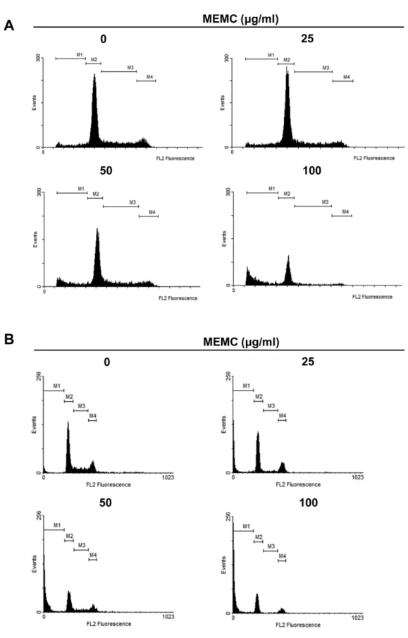
MEMC에 의한 세포주기 변화 분석
현재까지다양한암세포에서정상세포와는다르게세포주 기중 G1기및 G2기의 checkpoint 자체의변이또는이를조 절하는상류시그널의조절등에의하여비정상적인무한세 포증식이일어난다는것이보고되어있다[18, 29]. 또한이 러한암세포의특징을이용하여암세포의비정상적인세포 주기진행을저해하는항암소재의개발이활발하게진행되 고있다[34, 44]. 따라서본연구에서는 MEMC의암세포사 멸효과가세포주기에영향을주는지확인하기위하여 A549 와 HepG2를이용하여적정농도의 MEMC를 48시간처리한 다음세포주기변화를관찰하였다. 그결과, A549와 HepG2 공히세포주기조절에의한항암활성에서주로보이는 G1기
또는 G2/M기세포의증가는관찰되지않았으며, MEMC 처
리농도증가에따라 G1기, S기, G2/M기의세포비율이감소 하였다. 반면 MEMC 처리농도가증가할수록 apoptosis가 유발되었을것이라예상되는 subG1기의세포분포는증가하 였다(Fig. 2). Table 2에나타낸바와같이, A549의경우대 조군에서 9.92%였던 subG1기세포가최고농도인 100 µg/ml 처리군에서 44.07%로증가하였으며, HepG2의경우대조군 에서 4.33% subG1 세포가 100 µg/ml 처리군에서는 59.35%
로 증가하였다. 이와 같은 결과들은 MEMC가 A549와 HepG2의 apoptosis를유도하여세포증식을억제시키는가 능성을제시한다.
MEMC에 의한 Apoptosis 유도
세포에 apoptosis가유발되면세포막의유동성이증가되
며, 막인지질의비대칭성이소실되어지질이중막의내부에 Table 1. DPPH radical scavenging activity by methanol
extract of Machaerium cuspidatum.
Concentration (µg/ml)
Inhibition rate (%)
IC50 (µg/ml) MEMC
0.51 26.01 ± 1.45
1.66 2.56 68.59 ± 0.58
12.8 97.17 ± 0.07 Ascorbic acid
0.51 24.60 ± 0.24
1.23 2.56 97.31 ± 0.08
12.8 97.77 ± 0.06
존재하는포스파티딜세린(phosphatidyl serine)이세포표면 으로노출된다[12, 30]. 따라서 apoptosis가일어난세포의분 석을위해서포스파티딜세린과특이적으로결합하는 Annexin
V를표식자로널리사용하고있다. 세포주기분석결과로부
터 MEMC 처리에의해 subG1기세포가증가하였으므로,
Annexin V/7-Aminoactinoycin D (7-AAD) 염색방법을사용 Fig. 1. Effect of MEMC on cell growth and morphological changes in various cancer cell lines. (A) Human lung carcinoma A549 cells, human hepatocellular carcinoma HepG2 cells and human colon carcinoma HT29 cells were treated with indicated concentration of MEMC for 48 h. Cytotoxic effect of MEMC was determined by WST assay. Results are expressed as percentage of the control ± SD of three independent experiments. *p < 0.05 and **p < 0.005 compared with the control. (B-D) Morphological changes by MEMC in A549 (B), HepG2 (C) and HT29 (D) cells. Cell morphology was visualized by light microscopy. Scale bars, 200 µm.
Fig. 2. Accumulation of subG1-phase cells by MEMC treatment. A549 and HepG2 cells were treated with indicated concentrations of MEMC for 48 h, stained with propidium iodide for 10 min and analyzed by flow cytometry. DNA-fluorescence histograms of A549 (A) and HepG2 (B) are shown. M1, subG1 phase; M2, G1 phase; M3, S phase; M4, G2/M phase.
하여 MEMC에의한 A549와 HepG2의 apoptosis 유도를정 량적으로분석하였다. 그결과, Fig. 3에서보여지듯이 A549
와 HepG2 모두 MEMC 농도가증가할수록살아있는세포
비율은감소하는반면, Annexin V 양성세포비율이증가하
였다. 또한 저농도의 MEMC 처리시 early apoptotic 세포 (Annexin V+/7-AAD−)가증가되었으며, 농도가증가할수록 점차 late apoptotic 세포(Annexin V+/7-AAD+)가증가되어, 최고농도 100 µg/ml에서 Annexin V 양성세포는 A549의 경우 46.7%, HepG2의경우는 77.5%까지증가하였다. 이러 한결과로부터 MEMC에의해 A549와 HepG2의 apoptosis 가유도되며이들세포의증식이억제되는것을알수있었다.
MEMC에 의한 세포핵의 형태변화 분석
세포에 apoptosis가유도되면세포의수축, linker DNA 절 단에의한 chromosome의단편화, 염색질의응축등에의한
apoptotic body와 같은 전형적인 형태변화가 나타나며,
apoptosis 유도를확인하기위하여이러한특징을관찰하는
것은매우중요하다[11, 45]. 특히 apoptosis 유도에의해형 성되는 apoptotic body는염색시약에의해관찰이가능하므 로, A549와 HepG2 세포에 MEMC를처리한다음, DAPI 염 색을통하여 MEMC에의한 apoptosis 유도를확인하였다. 그결과, Fig. 4에서보여지듯이 0.1% DMSO를처리한대조
군의 경우, 완전한 핵의 형태가 관찰되는 반면, A549와
HepG2에서 공히 MEMC를 처리한 경우 핵이 응축된
apoptotic body가관찰되었다. 또한 MEMC 처리농도가증 가할수록 apoptotic body의형성도증가하여 apoptosis가일 어난세포의전형적인특징을관찰할수있었다.
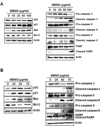
MEMC에 의한 apoptosis 유도의 분자 메커니즘
다음으로 MEMC에의한 A549 및 HepG2의 apoptosis 유
도기전분석을위하여 apoptosis 관련단백질의발현변화를
Western blot analysis로확인하였다. p53은 death receptor
(Fas, DR5)의발현을유도하여개시 caspase인 caspase-8과 실행 caspase인 caspase-3의활성화를통한 apoptosis의외 인성경로에관여할뿐아니라, Bcl-2 family 단백질의발현 Fig. 3. Apoptosis induction by MEMC in A549 and HepG2 cells. A549 (A) and HepG2 (B) cells were treated with indicated concentrations of MEMC for 48 h, stained with MuseTM Annexin V & Dead Cell Kit for 20 min and analyzed by MuseTM Cell Ana- lyzer. Dot plots represented four independent sections. Live cells (lower left): Annexin V−/7-AAD−, dead cells (upper left): Annexin V−/7-AAD+, early apoptotic cells (lower right): Annexin V+/7- AAD−, late apoptotic cells (upper right): Annexin V+/7-AAD+. Table 2. Cell cycle distribution of methanol extract of Mach-
aerium cuspidatum.
Cell line
MEMC (µg/ml)
% of cells SubG1
(M1)
G1 (M2)
S (M3)
G2/M (M4)
A549
0 9.92 57.79 21.97 10.31
25 12.15 59.46 20.28 7.94
50 17.65 50.42 21.70 9.80
100 44.07 40.18 10.44 3.94
HepG2
0 4.33 52.04 18.76 17.80
25 24.12 46.45 7.16 18.82
50 42.78 30.49 10.32 11.64
100 59.35 27.08 2.04 8.99
을조절하여내인성경로에의한 apoptosis에도중요한역할 을담당하는것으로알려져있다[15, 19]. 특히 Bcl-2 family 단백질의경우, 정상세포에서는 pro-apoptotic 단백질(Bax, Bak, Bid 등)과 anti-apoptotic 단백질(Bcl-2, Bcl-XL 등)이
균형을이루고있으나, 외부 자극으로증가된 p53에의해 pro-apoptotic 분자의발현이증가되고반면 anti-apoptotic 분자의발현이저해되어이러한세포내균형이깨어지게된 다. 증가된 pro-apoptotic 분자는 homodimer를 형성하여 cytochrome c를방출시키고, cytochrome c/Apaf-1/caspase- 9를포함하는 apoptosome 복합체를형성하여 caspase-3 활 성화에 따른 apoptosis가유도된다[3, 14]. 또한 최근에는 p21의 upregulation이 apoptosis의내인성경로를촉진시킨
다는연구결과가보고되어있다[22, 36]. 이러한외인성및
내인성경로를통하여활성화된 caspase는 PARP와같은여 러가지기질을분해하여 apoptosis를유도한다[27, 43].
A549와 HepG2에 MEMC를 농도별로 처리한 다음
Western blot analysis를수행한결과, Fig. 5에서나타난바 와같이 MEMC의농도가증가할수록 A549와 HepG2에서 종양억제유전자 p53, p21 및 pro-apoptotic 분자인 Bax의발 현이증가하였으며, anti-apoptotic 분자인 Bcl-2의발현이감 소하였다. 또한 MEMC 처리에의해 pro-caspase들이활성 화되어 cleaved caspase-3, -8, -9가증가하였으며, PARP의 분해가관찰되었다. 이러한결과로부터 MEMC 처리에의해 Fig. 5. Effect of MEMC on the expression of apoptotic pro- teins in A549 and HepG2 cells. MEMC-treated A549 (A) and HepG2 (B) cells were harvested and then proteins were isolated.
The expression of apoptotic proteins was estimated by Western blot analysis. Actin was used as an internal control.
Fig. 4. Apoptotic morphological changes of A549 and HepG2 cells by MEMC. A549 (A) and HepG2 (B) cells were exposed to various concentrations of MEMC for 48 h. The cells were fixed and stained with DAPI for 20 min. Stained cells were observed by fluorescence microscopic analysis and imaged using Axio Vision Program. Arrows indicate the apoptotic bodies. Scale bars, 100µm.