ATP Modulation of ATP-sensitive Potassium Channel ATP Sensitivity Varies with the Type of SUR Subunit*
Received for publication, November 1, 2000, and in revised form, November 29, 2000 Published, JBC Papers in Press, December 13, 2000, DOI 10.1074/jbc.M009959200
Dae-Kyu Song‡ and Frances M. Ashcroft§
From the University Laboratory of Physiology, Parks Road, Oxford OX1 3PT, United Kingdom
ATP-sensitive potassium (KATP) channels comprise Kir and SUR subunits. Using recombinant KATP chan- nels expressed in Xenopus oocytes, we observed that MgATP (100M) block of Kir6.2/SUR2A currents gradu- ally declined with time, whereas inhibition of Kir6.2/
SUR1 or Kir6.2⌬C36 currents did not change. The de- cline in Kir6.2/SUR2A ATP sensitivity was not observed in Mg2ⴙfree solution and was blocked by the phosphati- dylinositol (PI) 3-kinase inhibitors LY 294002 (10 M) and wortmannin (100 M), and by neomycin (100 M).
These results suggest that a MgATP-dependent synthe- sis of membrane phospholipids produces a secondary decrease in the ATP sensitivity of Kir6.2/SUR2A. Direct application of the phospholipids PI 4,5-bisphosphate and PI 3,4,5-trisphosphate in the presence of 100 M MgATP activated all three types of channel, but the response was faster for Kir6.2/SUR2A. Chimeric studies indicate that the different responses of Kir6.2/SUR2A and Kir6.2/SUR1 are mediated by the first six transmem- brane domains of SUR. The MgATP-dependent loss of ATP sensitivity of Kir6.2/SUR2A was enhanced by the actin filament disrupter cytochalasin and blocked by phalloidin (which stabilizes the cytoskeleton). Phalloi- din did not block the effect of PI 3,4,5-trisphosphate.
This suggests that MgATP may cause disruption of the cytoskeleton, leading to enhanced membrane phospho- lipid levels (or better targeting to the KATPchannel) and thus to decreased channel ATP sensitivity.
ATP-sensitive potassium (KATP)1 channels are widely dis- tributed, being found in pancreatic B-cells, cardiac, smooth, and skeletal muscles, and neurones (1). They play important functional roles in all these tissues by linking cellular metab- olism to electrical activity. Opening of the KATPchannel pro- duces a voltage-independent K⫹ current that hyperpolarizes the cell and reduces its electrical excitability; conversely, KATP channel closure usually decreases membrane excitability.
The KATP channel is an octamer, formed by the physical association of four inwardly rectifying potassium channel sub-
units (Kir6.2) and four regulatory sulfonylurea receptor sub- units (2). KATPchannels in different tissues are composed of different Kir and SUR subunits. In most tissues, Kir6.2 acts as the pore-forming subunit (3, 4). Two different sulfonylurea receptor genes (SUR1 and SUR2) have been identified, and further diversity is created by alternative splicing of SUR2 (5–9). In this paper, the major isoform of SUR2 (6) is called SUR2A. It shares 67% sequence identity with SUR1. There is evidence that SUR1 serves as the regulatory subunit of the KATPchannel in pancreatic B-cells and some types of neurone (5, 10), whereas a similar role is played by SUR2A in cardiac and skeletal muscle (6).
Binding sites for drugs and modulatory agents are found on both Kir6.2 and SUR subunits. The nucleotides ATP and ADP bind to an intracellular site on Kir6.2 and bring about KATP channel closure (11, 12), whereas a range of magnesium nucle- otides (including ATP and ADP) interact with the NBDs of SUR and thereby increase channel activity (13–16). The balance between these inhibitory and stimulatory effects results in channel inhibition by most concentrations of MgATP and high concentrations of MgADP (10 mM) and in channel activation by low micromolar concentrations of MgADP. It is thought that metabolic regulation of KATP channel activity is mediated, in part, by changes in the intracellular concentrations of these nucleotides.
Recent studies indicate that membrane phospholipids also play an important role in modulating KATPchannel activity.
Thus, phosphatidylinositol 4,5-bisphosphate (PIP2) increases the activity and reduces the ATP sensitivity of both native and cloned KATP channels (17–21). Overexpression of PI5-kinase, which enhances PIP2levels, reduces the ATP sensitivity of the channel in membrane patches (21), whereas breakdown of PIP2 by phospholipase C enhances the KATPchannel ATP sensitivity (22). This is in agreement with data indicating that PIP2binds directly to Kir channels (23). Interestingly, application of PIP3 and PI 3-P also enhances KATPchannel activity (19, 20, 24).
The rundown of both native and cloned KATPchannels that occurs in excised membrane patches is reversed by application of MgATP (25–27), and PIP2has also been implicated in this effect (25). It is postulated that PIP2 is produced in plasma membrane by serial phosphorylation of PI and that this process is sequentially catalyzed by PI 4-kinase and PI 5-kinase.
MgATP generation of phospholipids, therefore, has multiple but related effects on KATPchannel activity. It decreases the rate of channel rundown, it causes reactivation of the channel after removal of ATP, and it reduces the channel ATP sensitivity.
In this paper, we demonstrate that the time course of KATP channel inhibition by ATP is markedly different for Kir6.2/
SUR1 and Kir6.2/SUR2A. Whereas inhibition of Kir6.2/SUR1 currents by 100 M ATP does not change with time, that of Kir6.2/SUR2A decreases during prolonged nucleotide applica-
* This work was supported by the Wellcome Trust. The costs of publication of this article were defrayed in part by the payment of page charges. This article must therefore be hereby marked “advertisement”
in accordance with 18 U.S.C. Section 1734 solely to indicate this fact.
‡ Permanent address: Dept. of Physiology, Keimyung University School of Medicine, 194 Dongsang Dong, Choong Gu, Taegu, 700-712 Korea.
§ To whom correspondence should be addressed. E-mail:
1The abbreviations used are: KATP, ATP-sensitive potassium; NBD, nucleotide-binding domain; PIP2, and PI(4,5)P2, phosphatidylinositol 4,5-bisphosphate; PIP3, phosphatidylinositol 3,4,5-trisphosphate; PI, phosphatidylinositol; PI(3,4)P2, phosphatidylinositol 3,4-bisphosphate;
PI(4)P, phosphatidylinositol 4-phosphate; TM, transmembrane domain;
PI(3)P, phosphatidylinositol 3-phosphate.
© 2001 by The American Society for Biochemistry and Molecular Biology, Inc. Printed in U.S.A.
This paper is available on line at http://www.jbc.org
7143
by guest on December 20, 2016http://www.jbc.org/Downloaded from
tion. This ATP-dependent activation of Kir6.2/SUR2A currents by MgATP can be prevented by 10M LY 294002, a specific inhibitor of PI 3-kinase (28), suggesting that it results from MgATP-dependent production of PIP3or PI(3,4)P2rather than PI(4,5)P2 (PIP2). Both PIP3 and PIP2, however, are able to promote channel activity in excised patches. Studies with chi- meric SUR further suggest that the different responses of Kir6.2/SUR1 and Kir6.2/SUR2A channels are conferred by the first set of transmembrane domains of the sulfonylurea receptor.
EXPERIMENTAL PROCEDURES
Molecular Biology—Mouse Kir6.2 (GenBankTM accession number D50581; Refs. 3 and 4), rat SUR1 (GenBankTM accession number L40624; Ref. 5), and SUR2A (GenBankTMaccession number D83598;
Ref. 6) cDNAs were cloned in the pBF vector. A truncated form of Kir6.2 (Kir6.2⌬C), which lacks the C-terminal 36 amino acids and forms func- tional channels in the absence of SUR, was prepared as described previously (11). Chimeras between SUR1 and SUR2A were constructed as described previously (29). Capped mRNA was prepared using the mMESSAGE mMACHINE large scale in vitro transcription kit (Am- bion, Austin, TX), as previously described (30).
Oocyte Collection—Female Xenopus laevis were anesthetized with MS222 (2 g/liter added to the water). One ovary was removed via a mini-laparotomy, the incision was sutured, and the animal was allowed to recover. Immature stage V and VI oocytes were incubated for 60 min with 1.0 mg/ml collagenase (Sigma, type V) and manually defollicu- lated. Oocytes were either injected with⬃1 ng of Kir6.2⌬C mRNA or coinjected with⬃0.1 ng of Kir6.2 mRNA and ⬃2 ng of mRNA encoding either SUR1 or SUR2A. The final injection volume was 50 nl/oocyte.
Isolated oocytes were maintained in Barth’s solution and studied 1– 4 days after injection (30).
Electrophysiology—Patch pipettes were pulled from borosilicate glass and had resistances of 250 –500 k⍀ when filled with pipette solution. Macroscopic currents were recorded from giant excised inside- out patches at a holding potential of 0 mV and at 20 –24 °C using an EPC7 patch-clamp amplifier (List Electronik, Darmstadt, Germany;
Ref. 30). The pipette (external) solution contained 140 mMKCl, 1.2 mM
MgCl2, 2.6 mMCaCl2, 10 mMHEPES (pH 7.4 with KOH). The intracel- lular (bath) solution contained 107 mMKCl, 2 mMMgCl2, 1 mMCaCl2, 10 mMEGTA, 10 mMHEPES (pH 7.2 with KOH; final [K⫹],⬃140 mM).
The magnesium-free intracellular solution contained 140 mMKCl, 1 mM
EGTA, 10 mMHEPES (pH 7.2 with KOH). LY 294002 (CalBiochem), phalloidin, and cytochalasin (CalBiochem) were dissolved in Me2SO to make 10 mMstock solutions. Stock solutions (1 mM) of PIP2and PIP3
were made in magnesium-free intracellular solution and diluted to the desired concentration and sonicated (30 min on ice) immediately before use. Rapid exchange of solutions was achieved by positioning the patch in the mouth of one of a series of adjacent inflow pipes placed in the
bath. Test solutions were applied in random order unless otherwise stated.
In most experiments, currents were recorded in response to repeti- tive 3-s voltage ramps from⫺110 mV to ⫹100 mV. They were filtered at 10 kHz, digitized at 0.4 kHz using a Digidata 1200 Interface, and analyzed using pClamp software (Axon Instruments, Foster City, CA).
Records were stored on videotape and resampled at 20 Hz for presen- tation in the figures. The slope conductance was measured by fitting a straight line to the current-voltage relation between⫺20 and ⫺100 mV;
the average response to five consecutive ramps was calculated in each solution. In other experiments, macroscopic currents were recorded at a fixed holding potential of⫺50 mV in response to nucleotide or drug applications. Currents were sampled at 20 Hz and analyzed using Microcal Origin software (Microcal Software, Northampton, MA). Data are presented as the means⫾ S.E.
RESULTS
Macroscopic currents were recorded in inside-out patches from Xenopus oocytes expressing either Kir6.2/SUR1 or Kir6.2/
SUR2A. Current amplitudes were similar for both types of KATPchannel.
Time Course of ATP Inhibition—Fig. 1A shows that applica- tion of 100 M ATP to the intracellular membrane surface initially inhibited both Kir6.2/SUR1 and Kir6.2/SUR2A cur- rents by ⬃90%. Inhibition of Kir6.2/SUR1 currents did not change, or even slightly increased, over the course of a 10-min exposure to ATP. In contrast, there was a gradual decline in the ATP sensitivity of Kir6.2/SUR2A currents with time; despite the continued presence of nucleotide, a slow increase in current was observed that began about 2 min after the onset of ATP application and stabilized 8 –10 min later at around 75% block.
This decrease in ATP sensitivity was not reversed by a 1-min exposure to nucleotide-free solution (Fig. 1A). Mean data are shown in Fig. 1B.
The extent of initial block of both types of KATPcurrent by 100MATP is in agreement with that previously published for Kir6.2/SUR1 and Kir6.2/SUR2A channels. When expressed in oocytes and measured immediately after patch excision, half- maximal inhibition (Ki) of Kir6.2/SUR1 and Kir6.2/SUR2A channels is produced by 28 and 29MATP, respectively (31).
The Kivalue for Kir6.2/SUR1 is in good agreement with that found when the channel is expressed in mammalian cells (8 – 47
M; Refs. 21 and 32) and with what is found for the native-cell KATPchannel (26M; Ref. 33). Reported values for half-maxi- mal inhibition of cloned Kir6.2/SUR2A channels and for native cardiac KATPchannels vary widely, from 17 to 100M(6, 31, FIG. 1. Time course of current response in the presence of 100MMgATP. A, macroscopic currents recorded from oocytes coexpressing Kir6.2 and either SUR1 or SUR2A in response to a series of voltage ramps from⫺110 to ⫹100 mV. ATP was added to the intracellular solution as indicated by the bars. All solutions contained Mg2⫹. B, mean KATPconductance recorded for Kir6.2⌬C36 (Œ, n ⫽ 3), Kir6.2/SUR1 (f, n ⫽ 6), or Kir6.2/SUR2A (●, n⫽ 7) currents at different times after the addition of 100MMgATP to the intracellular solution. The slope conductance (G) is expressed as a fraction of the mean (Gc) of that obtained in control solution before exposure to ATP.
by guest on December 20, 2016http://www.jbc.org/Downloaded from
34). It seems possible that this may reflect, at least in part, the time at which the measurements were made during exposure to ATP. It may also reflect differences in the concentration of endogenous membrane phospholipids.
To confirm that the difference in nucleotide response be- tween Kir6.2/SUR1 and Kir6.2/SUR2A currents was conferred by the SUR subunit, we also measured the effect of continued exposure to ATP on a truncated form of Kir6.2 (Kir6.2⌬C), expressed in absence of SUR. Fig. 1B shows that Kir6.2⌬C currents resemble Kir6.2/SUR1 currents and show a slow de- cline with time in the presence of ATP, despite the lower ATP sensitivity. These data argue that the time-dependent activa- tion of Kir6.2/SUR2A currents is conferred by the SUR2A subunit.
Effect of Walker A Mutation on the ATP Sensitivity of SUR 2A—It is possible that the time-dependent stimulatory effect of MgATP is mediated via the NBDs of SUR2A, either directly or via hydrolysis of MgATP to MgADP. It is known that MgADP is able to stimulate both Kir6.2/SUR1 (13–16, 31) and Kir6.2/
SUR2A (31). Mutation of the lysine residue in the Walker A motif of NBD2 (but not NBD1) of SUR2A abolishes this stim- ulatory effect (35). We therefore examined the effect of MgATP
on channels containing this mutation (Kir6.2/SUR2A-K2A).
Fig. 2 shows that initial ATP sensitivity of Kir6.2/SUR2A- K2A currents is slightly greater than that found for the wild type channel; the mean block by 100MATP was 97.7⫾ 0.6%
(n⫽ 6) compared with 87.6 ⫾ 4.3% (n ⫽ 7) for Kir6.2/SUR2A- K2A and Kir6.2/SUR2A, respectively. This is not unexpected because a similar increase in ATP sensitivity is found when the equivalent residue is mutated in SUR1 (14). As in the case of SUR1, therefore, it may result from loss of MgATP activation mediated via the NBDs of SUR. Despite the enhanced ATP sensitivity, however, Kir6.2/SUR2A-K2A currents showed a time-dependent activation in the presence of MgATP that re- sembled that found for wild type channels; it began 1–2 min after exposure to ATP, and the current amplitude increased 3-fold within 5 min. This decrease in ATP sensitivity did not occur in the presence of 100MLY 294002, an inhibitor of PI 3-kinase (see below); rather the currents declined with time (as was also observed for Kir6.2/SUR2A; Fig. 3B). These results therefore suggest that the MgATP-dependent decline in ATP sensitivity is not mediated via nucleotide interaction with the NBDs of SUR.
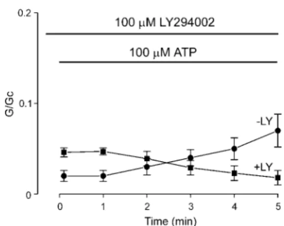
Mechanism of ATP-dependent Activation—Another mecha- nism by which the ATP sensitivity of the KATPchannel might be reduced is by the ATP-dependent generation of PIP2in the patch membrane caused by the action of endogenous lipid ki- nases (21). To test this hypothesis, we examined the effect of the lipid kinase inhibitor LY 294002 on the ATP sensitivity of Kir6.2/SUR2A currents (Fig. 3). LY 294002 is a potent and relatively specific inhibitor of PI 3-kinase with an IC50of 1.5– 4
M(27, 36). Thus, at a concentration of 10M, PI 3-kinase is totally inhibited, but there is little effect on PI 4-kinase, whereas at a concentration of 100 M, PI 4-kinase is also completely blocked (36). As shown in Fig. 3, 10MLY 294002 blocked the time-dependent decline in ATP sensitivity observed for Kir6.2/SUR2A currents and prevented the reduction in ATP sensitivity produced by preincubation with ATP (compare Figs.
1 and 3). It also slightly enhanced the ATP sensitivity of the channel. This suggests that production of PI(3)P, PI(3,4)P2, or PIP3 is required for the response. 100 M LY 294002 also blocked the ATP-dependent activation of Kir6.2/SUR2A cur- rents and further increased the extent of inhibition by 100M ATP. Wortmannin (100M, n⫽ 3), which also blocks PI 3-ki- nase (37), and neomycin (100M, n⫽ 3), which chelates phos- pholipids, produced an immediate decline in Kir6.2/SUR2A FIG. 2. Effects of an NBD mutation on the response to MgATP.
Mean conductance recorded for Kir6.2/SUR2A-K2A currents at differ- ent times after the addition of 100 MMgATP to the intracellular solution in the absence (●, n⫽ 6) or presence (f, n ⫽ 3) of 100MLY 294002. The slope conductance (G) is expressed as a fraction of the mean (Gc) of that obtained in control solution before exposure to ATP.
FIG. 3. Effects of LY 294002 on Kir6.2/SUR2A currents. A, macroscopic Kir6.2/SUR2A currents recorded in response to a series of voltage ramps from⫺110 to ⫹100 mV. ATP and LY 294002 (10M, above; 100M, below) were added to the intracellular solution as indicated by the bars.
All solutions contained Mg2⫹. B, mean conductance recorded for Kir6.2/SUR2A currents at different times after the addition of 100MMgATP to the intracellular solution in the presence of 10M(f, n⫽ 3) or 100M(Œ, n⫽ 3) LY 294002. The dashed line indicates the data obtained in control solution (as shown in Fig. 1B). The slope conductance (G) is expressed as a fraction of the mean (Gc) of that obtained in control solution before exposure to ATP. C, schematic illustrating the metabolism of phospholipids. The pathways inhibited by 10 and 100MLY 294002 are indicated.
by guest on December 20, 2016http://www.jbc.org/Downloaded from
channel activity and prevented the MgATP-dependent loss of ATP sensitivity (data not shown). As expected if a lipid kinase is involved, ATP was not effective at stimulating Kir6.2/SUR2A currents in the absence of Mg2⫹(data not shown).
Taken together, these results suggest that MgATP is used as a substrate by PI 3-kinase in the oocyte membrane to generate PIP3or other membrane lipids and that the gradual accumu- lation of PIP3causes a slow decline in the ATP sensitivity of Kir6.2/SUR2A currents. Because PI 4-kinase is not blocked by 10 M LY 294002, this activation cannot be mediated by PI(4,5)P2(Fig. 3C). Earlier studies have shown that KATPchan- nel is very sensitive to the level of PI 5-kinase activity (21), which favors the possibility that the effects of MgATP are mediated via production of PIP3 rather than PI(3)P or PI(3,4)P2. Although we cannot formally exclude a role for the latter two phospholipids, for simplicity, we will simply refer to PIP3(rather than PIP3,PI(3)P, or PI(3,4)P2) in the rest of this paper.
The time lag observed before ATP induces activation of Kir6.2/SUR2A currents may therefore reflect the time taken for sufficient PIP3to accumulate within the membrane to cause a measurable reduction in ATP sensitivity. Accumulation of PIP3 within the membrane may also explain why the ATP sensitivity of Kir6.2/SUR2A currents is not immediately re- stored on return to control solution. Clearly, if the lipid is not rapidly removed from the membrane, then subsequent ATP application will produce a smaller inhibitory response.
Mechanism of Action of PIP3—Although exogenously applied PIP3is known to modulate-cell KATPchannel activity (20, 38), it need not necessarily interact directly with the channel. It might also exert its effect indirectly, by modulating a protein that regulates the KATP channel. It is well established that PIP2and other membrane lipids influence the cell cytoskeleton by inhibiting the activity of actin-binding proteins that sever and cap actin filaments (39). Likewise, PIP3associates with the Rho family of GTPases (40), which are key regulators of actin filament structure (41). Agents that modulate the cytoskeleton also influence the activity of native KATPchannels in cardiac membranes in both the presence and absence of ATP (42, 43).
We therefore examined the effects of phalloidin, which stabi- lizes the cytoskeleton, and of cytochalasin, which disrupts the cytoskeleton, on the ATP-dependent activation of Kir6.2/
SUR2A currents. Cytochalasin (10M) increased the time-de- pendent current activation produced by 100MATP, whereas phalloidin (20M) diminished the extent of activation (Fig. 4A).
These results argue that the ATP-dependent activation of Kir6.2/SUR2A currents involves the cell cytoskeleton and that disruption of the cytoskeleton enhances the ATP-dependent decline in the channel ATP sensitivity.
The effect of cytochalasin might be mediated subsequent to PIP3production, or it might affect generation of the phospho- lipid. To distinguish between these possibilities, we examined whether LY 294002 was able to inhibit the response to cytocha- lasin; if cytochalasin influences PIP3 production, then LY 294002 should block the response whereas, conversely, if cy- tochalasin has an effect downstream of the phospholipid it should still be effective in the presence of LY 294002. As shown in Fig. 4 (A and B), both 10 and 100MLY 294002 completely abolished the response to cytochalasin. The simplest explana- tion of this result is that changes in the cell cytoskeleton do not modify the ATP sensitivity of Kir6.2/SUR2A currents directly.
Rather, disruption of the cytoskeleton enhances MgATP-de- pendent production of PIP3 (or its metabolites) or facilitates PIP3targeting to the KATPchannel and thereby enhances the loss of ATP sensitivity.
In contrast to Kir6.2/SUR2A channels, KATPchannels con-
taining the SUR1 subunit were not activated by cytochalasin in the presence of ATP (Fig. 4C). Thus, like the ATP-dependent activation itself, this response is specific to SUR2A.
Differential Sensitivity of Kir6.2/SUR1 and Kir6.2/SUR2A Currents to PIP3—It is striking that whereas Kir6.2/SUR2A currents show an ATP-dependent activation that appears to be mediated by PIP3production, this is not the case for Kir6.2/
SUR1 currents. In some patches, however, we observed a slow, time-dependent activation of Kir6.2/SUR1 currents when ex- posed to 1 mMATP (data not shown). This suggests that Kir6.2/
SUR1 channels may simply be less sensitive to PIP3 than Kir6.2/SUR2A channels.
We therefore tested the effect of direct application of PIP2or PIP3on Kir6.2/SUR1 and Kir6.2/SUR2A currents in the pres- ence of 100MATP. Fig. 5 (A and B) shows that (in the absence of Mg2⫹) PIP3 produces a time-dependent decline in the ATP sensitivity of both types of channel but that this effect was more rapid and pronounced in the case of Kir6.2/SUR2A than Kir6.2/SUR1. The magnitude and time course of the PIP3ac- tivation of Kir6.2⌬C36 was similar to that observed for Kir6.2/
SUR1 (Fig. 5B), providing additional support for the idea that FIG. 4. Effects of cytoskeletal agents. A, mean KATPconductance recorded for Kir6.2/SUR2A currents at different times after the addi- tion of 100MMgATP to the intracellular solution in the presence of 10
Mcytochalasin (●, n⫽ 4) or 20Mphalloidin (f, n⫽ 3) or of 10M cytochalasin plus 10MLY 294002 (E, n⫽ 3). The slope conductance (G) is expressed as a fraction of the mean (Gc) of that obtained in control solution before exposure to ATP. The dashed line indicates the control response in the absence of either agent. B, mean conductance recorded for Kir6.2/SUR2A currents at ⫺50 mV at different times after the addition of 100MMgATP (E, n⫽ 5), 100MMgATP plus 10M cytochalasin (●, n⫽ 4), 100MMgATP and 10Mcytochalasin plus 10
M(f, n⫽ 5), or 100MLY 294002 (Œ, n⫽ 5). The dashed line indicates the initial current level. The conductance (G) is expressed as a fraction of the mean (Gc) of that obtained at⫺50 mV in control solution before exposure to ATP. The patch was held at ⫺50 mV throughout the experiment. C, mean conductance recorded for Kir6.2/SUR1 currents at different times after the addition of 100MMgATP to the intracellular solution in presence of 10 Mcytochalasin (n⫽ 3). The dashed line indicates the response in the absence of cytochalasin. The slope con- ductance (G) was measured using a ramp protocol and is expressed as a fraction of the mean (Gc) of that obtained in control solution before exposure to ATP.
by guest on December 20, 2016http://www.jbc.org/Downloaded from
the differential action of the phospholipid on Kir6.2/SUR1 and Kir6.2/SUR2A is mediated by the SUR2A subunit. Similar results were found with PIP2(n⫽ 2–3 in each case, date not shown). Because the effects of PIP3were tested in the absence of Mg2⫹, we can be certain that current activation is the result of the phospholipid rather than ATP itself. It has previously been reported that the ATP sensitivity of both types of channel is reduced by PIP2in excised patches (18 –20, 22, 44), but the difference in sensitivity has not been remarked.
Fig. 5C shows that neither LY 294002 nor phalloidin is able to block the effect of PIP3on Kir6.2/SUR2A currents. Similar results were found with PIP2 (data not shown). The lack of effect of phalloidin is consistent with the idea that stabilization of the cell cytoskeleton is involved in the MgATP-dependent generation of phospholipids rather than directly affecting the KATPchannel. The data also indicate that PIP2does not have to be metabolized to PIP3to mediate its effect, because its effect was not blocked by LY 294002.
Molecular Basis of the Differential Sensitivity of Kir6.2/
SUR1 and Kir6.2/SUR2A Currents to Phospholipids—To de- termine the molecular basis of the differential sensitivity of Kir6.2/SUR1 and Kir6.2/SUR2A currents to phospholipids, we made chimeras between SUR1 and SUR2A and investigated the ability of 100 M MgATP to produce a time-dependent activation of KATPchannels containing chimeric SUR. These results are summarized in Fig. 6. SUR1 containing both NBDs, or just the last 42 amino acids of SUR2A, behaved like SUR1.
Likewise, SUR2A containing the last 42 amino acids, or NBD2, of SUR1 behaved like the parent channel. When the first six TMs of SUR1 were transferred into SUR2A, however, the abil- ity of MgATP to stimulate KATPchannel activity was abolished (Fig. 6A). Thus, it appears that this region of SUR is critical for the different response of Kir6.2/SUR1 and Kir6.2/SUR2A chan- nels to MgATP.
DISCUSSION
Our results suggest that the gradual loss of ATP sensitivity of Kir6.2/SUR2A currents is due to a MgATP-dependent syn- thesis of membrane phospholipids, which causes a secondary decrease in the channel ATP sensitivity. This hypothesis is supported by the facts that LY 294002, wortmannin, neomycin,
and Mg2⫹-free solutions prevent this effect and that the effect of MgATP is mimicked by the phospholipids PIP2and PIP3.
In the cell membrane, PI 4-kinase phosphorylates PI to give PI(4)P, which is subsequently phosphorylated by PI 5-kinase to FIG. 5. Time course of current re-
sponse in the presence of 5MPIP3
and 100MMgATP. A, macroscopic cur- rents recorded from oocytes coexpressing Kir6.2 and either SUR2A (above) or SUR1 (below) in response to a series of voltage ramps from⫺110 to ⫹100 mV. ATP and PIP3were added to the intracellular solu- tion as indicated by the bars. All solutions were Mg2⫹-free. B, mean KATPconduct- ance recorded for Kir6.2⌬C36 (Œ, n ⫽ 3);
Kir6.2/SUR1 (f, n⫽ 3) or Kir6.2/SUR2A (●, n⫽ 3) currents at different times after the addition of 5MPIP3and 100MATP to the intracellular solution. The slope conductance (G) is expressed as a fraction of the mean (Gc) of that obtained in con- trol solution before exposure to ATP. All solutions were Mg2⫹-free. C, mean con- ductance recorded for Kir6.2/SUR2A cur- rents at different times after the addition of 5MPIP3and 100MATP to the in- tracellular solution in the presence of 20
Mphalloidin (●, n ⫽ 3) or 10 M LY 294002 (f, n⫽ 3). The dashed line indi- cates the response in the absence of either agent. The slope conductance (G) is ex- pressed as a fraction of the mean (Gc) of that obtained in control solution before exposure to ATP.
FIG. 6. Effects of chimeras. A, mean conductance recorded from an inside-out patch excised from an oocyte coexpressing Kir6.2 and SUR- 215, at different times after the addition of 100MMgATP to the intracellular solution. The slope conductance (G) is expressed as a fraction of the mean (Gc) of that obtained in control solution before exposure to ATP. The dashed lines indicate the response of Kir6.2/SUR1 or Kir6.2/SUR2A currents. B, macroscopic conductance recorded from patches excised from oocytes coexpressing Kir6.2 and either SUR1, SUR2A, or the SUR chimera indicated, after 5 min of exposure to MgATP. Mean conductance in the presence of the test solution (G) is expressed relative to the mean conductance in the control (nucleotide- free) solution before nucleotide addition (Gc). The number of patches is given by each bar.
by guest on December 20, 2016http://www.jbc.org/Downloaded from
PIP2 (PI(4,5)P2). All three phospholipids are also phosphoryl- ated by PI 3-kinase to produce PI(3)P, PI(3,4)P2, and PIP3 respectively (Fig. 3C). Biochemical studies have shown that 10
MLY 294002 specifically inhibits PI 3-kinase and is without effect on PI 4-kinase but that at a concentration of 100M, LY 294002 also totally blocks PI 4-kinase (36). Because 10MLY 294002 abolished the magnesium-dependent loss of ATP sen- sitivity, we conclude that either PI(3)P, PI(3,4)P2, and/or PIP3 is involved the response. At this concentration, LY 294002 does not prevent PI(4)P or PI(4,5)P2formation, suggesting that nei- ther of these lipids is responsible for the magnesium-dependent decrease in ATP sensitivity.
When applied directly to the membrane patch, both PIP2and PIP3were able to reduce the ATP sensitivity of Kir6.2/SUR2A, although no MgATP-dependent activation of the channel was observed in the presence of 10 M LY 294002. This suggests that PIP3is a more potent regulator of the channel and that, in the presence of LY 294002, PIP2 generated by addition of MgATP does not accumulate to a concentration sufficient to reduce the channel ATP sensitivity. This may be due to the action of endogenous phospholipase C or lipid phosphatases. It is also worth pointing out that because many proteins bind PIPs, they may influence KATPchannel activity simply by se- questering the amount of PIPs available for interaction with the channel.
Mechanism of Action of PIP3—Our results suggest that the effect of PIP3on the ATP sensitivity of the KATPchannel is not mediated via the cell cytoskeleton, because it was not blocked by phalloidin. We did observe, however, that agents that per- turb the cytoskeleton have a marked effect on the MgATP-de- pendent decline in ATP sensitivity. Notably, disruption of actin microfilaments by cytochalasin enhanced the response, whereas stabilization of microfilaments by phalloidin de- creased the response. It is possible that the effect of cytochala- sin may be mediated upstream of PI 3-kinase because it can be blocked by 10MLY 294002. This might suggest that disrup- tion of the cytoskeleton enhances PIP3 levels (or targeting to the KATP channel), whereas, conversely, stabilization of the cytoskeleton by phalloidin reduces PIP3 levels. Furthermore, the ability of phalloidin to inhibit the ATP-dependent loss of ATP sensitivity suggests that MgATP may mediate its effect by inducing cytoskeletal disruption. This idea is supported by the fact that phalloidin did not prevent the loss of ATP sensitivity produced by direct application of PIP3to the intracellular mem- brane surface. In contrast, the stimulatory action of PIP3 on native-cell KATPchannels is prevented by phalloidin (24, 45).
Presumably, this difference reflects the different cell types, which may metabolize PIP3differently.
Interestingly, cytochalasin reduced the activity of native car- diac KATPchannels in the absence of ATP (42) but enhanced it when ATP was present (43). It seems plausible that the latter effect is due to an increase in MgATP-mediated PIP3genera- tion, as in the case for the cloned channel Kir6.2/SUR2A. A different mechanism may underlie the action of cytochalasin in the absence of ATP, which promotes KATP channel rundown (42).
Differential Sensitivity of Kir6.2/SUR1 and Kir6.2/SUR2A Channels—In contrast to Kir6.2/SUR2A currents, Kir6.2/SUR1 currents were not activated by 100MMgATP. Moreover, when PIP3was added directly to the patch in the presence of 100M MgATP, the phospholipid produced a much faster and more marked activation of Kir6.2/SUR2A than of Kir6.2/SUR1. Our results indicate that these different responses are mediated by the first set of transmembrane domains of SUR (TMs 1– 6). It is noteworthy that this region of SUR confers the different gating kinetics of Kir6.2/SUR1 and Kir6.2/SUR2A channels (46, 47).
This region of SUR may therefore be involved in transducing conformational changes in SUR into gating of the channel pore.
Both Kir6.2/SUR1 and Kir6.2⌬C channels have shorter bursts of openings and a lower open probability than Kir6.2/
SUR2A channels. Loussouarn and colleagues (48) have shown that there is an exponential correlation between the burst duration (or open probability) of the KATPchannel and its Kifor ATP inhibition, and it has been suggested that PIP2mediates its effect on KATPchannel ATP sensitivity, at least in part, via changes in the channel open probability (49). Whether or not a given increase in open probability (Po) results in a change in ATP sensitivity will therefore depend on the initial Po. Because this is lower for Kir6.2/SUR1 and Kir6.2⌬C currents, the like- lihood of observing a change in ATP sensitivity will be less.
It is therefore possible that the different responses of Kir6.2/
SUR2A and Kir6.2/SUR1 are due to their different single- channel kinetics; the higher Po of Kir6.2/SUR2A channels means that a further increase in Poproduced by PIP2is more likely to reduce the channel ATP sensitivity than in the case of Kir6.2/SUR1 channels. An alternative interpretation of our results, however, is that PIP3is able to interact directly with the SUR2A subunit of the KATPchannel to decrease the chan- nel ATP sensitivity (in addition to its effect on Kir6.2) and that the functional effect of this interaction involves the first six TMs of SUR.
Acknowledgments—We thank Phillippa Jones for expert technical assistance and Colin Nichols and Cathy Cukras for advice on the use of phospholipids.
REFERENCES
1. Ashcroft, F. M., and Ashcroft, S. J. H. (1990) Cell. Signal. 2, 197–214 2. Aguilar-Bryan, L., and Bryan, J. (1999) Endocr. Rev. 20, 101–135
3. Inagaki, N., Gonoi, T., Clement, J. P. IV., Namba, N., Inazawa, J., Gonzalez, G., Aguilar-Bryan, L., Seino, S., and Bryan, J. (1995) Science 270, 1166 –1170
4. Sakura, H., A¨ mma¨la¨, C., Smith, P. A., Gribble, F. M., and Ashcroft, F. M.
(1995) FEBS Lett. 377, 338 –344
5. Aguilar-Bryan, L., Nichols, C. G., Wechsler, S. W., Clement, J. P. IV., Boyd, A. E., Gonzalez, G., Herrera-Sosa, H., Nguy, K., Bryan, J., and Nelson, D. A.
(1995) Science 268, 423– 425
6. Inagaki, N., Gonoi, T., Clement, J. P., IV, Wang, C. Z., Aguilar-Bryan, L., Bryan, J., and Seino, S. (1996) Neuron 16, 1011–1017
7. Isomoto, S., Kondo, C., Yamada, M., Matsumoto, S., Higashiguchi, O., Horio, Y., Matsuzawa, Y., and Kurachi, Y. (1996) J. Biol. Chem. 271, 24321–24324 8. Chutkow, W. A., Simon, M. C., Le, Beau, M. M., and Burant, C. F. (1996)
Diabetes 45, 1439 –1445
9. Chutkow, W. A., Makielski, J. C., Melson, D. J., Burant, C. F., and Fan, Z.
(1996) J. Biol. Chem. 274, 13656 –13665
10. Liss, B., Bruns, R., and Roeper, J. (1999) EMBO J. 18, 833– 846
11. Tucker, S. J., Gribble, F. M., Zhao, C., Trapp, S., and Ashcroft, F. M. (1997) Nature 387, 179 –183
12. Tanabe, K., Tucker, S. J., Matsuo, M., Proks, P., Ashcroft, F. M., Seino, S., Amachi, T., and Ueda, K. (1999) J. Biol. Chem. 274, 3931–3933 13. Nichols, C. G., Shyng, S. L., Nestorowicz, A., Glaser, B., Clement, J. P. IV.,
Gonzalez, G., Aguilar-Bryan, L., Permutt, M. A., and Bryan, J. (1996) Science 272, 1785–1787
14. Gribble, F. M., Tucker, S. J., and Ashcroft, F. M. (1997) EMBO J. 16, 1145–1152
15. Gribble, F. M., Tucker, S. J., Haug, T., and Ashcroft, F. M. (1998) Proc. Natl.
Acad. Sci. U. S. A. 95, 7185–7190
16. Shyng, S., Ferrigni, T., and Nichols, C. G. (1997) J. Gen. Physiol. 110, 643– 654 17. Hilgeman, D. W., and Ball, R. (1996) Science 273, 956 –959
18. Fan, Z., and Makielski, J. C. (1997) J. Biol. Chem. 272, 5388 –5395 19. Baukrowitz, T., Schulte, U., Oliver, D., Herlitze, S., Krauter, T., Tucker, S. J.,
Ruppersberg, J. P., and Fakler, B. (1998) Science 282, 1141–1144 20. Shyng, S. L., and Nichols, C. G. (1998) Science 282, 1138 –1141
21. Shyng, S. L., Barbieri, A., Gumusboga, A., Cukras, C., Pike, L., Davis, J. N., Stahl, P. D., and Nichols, C. G. (2000) Proc. Natl. Acad. Sci. U. S. A. 97, 937–941
22. Xie, L. H., Horie, M., and Takano, M. (1999) Proc. Natl. Acad. Sci. U. S. A. 96, 15292–15297
23. Huang, C. L., Feng, S., and Hilgemann, D. W. (1998) Nature 391, 803– 806 24. Harvey, J., McKay, N. G., Walker, K. S., Kay, J. V. D., Downes, C. P., and
Ashford, M. L. J. (2000) J. Biol. Chem. 275, 4660 – 4669
25. Xie, L. H., Takano, M., Kakei, M., Okamura, M., and Noma, A. (1999) J. Physiol. (Lond.) 514, 655– 665
26. Findlay, I., and Dunne, M. J. (1986) Pfluegers Arch. Eur. J. Physiol. 407, 238 –240
27. Ohno-Shosaku, T., Zunkler, B. J., and Trube, G. (1987) Pfluegers Arch. Eur. J.
Physiol. 408, 133–138
28. Vlahos, C. J., Matter, W. F., Hui, K. Y., and Brown, R. F. (1994) J. Biol. Chem.
by guest on December 20, 2016http://www.jbc.org/Downloaded from
269, 5241–5248
29. Ashfield, R., Gribble, F. M., Ashcroft, S. J. H., and Ashcroft, F. M. (1999) Diabetes 48, 1341–1347
30. Gribble, F. M., Ashfield, R., A¨ mma¨la¨, C., and Ashcroft, F. M. (1997) J. Physiol.
(Lond.) 498, 87–98
31. Gribble, F. M., Tucker, S. J., Seino, S., and Ashcroft, F. M. (1998) Diabetes 47, 1412–1418
32. Aguilar-Bryan, L., Clement, J. P. I. V., Gonzalez, G., Kunjilwar, K., Babenko, A., and Bryan, J. (1998) Physiol. Rev. 78, 227–245
33. Ashcroft, F. M., and Kakei, M. (1989) J. Physiol. (Lond.) 416, 349 –367 34. Babenko, A. P., Gonzalez, G., Aguilar-Bryan, L., and Bryan, J. (1998) Circ. Res.
83, 1132–1143
35. Gribble, F. M., Reimann, F., Ashfield, R., and Ashcroft, F. M. (2000) Mol.
Pharmacol. 57, 1256 –1261
36. Rosado, J. A., and Sage, S. O. (2000) J. Biol. Chem. 275, 9110 –9113 37. Powis, G., Bonjouklian, R., Berggren, M. M., Gallegos, A., Abraham, R., Ash-
endel, C., Zalkow, L., Matter, W. F., Dodge, J., and Grindey, G. (1994) Cancer Res. 54, 2419 –2423
38. Harvey, J., Hardy, S. C., Irving, I., and Ashford, M. L. J. (2000) J. Physiol.
(Lond.) 527, 95–107
39. Jammey, P. A. (1995) Chem. Biol. 2, 61– 65
40. Missy, K., Van, Poucke, V., Raynal, P., Viala, C., Mauco, G., Plantavid, M., Chap, H., and Payrastre, B. (1998) J. Biol. Chem. 273, 30279 –30286 41. Settleman, J. (2000) Nat. Cell Biol. 2, E7–E9
42. Furukawa, T., Yamane, T., Terai, T., Katayama, Y., and Hiraoka, M. (1996) Pfluegers Arch. Eur. J. Physiol. 431, 504 –512
43. Terzic, A., Jahangir, A., and Kurachi, Y. (1995) Am. J. Physiol. 269, C525–C545
44. Fan, Z., and Makielski, J. C. (1999) J. Gen. Physiol. 114, 251–269 45. Harvey, J., and Ashford, M. L. (1998) J. Physiol. (Lond.) 511, 695–706 46. Babenko, A. P., Gonzalez, G., and Bryan, J. (1999) J. Biol. Chem. 274,
11587–11592
47. Proks, P., Ashfield, R., and Ashcroft, F. M. (1999) J. Biol. Chem. 274, 25393–25397
48. Loussouarn, G., Makhina, E. N., Rose, T., and Nichols, C. G. (2000) J. Biol.
Chem. 275, 1137–1144
49. Enkvetchakul, D., Loussouarn, G., Makhina, E., Shyng, S. L., and Nichols, C. G. (2000) Biophys. J. 78, 2334 –2348
by guest on December 20, 2016http://www.jbc.org/Downloaded from
Dae-Kyu Song and Frances M. Ashcroft the Type of SUR Subunit
ATP Modulation of ATP-sensitive Potassium Channel ATP Sensitivity Varies with
doi: 10.1074/jbc.M009959200 originally published online December 13, 2000 2001, 276:7143-7149.
J. Biol. Chem.
10.1074/jbc.M009959200 Access the most updated version of this article at doi:
Alerts:
When a correction for this article is posted
•
When this article is cited
•
to choose from all of JBC's e-mail alerts Click here
http://www.jbc.org/content/276/10/7143.full.html#ref-list-1
This article cites 49 references, 30 of which can be accessed free at
by guest on December 20, 2016http://www.jbc.org/Downloaded from