125
Open Access
Left Ventricular Function in Children and Adolescents With Type 1 Diabetes Mellitus
Eun Ha Kim, MD and Yeo Hyang Kim, MD
Department of Pediatrics, Keimyung University School of Medicine, Daegu, Korea ABSTRACT
Background and Objectives: Adult studies have reported that patients with diabetes mellitus (DM) show ultra- structural and functional myocardial deterioration. The aim of this study was to assess whether cardiac functional deterioration can be detected in pediatric patients with type I DM and whether or not a relatively short duration of DM and hyperglycemia influences cardiac function. Subjects and Methods: Forty-seven children and adoles- cents with DM and 38 healthy subjects (control group) were enrolled. Glycosylated hemoglobin (HbA1c), DM- induced complications, and left ventricular (LV) function as assessed using conventional and unconventional echo- cardiography {tissue Doppler imaging (TDI) and vector velocity imaging (VVI)} were evaluated. Results: The con- ventional echocardiographic parameters, with the exception of early peak mitral inflow velocity, the findings of pulsed wave TDI at the annular level, and regional ventricular function by VVI, were not significantly different between the two groups. Using the conventional and unconventional indices of systolic and diastolic function, no significant relationship was found between the duration of DM and the echocardiographic parameters. The de- celeration time (DT) and E’/A’ had an inverse correlation with HbA1c (p=0.042 and p=0.016, respectively).
Conclusion: Patients with DM in childhood and early adolescence rarely have insight on the significance of DM, and their diet is difficult to control. An alteration of myocardial function induced by DM may begin earlier than generally thought, and these changes are accelerated when glycemic control is poor. We recommend the early institution of close observation of patients with diabetes for alterations in cardiac function, in addition to other diabetic complications. (Korean Circ J 2010;40:125-130)
KEY WORDS:Child; Diabetes mellitus; Hyperglycemia; Ventricular function.
Introduction
Studies in adults have reported that patients with dia- betes mellitus (DM) show ultrastructural and function- al deterioration of the myocardium.1)2) These patients are susceptible to heart failure and have a higher pre- valence of coronary heart disease, hypertension, and car- diomyopathy.3-6)
Deterioration of left ventricular (LV) function in pa- tients with type 1 DM can exist in the absence of other combined cardiac problems.4)
Echocardiography can be used for the diagnosis of diabetic cardiomyopathy or diabetes-induced myocar- dial dysfunction. In adults with type 1 DM, numerous studies have reported controversial results. Several in- vestigators have shown normal LV systolic function at rest and abnormal contractile responses during exer- cise.2)7) With respect to LV diastolic function, several studies have reported an abnormal pattern,8)9) whereas another study found no evidence of dysfunction at rest.10) However, no studies have been conducted involving pa- tients of a younger age with a shorter duration of DM and hyperglycemia.
The aim of this study was to assess whether or not cardiac functional deterioration can be detected in pe- diatric patients with type I DM and whether or not DM of a relatively short duration and hyperglycemia influence cardiac function.
Received: June 3, 2009 Revision Received: July 9, 2009 Accepted: August 4, 2009
Correspondence: Yeo Hyang Kim, MD, Department of Pediatrics, Kei- myung University School of Medicine, 197 Dongsan-dong, Jung-gu, Daegu 700-712, Korea
Tel: 82-53-250-7524, Fax: 82-53-250-7783 E-mail: [email protected]
○ ○
○ cc This is an Open Access article distributed under the terms of the Creative Commons Attribution Non-Commercial License (http://creativecommons.
org/licenses/by-nc/3.0) which permits unrestricted non-commercial use, distribution, and reproduction in any medium, provided the original work is properly cited.
Subjects and Methods
Study population
We studied 38 children and adolescents who parti- cipated in the 2007 Diabetes Summer Education Camp in Daegu and 9 pediatric patients who were attending the Diabetes Clinic for Children of the Keimyung Uni- versity Dongsan Medical Center. All patients were diag- nosed with type I DM and received insulin therapy only.
The patients consisted of 16 boys and 31 girls, with a mean age of 12.8 years (range, 7.1-16.8 years). The mean duration of disease was 3.4 years (range, 0.3-7.7 years).
None of the patients had definite DM-induced compli- cations, including renal impairment, retinopathy, neu- ropathy, and clinical symptoms of cardiac dysfunction or cardiomyopathy. The conventional laboratory che- mistry results were normal, except for the glycosylated hemoglobin (HbA1c) level. The control group consist- ed of 38 children (18 boys and 20 girls), with a mean age of 11 years (range, 6.9-17.1 years). They were referred to us with a chief complaint of heart murmurs or chest pain, and did not show abnormal findings on labora- tory testing, electrocardiograms, or echocardiography.
Echocardiography
An echocardiography was performed using an Acu- son Sequire 512 (Siemens Medical Solution, Mountain View, CA, USA) with a 4 or 8 MHz transducer. All of the tests which were performed were saved on a hard drive and magneto-optical (MO) disk of echocardiogra- phic equipment. On the parasternal long axis view, with the use of M-mode and conventional echocardiography, several parameters, including fractional shortening (FS), ejection fraction (EF), LV dimension, and volume per surface area, were measured.
For diastolic functional analysis, the mitral inflow sig- nal was acquired in an apical four-chamber view. The pulsed Doppler sample volume was 2 mm and it was placed at the mitral valve tip. The early peak flow velo- city (E) and atrial filling velocity (A) were measured three times and averaged, and the E/A ratio was calcu- lated. The time elapsed until the maximal E reached the baseline {the early filling deceleration time (DT)}, and the time from aortic valve closure to mitral valve opening {the isovolumic relaxation time (IVRT)}, were measured.
Tissue Doppler imaging (TDI) measurements were performed on the basal septum and mitral annulus.
The maximal systolic myocardial velocity (S’) and early and late diastolic myocardial velocity (E’, A’) were meas- ured. In this study, the sample volume was set at 3 mm.
The time between mitral valve closure and opening (a’) and the systolic time (b’) were measured. The value of (a’-b’) was divided by b’ and the modified myocardial performance index (modified Tei index) was obtained.
To analyze the vector velocity imaging (VVI), an ul-
trasound window of the images was controlled in such a manner that the number of frames should be >70 frames per single cardiac beat on an apical 4-chamber view and 2D images were acoustically captured. Secured images were saved on a MO disk for off-line analysis.
The contour of the endocardium was manually traced before the analysis was performed on the saved images with the use of an offline analysis program {Syngo US Workplace 3.0 (Simens Medical Solution, Mountain View, CA, USA)}. The tracing was performed several times to achieve the most appropriate endocardial con- tour, which was confirmed by the visual display mode of the velocity images. In the septum and lateral wall of the LV, the basal and middle parts were subjected to the measurement of maximal velocity, strain, strain rate (SR), and displacement.
All of the echocardiographic parameters obtained for pulsed wave Doppler and TDI were measured by a sin- gle investigator from three cardiac cycles and averaged.
The parameters which were obtained from the VVI were the average value of the measurement performed twice.
Statistical analysis
All of the results which were measured using echocar- diography were expressed as the mean±SD. Statistical analysis was performed using Statistical Package for the Social Sciences (SPSS) for Windows, version 14.0 (SPSS Inc., Chicago, IL, USA). A comparison of echocardio- graphic measurements in the septum and lateral wall of the LV between the patient and control groups was made with the use of an independent t-test. The correlation between echocardiographic parameters and the dura- tion of DM or the HbA1c level was assessed using cor- relation analysis. A p<0.05 was considered statistically significant.
Results
Patients
The clinical data of the patients and controls are listed
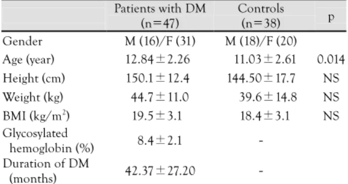
Table 1. Clinical characteristics of patients with diabetes melli- tus and controls in the study groups
Patients with DM (n=47)
Controls (n=38) p Gender M (16)/F (31) M (18)/F (20) Age (year) 12.84±2.26 11.03±2.61 0.014 Height (cm) 150.1±12.4 144.50±17.7 NS Weight (kg) 44.7±11.0 39.6±14.8 NS BMI (kg/m2) 19.5±3.1 18.4±3.1 NS Glycosylated
hemoglobin (%) 8.4±2.1 - Duration of DM
(months) 42.37±27.20 -
Continuous variables are expressed as the mean±standard deviat- ion. DM: diabetes mellitus, M: male, F: female, BMI: body mass in- dex, NS: non-specific
in Table 1. The age of patients was higher than the con- trols (p=0.014). The number of patients with a dura- tion of disease <36 months was 19 and the number of patients with a duration of disease >48 months was 18.
Analysis of the cardiac function using 2-dimensional and Doppler methods
The data derived from conventional echocardiogra- phic analysis are listed in Table 2. The E velocity of the patients was significantly lower than that of controls (p=0.028). However, the other conventional echocar- diographic parameters in the patient group, including mitral flow patterns, were not significantly different from
the controls. Although the findings of pulsed wave TDI at the annular level showed a lower S’, E’, and E’/A’, and a higher Tei index in the patient group, these differences were not significant (Table 3).
An analysis of the cardiac function using vector velocity imaging
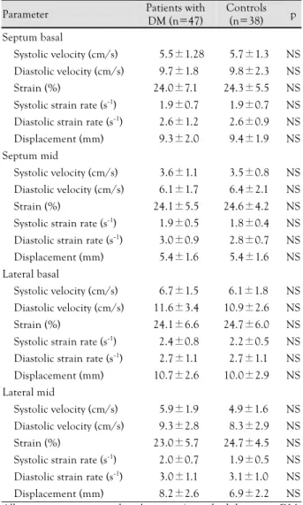
The regional myocardial velocity, strain, systolic and diastolic SR, and displacement findings evaluated at the basal and mid-septal, and basal and mid-lateral wall levels are listed in Table 4. These parameters also did not show significant differences between the patient and control groups.
Relationship between duration of diabetes mellitus and echocardiographic parameters
Characteristic and conventional echocardiographic data according to duration of DM are listed in Table 5.
The group with a duration of DM >4 years had a higher HbA1c than the group with a duration of DM <3 years (p=0.01). The group with a duration of DM >4 years
Table 2. Echocardiographic parameters derived from two-di- mensional and Doppler imaging in the study groups
Parameter Patients with DM (n =47)
Controls (n=38) p End-diastolic diameter (mm) 4.2±0.4 4.1±0.5 NS End-systolic diameter (mm) 2.6±0.3 2.6±0.4 NS Diastolic septal thickness (mm) 0.8±0.2 0.8±0.2 NS Diastolic posterior wall
thickness (mm) 0.8±0.1 0.8±0.1 NS End-diastolic volume (cm3) 79.6±19.5 76.8±22.2 NS End-systolic volume (cm3) 24.6±7.4 25.4±9.0 NS Stroke volume (mL) 55.0±15.6 51.4±16.3 NS Cardiac output (L/min) 4.5±1.6 4.5±1.5 NS Fractional shortening (%) 38.6±5.4 36.9±5.4 NS Ejection fraction (%) 69.0±6.8 66.8±7.2 NS E (cm/s) 99.2±16.5 107.6±17.4 0.028 A (cm/s) 50.5±11.0 51.9±10.3 NS
E/A ratio 2.1±0.6 2.1±0.5 NS
Mitral deceleration time (ms) 144.6±21.9 134.1±27.2 NS Isovolumic relaxation
time (ms) 59.2±9.6 59.6±10.2 NS All measures are expressed as the mean±standard deviation. DM:
diabetes mellitus, NS: non-specific
Table 4. Vector velocity imaging findings in the study groups Parameter Patients with
DM (n=47)
Controls (n=38) p Septum basal
Systolic velocity (cm/s) 5.5±1.28 5.7±1.3 NS Diastolic velocity (cm/s) 9.7±1.8 9.8±2.3 NS Strain (%) 24.0±7.1 24.3±5.5 NS Systolic strain rate (s-1) 1.9±0.7 1.9±0.7 NS Diastolic strain rate (s-1) 2.6±1.2 2.6±0.9 NS Displacement (mm) 9.3±2.0 9.4±1.9 NS Septum mid
Systolic velocity (cm/s) 3.6±1.1 3.5±0.8 NS Diastolic velocity (cm/s) 6.1±1.7 6.4±2.1 NS Strain (%) 24.1±5.5 24.6±4.2 NS Systolic strain rate (s-1) 1.9±0.5 1.8±0.4 NS Diastolic strain rate (s-1) 3.0±0.9 2.8±0.7 NS Displacement (mm) 5.4±1.6 5.4±1.6 NS Lateral basal
Systolic velocity (cm/s) 6.7±1.5 6.1±1.8 NS Diastolic velocity (cm/s) 11.6±3.4 10.9±2.6 NS Strain (%) 24.1±6.6 24.7±6.0 NS Systolic strain rate (s-1) 2.4±0.8 2.2±0.5 NS Diastolic strain rate (s-1) 2.7±1.1 2.7±1.1 NS Displacement (mm) 10.7±2.6 10.0±2.9 NS Lateral mid
Systolic velocity (cm/s) 5.9±1.9 4.9±1.6 NS Diastolic velocity (cm/s) 9.3±2.8 8.3±2.9 NS Strain (%) 23.0±5.7 24.7±4.5 NS Systolic strain rate (s-1) 2.0±0.7 1.9±0.5 NS Diastolic strain rate (s-1) 3.0±1.1 3.1±1.0 NS Displacement (mm) 8.2±2.6 6.9±2.2 NS All measures are expressed as the mean±standard deviation. DM:
diabetes mellitus, NS: non-specific Table 3. Tissue Doppler imaging findings in the study groups
Parameter Patients with DM (n=47) Controls (n=38) p Septum
S’ (cm/s) 9.3±1.2 9.5±1.2 NS
E’ (cm/s) 16.0±2.0 16.1±1.9 NS
A’ (cm/s) 7.2±1.4 6.6±1.6 NS
E’/A’ 2.3±0.5 2.6±1.7 NS E/E’ 6.3±1.3 6.7±1.2 NS
Tei index 0.5±0.1 0.4±0.2 NS
Lateral
S’ (cm/s) 12.2±2.5 11.3±2.5 NS
E’ (cm/s) 21.5±2.9 20.8±2.6 NS
A’ (cm/s) 6.9±1.4 6.6±1.5 NS
E’/A’ 3.2±0.7 3.3±0.7 NS E/E’ 4.7±0.7 5.2±1.0 NS
Tei index 0.5±0.1 0.4±0.1 NS
All measures are expressed as the mean±standard deviation. DM:
diabetes mellitus, NS: non-specific
had a lower EF, FS, E velocity, and prolonged IVRT com- pared to the group with a duration of DM <3 years; how- ever, these results were not significantly different be- tween the two groups. The findings of pulsed wave TDI at the annular level did not show significant differences.
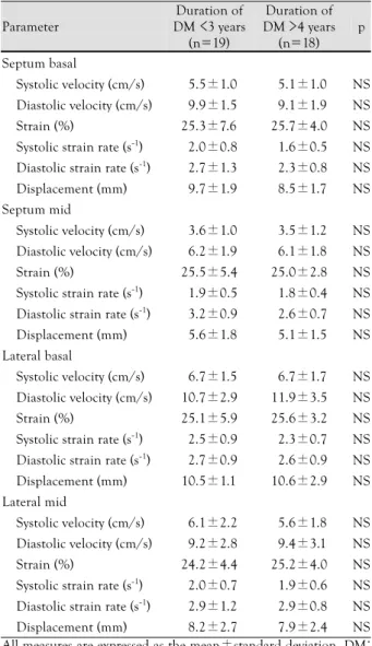
The regional myocardial systolic and diastolic velocities evaluated at the basal septum using VVI were lower in the group with a duration of DM >4 years than the group with a duration of DM <3 years and the control group
(Table 6). Also, the regional myocardial systolic and dia- stolic SRs evaluated at the basal and mid-septum were lower in the group with a duration of DM >4 years; how- ever, none of the values were significantly different be- tween the two groups.
Based on the conventional and unconventional in-
Table 5. Baseline characteristics and echocardiographic para- meters according to duration of DM
Parameter
Duration of DM <3 years
(n=19)
Duration of DM >4 years
(n=18) p
Gender M (6)/F (13) M (6)/ F (12) Duration of DM (months) 20 (3-29) 66(48-92) Glycosylated hemoglobin
(%) 7.6±1.9 9.4±2.2 0.01
Conventional echocardiography
Fractional shortening (%) 38.7±5.1 37.8±4.9 NS Ejection fraction (%) 69.0±6.5 68.1±6.1 NS E (cm/s) 99.7±16.4 98.4±18.7 NS A (cm/s) 48.9±8.6 52.7±14.0 NS Mitral deceleration time
(ms) 148.5±20.3 143.6±26.0 NS Isovolumic relaxation time
(ms) 58.1±9.6 60.3±9.2 NS
Tissue Doppler imaging
Septum
S’ (cm/s) 9.3±1.0 9.4±1.5 NS
E’ (cm/s) 16.4±1.8 16.2±2.3 NS
A’ (cm/s) 6.9±1.4 7.5±1.5 NS
E’/A’ 6.2±1.4 6.1±1.2 NS
Tei index 0.5±0.1 0.5±0.1 NS
Lateral S’ (cm/s) 12.5±2.6 11.9±2.4 NS E’ (cm/s) 22.5±2.4 21.2±3.4 NS
A’ (cm/s) 6.7±0.9 7.1±1.5 NS
E’/A’ 4.5±0.9 4.6±0.8 NS
Tei index 0.5±0.1 0.5±0.2 NS
All measures are expressed as the mean±standard deviation. DM:
diabetes mellitus, M: male, F: female, NS: non-specific
Table 6. Echocardiographic parameters derived from vector ve- locity imaging according to duration of DM
Parameter
Duration of DM <3 years
(n=19)
Duration of DM >4 years (n=18)
p
Septum basal
Systolic velocity (cm/s) 5.5±1.0 5.1±1.0 NS Diastolic velocity (cm/s) 9.9±1.5 9.1±1.9 NS Strain (%) 25.3±7.6 25.7±4.0 NS Systolic strain rate (s-1) 2.0±0.8 1.6±0.5 NS Diastolic strain rate (s-1) 2.7±1.3 2.3±0.8 NS Displacement (mm) 9.7±1.9 8.5±1.7 NS Septum mid
Systolic velocity (cm/s) 3.6±1.0 3.5±1.2 NS Diastolic velocity (cm/s) 6.2±1.9 6.1±1.8 NS Strain (%) 25.5±5.4 25.0±2.8 NS Systolic strain rate (s-1) 1.9±0.5 1.8±0.4 NS Diastolic strain rate (s-1) 3.2±0.9 2.6±0.7 NS Displacement (mm) 5.6±1.8 5.1±1.5 NS Lateral basal
Systolic velocity (cm/s) 6.7±1.5 6.7±1.7 NS Diastolic velocity (cm/s) 10.7±2.9 11.9±3.5 NS Strain (%) 25.1±5.9 25.6±3.2 NS Systolic strain rate (s-1) 2.5±0.9 2.3±0.7 NS Diastolic strain rate (s-1) 2.7±0.9 2.6±0.9 NS Displacement (mm) 10.5±1.1 10.6±2.9 NS Lateral mid
Systolic velocity (cm/s) 6.1±2.2 5.6±1.8 NS Diastolic velocity (cm/s) 9.2±2.8 9.4±3.1 NS Strain (%) 24.2±4.4 25.2±4.0 NS Systolic strain rate (s-1) 2.0±0.7 1.9±0.6 NS Diastolic strain rate (s-1) 2.9±1.2 2.9±0.8 NS Displacement (mm) 8.2±2.7 7.9±2.4 NS All measures are expressed as the mean±standard deviation. DM:
diabetes mellitus, NS: non-specific
DT
200 150 100 50
0
0 5 10 15 20 HbA1c
r=-0.357 p=0.016
TDI E’/A’
6 5 4 3 2 1 0
0 5 10 15 20 HbA1c
r=-0.301 p=0.042
Fig. 1. Correlation between echocardiographic parameters and glycosylated hemoglobin (%). A: correlation between deceleration time (DT) and glycosylated hemoglobin (%). B: correlation between tissue Doppler imaging (TDI) E’/A’ ratio and glycosylated hemoglobin (%).
A B
dices of systolic and diastolic function, no significant re- lationship existed between the duration of DM and echo- cardiographic parameters. DT and E’/A’ had significant inverse correlations with HbA1c (Fig. 1) (p=0.042 and p=0.016, respectively). No significant relationship exist- ed between the duration of DM or HbA1c and several parameters obtained from the VVI.
Discussion
This study yielded the following results: 1) no change of LV systolic function when evaluated by new sensi- tive echocardiographic techniques, such as VVI, in the presence of normal systolic function during conventio- nal and unconventional echocardiographic examina- tions; 2) the change in LV diastolic function when ev- aluated by conventional echocardiographic (transmi- tral) examination in the presence of normal diastolic function during unconventional echocardiographic ex- amination; 3) significant inverse correlations between conventional/unconventional echocardiographic para- meters and HbA1c level; and 4) mild progression of LV systolic and diastolic functions from normal to dys- function according to the duration of DM in spite of no significant differences between the groups.
In the current study, LV systolic function was eva- luated during the resting state and did not show a sig- nificant difference between the two groups. The results of the current study, which documented normal con- traction at rest, were the same as previous studies.2)7)
LV diastolic dysfunction in patients with DM may be caused by increased LV diastolic stiffness, deposition of advanced glycation end products, and cardiac fibrosis, all as a consequence of DM.11)12) Previous studies have reported a change in early diastolic ventricular func- tion in young adults with type I DM with normal LV EF.8)9) The study to determine the relationship between LV diastolic dysfunction and the duration of DM de- monstrated that a duration of DM >4 years was corre- lated with significant LV diastolic dysfunction.13) In com- parison to the previous study, although LV diastolic func- tion using mitral inflow Doppler analysis showed a sig- nificant change in the DM group, LV diastolic function evaluated by TDI and VVI analysis, which were more sensitive for the detection of LV functional alteration, did not show a significant difference between the groups.
The present data showed that there was no alteration in LV diastolic function in pediatric patients with type I DM. Indeed, the subjects in the current study were younger (mean age, 12 years and 5 months) and of sh- orter disease duration (mean duration, 3 years and 5 months). However, the striking finding involved the demonstration that LV functional alteration was based on the duration of DM. Although there were no signi- ficant differences between the duration of DM in the
>3 and >4 years groups, several echocardiographic pa- rameters were lower in the group with a duration of DM
>4 years. We therefore suggest that LV systolic and dia- stolic function may progress from normal to subtle ch- anges or dysfunction according to the duration of DM.
In the current study, a higher HbA1c was associated with a lower DT and E’/A’. When LV compliance de- creases and relaxation is abnormal, the DT becomes shorter and the E’/A’ ratio becomes <1.14)15) The present data suggest that hyperglycemia induces changes in LV diastolic function, especially decreased compliance and relaxation. However, of the subjects in the present study, decreased DT and E’/A’ according to hyperglycemia, were not significantly different in comparison with the control group and do not necessarily indicate definite LV diastolic dysfunction. The influence of hyperglyce- mia on LV function suggest that hyperglycemia may de- crease the expression of sarco(endo)plasmic reticulum Ca2-ATPase (SERCA2a) and the SERCA2a-to-phosph- olamban ratio.16)17) These reports imply that active con- trol of hyperglycemia and lowering blood glucose levels may attenuate these processes. Although it is difficult to clinically identify whether hyperglycemia is associated with myocardiac dysfunction in patients with DM, re- cent echocardiographic studies have shown that correla- tions exist between the state of glycemic control and ven- tricular function. Recent studies involving adult patients with type 2 DM showed that hyperglycemia influences on LV systolic function, especially with respect to ven- tricular long axis function.18)19) A study using magnetic resonance imaging indicates that there is a correlation between hyperglycemia and LV function in type 1 DM as well.20) The other study demonstrated that appro- priate control of hyperglycemia improves LV function and reduced LV mass in type 1 DM patients,21) and that there is a significant correlation between hyperglyce- mic control and changes in LV function. In the cur- rent study, LV diastolic function showed a significant and inverse correlation with HbA1c. Of importance, this finding was demonstrated in a very early stage of type I DM. We suggest that to maintain normal ventri- cular function in patients with DM apart from the dur- ation of DM, more aggressive control of blood glucose levels are needed and must be started as early as possible.
The limitations of the present study were that the number of enrolled patients was relatively small and that it was a cross-sectional study in which a comparison was made without baseline data.
Children and young adolescents rarely have insight on regarding their disease, and their diet is accordingly difficult to control. Therefore, alteration of myocardiac function induced by DM may begin earlier than is ge- nerally thought and these changes may be accelerated when glycemic control is poor. We recommend that close observation should begin early and should include de-
tection of diabetic cardiac alterations, as well as other diabetic complications.
Acknowledgments
This work was supported by the Korea Research Foundation Grant funded by the Korean Government (MOEHRD) (KRF-2007-531- E00039).
REFERENCES
1) Di Bello V, Talarico L, Picano E, et al. Increased echodensity of myocardial wall in the diabetic heart: an ultrasound tissue ch- aracterization study. J Am Coll Cardiol 1995;25:1408-15.
2) Fraser GE, Luke R, Thompson S, Smith H, Carter S, Sharpe N.
Comparison of echocardiographic variables between type 1 dia- betics and normal controls. Am J Cardiol 1995;75:141-5.
3) Kannel W, McGee D. Diabetes and cardiovascular disease: the Framingham study. JAMA 1979;241:2035-8.
4) Trost S, LeWinter M. Diabetic cardiomyopathy. Curr Treat Op- tions Cardiovasc Med 2001;3:481-92.
5) Cho KI, Park JH, Lee CK, et al. Isolated and combined influ- ences of diabetes and hypertension on the myocardial function and geometry. Korean Circ J 2006;36:411-7.
6) Choi EK, Park TB, Oh S, et al. The relationship of coronary ar- terial lesion with clinical factors in type II diabetes. Korean Circ J 2002;32:106-17.
7) Palmieri V, Capaldo B, Russo C, et al. Left ventricular chamber and myocardial systolic function reserve in patients with type 1 diabetes mellitus: insight from traditional and Doppler tissue im- aging echocardiography. J Am Soc Echocardiogr 2006;19:848-56.
8) Di Cori AD, Di Bello V, Miccoli R, et al. Left ventricular func- tion in normotensive young adults with well-controlled type 1 diabetes mellitus. Am J Cardiol 2007;99:84-90.
9) de Simone G, Mureddu GF, Vaccaro O, et al. Cardiac abnorma- lities in type 1 diabetes. Ital Heart J 2000;1:493-9.
10) Romanens M, Fankhauser S, Saner B, Michaud L, Saner H. No evidence for systolic or diastolic left ventricular dysfunction at rest in selected patients with long-term type I diabetes mellitus. Eur J Heart Fail 1999;1:169-75.
11) van Heerebeek L, Hamdani N, Handoko ML, et al. Diastolic stiffness of the failing diabetic heart: importance of fibrosis, ad- vanced glycation end products, and myocyte resting tension. Cir- culation 2008;117:43-51.
12) van Heerebeek L, Somsen A, Paulus WJ. The failing diabetic heart: focus on diastolic left ventricular dysfunction. Curr Diab Rep 2009;9:79-86.
13) From AM, Scott CG, Chen HH. Changes in diastolic dysfunction in diabetes mellitus over time. Am J Cardiol 2009;103:1463-6.
14) Ohno M, Cheng CP, Little WC. Mechanism of altered patterns of left ventricular filling during the development of congestive heart failure. Circulation 1994;89:2241-50.
15) Sohn DW, Chai IH, Lee DJ, et al. Assessment of mitral annulus velocity by Doppler tissue imaging in the evaluation of left ven- tricular diastolic function. J Am Coll Cardiol 1997;30:474-80.
16) Trost SU, Belke DD, Bluhm WF, Meyer M, Swanson E, Dill- mann WH. Overexpression of the sarcoplasmic reticulum Ca2+- ATPase improves myocardial contractility in diabetic cardio- myopathy. Diabetes 2002;51:1166-71.
17) Bidasee KR, Zhang Y, Shao CH, et al. Diabetes increases forma- tion of advanced glycation end products on Sarco(endo)plasmic reticulum Ca2+-ATPase. Diabetes 2004;53:463-73.
18) Govind S, Brodin LA, Nowak J, et al. Isolated type 2 diabetes mellitus causes myocardial dysfunction that becomes worse in the presence of cardiovascular diseases: results of the myocardial Dop- pler in diabetes (MYDID) study 1. Cardiology 2005;103:189-95.
19) Fang ZY, Schull-Meade R, Downey M, Prins J, Marwick TH.
Determinants of subclinical diabetic heart disease. Diabetologia 2005;48:394-402.
20) Chung J, Abraszewski P, Yu X, et al. Paradoxical increase in ventricular torsion and systolic torsion rate in type I diabetic pa- tients under tight glycemic control. J Am Coll Cardiol 2006;47:
384-90.
21) Aepfelbacher FC, Yeon SB, Weinrauch LA, D’Elia J, Burger AJ.
Improved glycemic control induces regression of left ventricular mass in patients with type 1 diabetes mellitus. Int J Cardiol 2004;
94:47-51.