This is an Open Access article distributed under the terms of the Creative Commons At- tribution Non-Commercial License (http://creativecommons.org/licenses/by-nc/3.0/) which permits unrestricted non-commercial use, distribution, and reproduction in any medium, provided the original work is properly cited.
Risk Factors Associated with Left Ventricular Diastolic Dysfunction in Type 2 Diabetic Patients without
Hypertension
Jung Hyun Noh1,2, Joon Hyung Doh1, Sung Yun Lee1, Tae Nyun Kim3, Hyuk Lee1, Hwa Young Song1, Jeong Hyun Park1, Kyung Soo Ko1, Byoung Doo Rhee1, Dong Jun Kim1
1Department of Internal Medicine, Inje University College of Medicine, Goyang,
2Clinical Research Center, Inje University Ilsan Paik Hospital, Goyang,
3Department of Internal Medicine, Korea University College of Medicine, Seoul, Korea
Background: Hypertension and age are recognized as important risk factors for left ventricular (LV) diastolic dysfunction. Some studies have shown that diabetes itself may also be an independent risk factor for LV diastolic dysfunction, although this is con- troversial. The aim of this study was to determine the factors associated with LV diastolic dysfunction in patients with type 2 dia- betes in the absence of hypertension or ischemic heart disease (IHD).
Methods: Participants in this study consisted of 65 type 2 diabetes patients (M : F = 45 : 20; mean age 51 [26 to 76] years; mean body mass index [BMI] 25.0 ± 2.5 kg/m2) without hypertension, heart disease, or renal disease. Individuals with ischemic elec- trocardiographic changes were excluded. LV diastolic function was evaluated by Doppler echocardiographic studies.
Results: Fifteen patients (23.1%) showed LV diastolic dysfunction on Doppler echocardiographic studies. Patients with LV dia- stolic dysfunction were older than those without diastolic dysfunction (60.0 ± 2.5 vs. 50.5 ± 1.9 years; P < 0.01). After adjusting for age and sex, BMI was higher (26.6 ± 0.7 vs. 24.6 ± 0.3 kg/m2; P < 0.01) and diabetes duration was longer (9.65 ± 1.48 vs. 4.71
± 0.78 years; P < 0.01) in patients with LV diastolic dysfunction than in those without diastolic dysfunction. There were no differ- ences in sex, smoking, blood pressure, lipid profiles, hemoglobin A1C, fasting glucose, fasting insulin, or diabetic microvascular complications between the LV diastolic dysfunction group and the normal diastolic function group. After adjusting for age, sex, and BMI, diabetes duration was found to be independently associated with LV diastolic dysfunction (odds ratio 1.38; confidence interval 1.12 to 1.72; P = 0.003).
Conclusion: These results suggest that diabetes duration may be a risk factor for LV diastolic dysfunction in type 2 diabetic pa- tients without hypertension or IHD.
Keywords: Diabetes duration; Diabetes mellitus; Left ventricular diastolic dysfunction
Corresponding author: Dong Jun Kim
Department of Internal Medicine, Inje University Ilsan Paik Hospital, 2240 Daehwa-dong, IlsanSeo-gu, Goyang, Gyeonggi 411-706, Korea E-mail: [email protected]
INTRODUCTION
Heart failure is a common comorbidity and fatal complication of diabetes mellitus. The Framingham heart study demonstrat- ed an increased risk of heart failure in patients with diabetes: a two-fold higher incidence in men and a five-fold higher inci-
dence in women with diabetes compared with age-matched non-diabetic subjects [1]. Many epidemiological studies have confirmed a significantly increased prevalence of cardiac dys- function in diabetic patients, independent of the influence of relevant covariates [2-4]. Left ventricular (LV) diastolic dys- function is thought be an early preclinical manifestation of pISSN 1976-9180 · eISSN 2093-2650
heart failure [5]. The incidence of diastolic dysfunction in dia- betic patients has been demonstrated to be 30-75% in recent studies [6-8].
Considering the high prevalence and significant morbidity and mortality of heart failure in patients with type 2 diabetes, identification of risk factors for LV diastolic dysfunction and an index of early-stage diabetic cardiomyopathy are necessary to delay or prevent the onset of heart failure. Age, hypertension, and ischemic heart disease (IHD) are thought to be important risk factors for diastolic dysfunction in both patients with dia- betes and non-diabetics [9-11]. Although several studies have shown that poor glycemic control and longer duration of dia- betes may be associated with early diastolic dysfunction in type 2 diabetes [12-14], there have been few studies on the factors associated with LV diastolic dysfunction in type 2 diabetes with- out hypertension or IHD. Therefore, we sought to determine the risk factors associated with subclinical LV diastolic dysfunc- tion in type 2 diabetic patients without hypertension or IHD.
METHODS
Study participants
This study was approved by the Institutional Review Board of Ilsan-Paik Hospital. The study subjects consisted of 65 type 2 diabetic patients without hypertension or IHD (M : F = 45 : 20;
mean age 51 [26-76] years; mean body mass index [BMI] 25.0
± 2.5 kg/m2) who initially visited the outpatient diabetes clinic at Inje University Ilsan-Paik Hospital between January 2006 and April 2006. Inclusion criteria were: i) normal arterial blood pressure (< 130/85 mm Hg) without antihypertensive medica- tion, ii) no symptoms or signs of heart disease, iii) no history of coronary heart disease (stable angina, unstable angina, myo- cardial infarction, or revascularization) or valvular heart dis- ease, iv) sinus rhythm and no evidence of IHD on resting 12- lead ECG, v) no evidence of severe medical illness including liver cirrhosis, end-stage renal disease, or cancer. Patients were excluded from participation in the study if either of the follow- ing criteria applied: i) diabetes diagnosed before the age of 26, or ii) history of type 1 diabetes or diabetic ketoacidosis.
Data collection and clinical evaluation
Height and weight were measured in the morning with the subjects wearing light clothing but no shoes. Blood pressure was measured with a mercury sphygmomanometer on the right arm with the subjects in a sitting position after a 5-min-
ute rest. BMI was calculated as weight in kilograms divided by the square of the height in meters. Diabetes duration and dia- betic complications were ascertained from review of medical records. Diabetic complications were recorded as retinopathy, neuropathy (absence of ankle jerks and reduced vibratory sen- sation, or impairment on nerve conduction studies or auto- nomic function tests), overt proteinuria (24 hour urine protein
≥ 300 mg/day), or microalbuminuria. Fasting plasma glucose (FPG), total cholesterol, high-density lipoprotein (HDL)-cho- lesterol, low-density (LDL)-cholesterol, lipoprotein a [Lp(a)], triglycerides, and uric acid were measured with an autoana- lyzer (Beckman Coulter, Miami, FL, USA). Level of hemoglo- bin A1C (HbA1C) (high performance liquid chromatography;
Tosoh, Tokyo, Japan), serum insulin (two-site chemilumines- cent immunometric assay; Roche, Basel, Switzerland), and high- sensitivity C-reactive protein (hs-CRP, enzyme-linked immu- nosorbent assay; DRG Diagnostics, Marburg, Germany) were also measured.
Echocardiographic study
All echocardiographic examinations were performed on a GE VingMed Vivid 5® Echocardiography System (GE Medical Sys- tems, Milwaukee, WI, USA) with a 2.5 MHz transducer. All smokers were prevented from smoking for at least 30 minutes before the examination. The examination was performed while the patient was in a period of quiet respiration. Echocardio- grams were stored digitally and analyzed by one examiner. All recordings were performed at a high sweep speed (100 mm/
sec) with simultaneous electrocardiographic (ECG) recording and included complete M-mode, 2-dimensional, and Doppler echocardiographic examinations, with emphasis on evaluation of LV diastolic function [10-12]. For evaluation of diastolic myocardial function, mitral inflow velocities (E- and A-waves, cm/sec), deceleration time (DT), isovolumic relaxation time (IVRT), systolic (S) and diastolic (D) pulmonary venous for- ward flow, pulmonary venous atrial reversal velocity (Ar), and early (E’) and late (A’) diastolic velocity of mitral annulus were measured in compliance with the standard protocols [10-12].
Left ventricular systolic function was determined by estima- tion of left ventricular ejection fraction (LVEF). The normal range of LVEF was 65 ± 10%.
The following criteria were used for the diagnosis of LV dia- stolic function [15]: impaired relaxation pattern was defined as E/A ratio < 1.0 and DT > 200 ms; pseudonormal pattern as E/A ratio from 1.0 - 2.0 with at least two of the following: S/D
ratio < 1, or Ar ≥ 35 cm/sec, or E’ < A’ and E/E’ ratio > 10; and restrictive pattern as E/A ratio > 2.0 and DT < 150 ms. All dia- stolic dysfunctions in this study were impaired relaxation pat- tern.
Statistical analysis
Data are presented as mean ± SEM. Statistical analysis was performed using SPSS for Windows (SPSS Inc., Chicago, IL, USA). Differences in variables between patients with and without LV diastolic dysfunction were analyzed using Mann- Whitney test and Fisher’s exact test. Analysis of covariance (ANCOVA) test was used for age and sex-adjusted character- istics according to the presence of diastolic dysfunction (Table
1). ANCOVA test was also used to assess the independent asso- ciation of duration of diabetes with the presence of diastolic dys- function, with age, sex, and BMI as covariates (Fig. 1). Logistic regression analysis was used to examine independent determi- nants for diastolic dysfunction, with age, sex, BMI, and dura- tion of diabetes as covariates. All probability values were two- tailed, and statistical significance was defined as P < 0.05.
RESULTS
Characteristics of participants
The median duration of diabetes for the study subjects was 5 years (range, 0 to 26). Mean HbA1C was 8.0 ± 2.1%. Fifteen pa- Table 1. Age, sex and age and sex-adjusted characteristics according to the presence of diastolic dysfunction in type 2 diabetic patients without hypertension
Normal left ventricular diastolic function
(n = 50) Left ventricular diastolic dysfunction
(n = 15) P value
Age, yr 51.2 ± 1.8 60.0 ± 2.5 <0.01
Men/Total 36/50 9/15 0.52
BMI, kg/m2 24.6 ± 0.3 26.8 ± 0.8 0.01
Current smoking, % 26 20 0.74
Systolic blood pressure, mm Hg 119.9 ± 1.4 120.9 ± 2.9 0.51
Diastolic blood pressure, mm Hg 71.6 ± 1.0 72.1 ± 1.8 0.81
Pulse pressure, mm Hg 48.3 ± 1.1 48.8 ± 2.1 0.85
LDL-C, mg/dL 120.8 ± 6.2 120.6 ± 11.6 0.99
Triglyceride, mg/dL 158.2 ± 15.6 189.6 ± 29.3 0.36
HDL-C, mg/dL 41.9 ± 1.5 44.4 ± 2.9 0.46
Lp(a), mg/dL 18.5 ± 3.3 25.8 ± 6.2 0.31
Duration of diabetes, yr 4.77 ± 0.79 9.73 ± 1.49 <0.01
Fasting glucose, mg/dL 161.0 ± 6.1 157.0 ± 11.4 0.76
HbA1C, % 8.16 ± 0.29 7.63 ± 0.54 0.39
Fasting insulin, µU/mL 6.47 ± 0.65 6.79 ± 1.21 0.82
HOMA-IR 2.64 ± 0.29 2.57 ± 0.55 0.91
Diabetic retinopathy, % 14.4 ± 5.6 32.1 ± 10.4 0.15
Overt proteiunuria, % 4.0 13.3 0.23
Microalbuminuria, % 8.0 13.3 0.33
Diabetic neuropathy, % 8.0 20.0 0.13
WBC, /mm3 6,110 ± 288 7,141 ± 557 0.11
hs-CRP, mg/L 0.59 ± 0.41 0.33 ± 0.78 0.77
Uric acid, mg/dL 4.71 ± 0.19 5.37 ± 0.36 0.12
Data are expressed as mean ± SEM.
BMI, body mass index; LDL-C, low-density lipoprotein cholesterol; HDL-C, high-density lipoprotein cholesterol; Lp(a), lipoprotein a; HOMA- IR, homeostasis model assessment-insulin resistance, hs-CRP, high-sensitivity C-reactive protein.
tients (23.1%) showed diastolic dysfunction on Doppler echo- cardiographic studies. The characteristics of the patients with and without prevalent diastolic dysfunction are compared in Table 1. Patients with LV diastolic dysfunction were older than patients without LV diastolic dysfunction (60.0 ± 2.5 vs. 50.5 ± 1.9 years; P < 0.01). There was no difference in the sex ratio between the two groups. After adjusting for age and sex, BMI was found to be higher (26.6 ± 0.7 vs. 24.6 ± 0.3 kg/m2; P < 0.01) and diabetes duration was found to be longer (9.65 ± 1.48 vs.
4.71 ± 0.78 years; P < 0.01) in patients with LV diastolic dysfunc- tion than in those without LV diastolic dysfunction. There were no differences in smoking, blood pressure, lipid profiles, HbA1C, fasting plasma glucose, fasting serum insulin, or diabetic mi- crovascular complications between the LV diastolic dysfunc- tion group and the normal LV diastolic function group. There was no difference between the two groups in medications for hyperglycemia or dyslipidemia, including thiazolidinediones, statins, and insulin (data not shown).
Echocardiologic parameters
No subject had LV systolic dysfunction and no differences in LVEF were found between the LV diastolic dysfunction group and the normal LV diastolic function group (P = 0.18). The E/
A ratio was lower in patients with LV diastolic dysfunction (0.70
± 0.04 vs. 1.07 ± 0.29; P < 0.001). There were no significant dif- ferences in other parameters between the LV diastolic dysfunc- tion group and the normal LV diastolic function group (Table 2).
Diabetes duration as an independent determinant for diastolic dysfunction
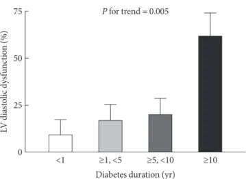
Logistic regression analysis for diastolic dysfunction with age, sex, BMI, and diabetes duration as covariates showed that dia- betes duration and BMI were independent determinants (Table 3). When participants were classified into four groups based on the duration of diabetes (≤ 1, >1 and ≤ 5, > 5 and ≤ 10, > 10 years), the frequency of LV diastolic dysfunction increased with increasing duration of diabetes after adjusting for age, sex, and BMI (P for trend = 0.005; Fig. 1).
DISCUSSION
In the present study, we found that duration of diabetes was strongly associated with the presence of LV diastolic dysfunc- tion in type 2 diabetic patients without hypertension or IHD.
After adjusting for age, sex, and BMI, the frequency of LV dia- Table 2. Echocardiographic parameters
Normal left ventricular diastolic function
(n = 50)
Left ventricular diastolic dysfunction
(n = 15) P value E/A ratio 1.07 ± 0.29 0.70 ± 0.04 <0.001
DT, ms 221.9 ± 53.8 238 ± 27.5 0.14
IVRT, ms 101.0 ± 44.9 120.7 ± 71.5 0.37
E’, cm/s 11.2 ± 2.7 10.4 ± 2.3 0.51
A’, cm/s 7.9 ± 2.3 8.1 ± 3.4 0.80
E/E’ ratio 8.5 ± 2.7 9.5 ± 3.0 0.25
LVEF, % 67.5 ± 4.2 65.5 ± 5.9 0.18
Data are expressed as mean ± SD.
E/A, the ratio of early and late left ventricular diastolic filling; DT, the E-wave deceleration time; IVRT, the isovolumic relaxation time; E’
the early diastolic velocity by Tissue Doppler at lateral mitral annulus;
A’, the late diastolic velocity by Tissue Doppler at lateral mitral annu- lus; E/E’, the ratio of E and E’; LVEF, left ventricular ejection fraction.
Table 3. Logistic regression analysis of diastolic dysfunction in type 2 diabetic patients without hypertension with age, sex, BMI, and diabetes duration as covariates
Exp (B) (95% CI) P value
Age 1.04 (0.96 to 1.12) 0.385
Female 0.57 (0.09 to 3.75) 0.558
BMI 2.03 (1.21 to 3.41) 0.008
DM duration 1.39 (1.12 to 1.72) 0.003
BMI, body mass index; Exp (B), exponentiation of the B coefficient;
DM, diabetes mellitus; CI, confidence interval.
Fig. 1. Frequency of LV diastolic dysfunction according to di- abetes duration in type 2 diabetic patients without hyperten- sion, after adjusting for age, sex, and BMI. LV, left ventricular;
BMI, body mass index.
LV diastolic dysfunction (%)
P for trend = 0.005
Diabetes duration (yr)
<1 ≥1, <5 ≥5, <10 ≥10 75
50
25
0
stolic dysfunction correlated positively with the duration of diabetes.
Some previous studies demonstrated LV diastolic dysfunc- tion in normotensive patients with diabetes, and the existence of LV diastolic dysfunction in the absence of coronary artery disease and hypertension has been ascribed to diabetic cardio- myopathy [6-8]; however, there are very few studies on the fac- tors associated with LV diastolic dysfunction in type 2 diabetes without hypertension or IHD. Aging and duration of diabetes were related to LV dysfunction in normotensive type 2 diabet- ic patients in a previous study [9], which demonstrated a simi- lar results of the present study.
The proposed mechanisms of diabetic cardiomyopathy from animal studies are: i) excessive production of reactive oxygen species [16], ii) over-activation of poly-(ADP-ribose) polymer- ase [17], iii) increased activity of protein kinase C [18], iv) dys- functional calcium handling in cardiomyocytes [19], and v) enhanced activity of the renin-angiotensin-aldosterone system [20]. Several studies have suggested that hyperglycemia alters the metabolism of cardiac myocytes and could be the primary insult in the pathogenesis of diabetic cardiomyopathy [21,22].
Even in type 2 diabetic patients without cardiac involvement, uncontrolled hyperglycemia is known to provoke diastolic LV dysfunction [12,13]. Nichols et al. [23] demonstrated that a re- duction in HbA1C coupled with a lower baseline HbA1C was predictive of a decreased incidence of heart failure in a multi- variate model, emphasizing the importance of glycemic control for prevention of heart failure.
Some data have suggested that diabetic cardiomyopathy could be one of the microvascular complications of diabetes related to endothelial dysfunction [21,24,25]. Considering that duration of diabetes is the strongest predictor for diabetic mi- crovascular complications, the strong association of duration of diabetes with LV diastolic dysfunction observed in this study is plausible. In this study, the level of HbA1C was not associat- ed with LV diastolic dysfunction, whereas diabetes duration was a significant determinant, suggesting that cumulative ex- posure to hyperglycemia may be important for the development of LV diastolic dysfunction.
Several studies have reported that obesity is associated with heart failure and ventricular dysfunction [26-29]. In a middle- aged African-American cohort, obesity was associated with concentric hypertrophy, which was strongly associated with diastolic dysfunction [30]. Another study found that reduced LV diastolic function was apparent in 24% of severely obese
subjects, and that the risk was linearly associated with BMI [29].
However, the association of obesity with diastolic dysfunction, independent of other causes, remains controversial [5,31]. From the results of this study, we suggest that obesity may be associ- ated with LV diastolic dysfunction independent of other clini- cal parameters related to diastolic dysfunction in type 2 diabet- ic patients without hypertension or IHD.
In this study, the prevalence of LV diastolic dysfunction was lower than in other studies [6-8], which could be because of the relatively short duration of diabetes, and our selection cri- teria excluding patients with hypertension or IHD. We did not find a significant association of diabetic microvascular com- plications such as retinopathy, nephropathy, and neuropathy with LV diastolic dysfunction, although frequencies of each microvascular complication tended to increase with the pres- ence of LV diastolic dysfunction. This non-significant associa- tion may be explained by the earlier development of diabetic cardiomyopathy compared with other microvascular compli- cations [32-34], relatively well-controlled hyperglycemia, short duration of diabetes, and selection criteria excluding patients with hypertension or IHD.
One limitation of our study is that it was cross-sectional and unable to suggest a causal relationship. Other limitations were the uncertainty of ischemic heart disease and diabetes duration.
Our exclusion of IHD based on ECG and presence of symptoms might have not ruled out IHD completely. Besides, the onset of type 2 diabetes is not always clear, and previous medical re- cords related to initial diagnosis were not available. Although the good correlation of diabetes duration with diabetic retin- opathy (r = 0.516, P < 0.01) may lessen this concern, another prospective study for individuals with newly detected type 2 diabetes is needed to confirm the results of our study.
Despite the limitations of the study, our data showed that duration of diabetes could be a marker for LV diastolic dysfunc- tion independent of other diastolic dysfunction-related vari- ables in type 2 diabetic patients without hypertension or IHD.
ACKNOWLEDGEMENT
This work was supported by grant from Inje University, 2004.
REFERENCES
Kannel WB, McGee DL. Diabetes and cardiovascular disease:
1.
the Framingham study. JAMA 1979;241:2035-8.
Raev DC. Which left ventricular function is impaired earlier 2.
in the evolution of diabetic cardiomyopathy? An echocardio- graphic study of young type 1 diabetic patients. Diabetes Care 1994;17:633-9.
Candido R, Srivastava P, Cooper ME, Burrell LM. Diabetes 3.
mellitus: a cardiovascular disease. Curr Opin Investig Drugs 2003;4:1088-93.
Stamler J, Vaccaro O, Neaton JD, Wentworth D. Diabetes, other 4.
risk factors, and 12-yr cardiovascular mortality for men screened in the Multiple Risk Factor Intervention Trial. Diabetes Care 1993;16:434-44.
Persson H, Lonn E, Edner M, Baruch L, Lang CC, Morton JJ, 5.
Ostergren J, McKelvie RS; Investigators of the CHARM Echo- cardiographic Substudy-CHARMES. Diastolic dysfunction in heart failure with preserved systolic function: need for objec- tive evidence: results from the CHARM Echocardiographic Substudy-CHARMES. J Am Coll Cardiol 2007;49:687-94.
Poirier P, Bogaty P, Garneau C, Marois L, Dumesnil JG. Dia- 6.
stolic dysfunction in normotensive men with well-controlled type 2 diabetes: importance of maneuvers in echocardiograph- ic screening for preclinical diabetic cardiomyopathy. Diabetes Care 2001;24:5-10.
Zabalgoitia M, Ismaeil MF, Anderson L, Maklady FA. Preva- 7.
lence of diastolic dysfunction in normotensive, asymptomatic patients with well-controlled type 2 diabetes mellitus. Am J Cardiol 2001;87:320-3.
Boyer JK, Thanigaraj S, Schechtman KB, Pérez JE. Prevalence 8.
of ventricular diastolic dysfunction in asymptomatic, normo- tensive patients with diabetes mellitus. Am J Cardiol 2004;93:
870-5.
Masugata H, Senda S, Goda F, Yoshihara Y, Yoshikawa K, Fujita 9.
N, Daikuhara H, Okuyama H, Taoka T, Kohno M. Left ventric- ular diastolic dysfunction in normotensive diabetic patients in various age strata. Diabetes Res Clin Pract 2008;79:91-6.
Danielsen R. Factors contributing to left ventricular diastolic 10.
dysfunction in long-term type I diabetic subjects. Acta Med Scand 1988;224:249-56.
Nicolino A, Longobardi G, Furgi G, Rossi M, Zoccolillo N, Fer- 11.
rara N, Rengo F. Left ventricular diastolic filling in diabetes mel- litus with and without hypertension. Am J Hypertens 1995;8:
382-9.
von Bibra H, Hansen A, Dounis V, Bystedt T, Malmberg K, 12.
Rydén L. Augmented metabolic control improves myocardial diastolic function and perfusion in patients with non-insulin dependent diabetes. Heart 2004;90:1483-4.
Grandi AM, Piantanida E, Franzetti I, Bernasconi M, Maresca 13.
A, Marnini P, Guasti L, Venco A. Effect of glycemic control on left ventricular diastolic function in type 1 diabetes mellitus. Am J Cardiol 2006;97:71-6.
Celentano A, Vaccaro O, Tammaro P, Galderisi M, Crivaro M, 14.
Oliviero M, Imperatore G, Palmieri V, Iovino V, Riccardi G.
Early abnormalities of cardiac function in non-insulin-depen- dent diabetes mellitus and impaired glucose tolerance. Am J Cardiol 1995;76:1173-6.
Konduracka E, Gackowski A, Rostoff P, Galicka-Latala D, 15.
Frasik W, Piwowarska W. Diabetes-specific cardiomyopathy in type 1 diabetes mellitus: no evidence for its occurrence in the era of intensive insulin therapy. Eur Heart J 2007;28:2465-71.
Wold LE, Ceylan-Isik AF, Fang CX, Yang X, Li SY, Sreejayan N, 16.
Privratsky JR, Ren J. Metallothionein alleviates cardiac dysfunc- tion in streptozotocin-induced diabetes: role of Ca2+ cycling proteins, NADPH oxidase, poly(ADP-Ribose) polymerase and myosin heavy chain isozyme. Free Radic Biol Med 2006;40:
1419-29.
Minchenko AG, Stevens MJ, White L, Abatan OI, Komjáti K, 17.
Pacher P, Szabó C, Obrosova IG. Diabetes-induced overexpres- sion of endothelin-1 and endothelin receptors in the rat renal cortex is mediated via poly(ADP-ribose) polymerase activation.
FASEB J 2003;17:1514-6.
Way KJ, Isshiki K, Suzuma K, Yokota T, Zvagelsky D, Schoen 18.
FJ, Sandusky GE, Pechous PA, Vlahos CJ, Wakasaki H, King GL. Expression of connective tissue growth factor is increased in injured myocardium associated with protein kinase C beta2 activation and diabetes. Diabetes 2002;51:2709-18.
Vetter R, Rehfeld U, Reissfelder C, Weiss W, Wagner KD, Gün- 19.
ther J, Hammes A, Tschöpe C, Dillmann W, Paul M. Transgen- ic overexpression of the sarcoplasmic reticulum Ca2+ATPase improves reticular Ca2+ handling in normal and diabetic rat hearts. FASEB J 2002;16:1657-9.
Privratsky JR, Wold LE, Sowers JR, Quinn MT, Ren J. AT1 block- 20.
ade prevents glucose-induced cardiac dysfunction in ventricu- lar myocytes: role of the AT1 receptor and NADPH oxidase.
Hypertension 2003;42:206-12.
Du X, Matsumura T, Edelstein D, Rossetti L, Zsengellér Z, Szabó 21.
C, Brownlee M. Inhibition of GAPDH activity by poly (ADP- ribose) polymerase activates three major pathways of hyperg- lycemic damage in endothelial cells. J Clin Invest 2003;112:
1049-57.
Farhangkhoee H, Khan ZA, Mukherjee S, Cukiernik M, Barbin 22.
YP, Karmazyn M, Chakrabarti S. Heme oxygenase in diabetes-
induced oxidative stress in the heart. J Mol Cell Cardiol 2003;
35:1439-48.
Nichols GA, Hillier TA, Erbey JR, Brown JB. Congestive heart 23.
failure in type 2 diabetes: prevalence, incidence, and risk fac- tors. Diabetes Care 2001;24:1614-9.
Bell DS. Diabetic cardiomyopathy. A unique entity or a com- 24.
plication of coronary artery disease? Diabetes Care 1995;18:
708-14.
Okruhlicova L, Tribulova N, Weismann P, Sotnikova R. Ultra- 25.
structure and histochemistry of rat myocardial capillary en- dothelial cells in response to diabetes and hypertension. Cell Res 2005;15:532-8.
Kenchaiah S, Evans JC, Levy D, Wilson PW, Benjamin EJ, Lar- 26.
son MG, Kannel WB, Vasan RS. Obesity and the risk of heart failure. N Engl J Med 2002;347:305-13.
Taegtmeyer H, McNulty P, Young ME. Adaptation and malad- 27.
aptation of the heart in diabetes: part I: general concepts. Cir- culation 2002;105:1727-33.
He J, Ogden LG, Bazzano LA, Vupputuri S, Loria C, Whelton 28.
PK. Risk factors for congestive heart failure in US men and women: NHANES I epidemiologic follow-up study. Arch In- tern Med 2001;161:996-1002.
Wong CY, O’Moore-Sullivan T, Leano R, Byrne N, Beller E, 29.
Marwick TH. Alterations of left ventricular myocardial charac- teristics associated with obesity. Circulation 2004;110:3081-7.
Fox ER, Taylor J, Taylor H, Han H, Samdarshi T, Arnett D, 30.
Myerson M. Left ventricular geometric patterns in the Jackson cohort of the Atherosclerotic Risk in Communities (ARIC) Study: clinical correlates and influences on systolic and diastol- ic dysfunction. Am Heart J 2007;153:238-44.
Bajraktari G, Qirko S, Rexhepaj N, Bakalli A, Beqiri A, Elezi S, 31.
Ndrepepa G. Non-insulin dependent diabetes as an indepen- dent predictor of asymptomatic left ventricular diastolic dys- function. Croat Med J 2005;46:225-31.
Di Bonito P, Cuomo S, Moio N, Sibilio G, Sabatini D, Quattrin 32.
S, Capaldo B. Diastolic dysfunction in patients with non-insu- lin-dependent diabetes mellitus of short duration. Diabet Med 1996;13:321-4.
Vanninen E, Mustonen J, Vainio P, Lansimies E, Uusitupa M.
33.
Left ventricular function and dimensions in newly diagnosed non-insulin-dependent diabetes mellitus. Am J Cardiol 1992;
70:371-8.
Gough SC, Smyllie J, Barker M, Berkin KE, Rice PJ, Grant PJ.
34.
Diastolic dysfunction is not related to changes in glycaemic con- trol over 6 months in type 2 (non-insulin-dependent) diabetes mellitus. A cross-sectional study. Acta Diabetol 1995;32:110-5.