27 Accepted for Publication: March 22, 2009
*Corresponding author: Il-Suk Sohn, 134-727, Cardiovascular Center of Kyunghee East-West Neo Medical Center, Phone: +82-2-440-6108, FAX:
+82-2-442-6638, E-mail: [email protected] Chonnam Medical Journal
Vol. 45, No. 1, pp. 27∼32 DOI: 10.4068/cmj.2009.45.1.27
Impact of Diabetes on the Left Ventricular Long Axis and Midwall Function in Patients with Hypertension
Jeong-Hwan Park, Il-Suk Sohn*, Jin-Man Cho and Chong-Jin Kim Department of Cardiology, Kyunghee East-West Neo Medical Center, Seoul, Korea
Diabetes mellitus (DM) is a strong risk factor for cardiovascular morbidity and mortality. It is suggested that both hypertension and DM may synergistically affect left ventricular (LV) structure and function. To assess whether hypertensive patients with DM have more depressed LV function in terms of long axis and midwall function by use of tissue Doppler echocardiogram than do patients with hypertension only.
Hypertensive patients (n=200) and hypertensive patients with DM (n=60), all of whom had normal LV ejection fraction, were recruited for the study. Healthy age- and sex-matched subjects (n=20) served as the control group. Echocardiography was performed including LV wall thickness, dimension, Doppler, and tissue Doppler imaging. Blood pressure (BP) was measured in the supine position before and after echocardiographic examination. The hypertensive patients with or without DM had higher BP and LV wall thickness, had more decreased afterload measured by meridional end-systolic stress, and had more depressed LV diastolic, long axis, and global function than did the controls. There was no difference in LV function between the hypertensive patients with and without DM. The hypertensive patients with or without DM had depressed LV function compared with the controls, but there was no difference between the hypertensive patients with or without DM. DM does not seem to have additive adverse effects on the LV long axis and midwall function in persons with hypertension.
Key Words: Diabetes mellitus; Hypertension; Ventricular function, Left; Echocardiography
Introduction
Diabetes mellitus (DM) and hypertension both con- tribute to cardiovascular disease leading to atherosclero- sis, ischemic heart disease, and heart failure. Hyper- tension is often associated with impaired glucose toler- ance and insulin resistance and is not uncommonly combined with DM.1 Hypertension and DM are conti- nuously increasing in the general population and are
associated with adverse remodeling of the cardiovas- cular system.2 This high risk for progressive adverse effects on the heart from the preclinical phase has resulted in novel American College of Cardiology/
American Heart Association (ACC/AHA) guidelines for the classification of heart failure, which include sta- ge A and B before overt heart failure symptoms dev- elop.3
DM can affect cardiac structure and function in the absence of ischemic heart disease or hypertension.4 This condition, also known as diabetic cardiomyopathy,5 is often characterized by left ventricular (LV) hypertrophy and diastolic dysfunction, which can be easily detected
by using Doppler echocardiography. Elevated blood pressure itself typically affects the heart, and it is com- monly associated with increased LV mass, LV dila- tation, diastolic dysfunction, reduced chamber compli- ance, and progression to overt heart failure.6-8
Recent echocardiographic advances, especially in tissue Doppler imaging, can assess subtle changes in LV structure and function. This may provide useful in- formation about diastolic function in patients with DM, hypertension, or both.9,10 However, there are few data from tissue Doppler imaging on the impact of DM on LV function in patients with hypertension. Therefore, we tried to assess whether hypertensive patients with DM have more depressed LV function in terms of long axis and midwall function by use of tissue Doppler echocardiogram than do patients with hypertension only.
Materials and Methods
Patients with hypertension who had been treated with antihypertensive drugs and who had not changed during the previous 6 months were enrolled. Excluded from the study were those (1) who had a history of previous coronary artery disease or with endovascular intervention, bypass surgery, or who had regional wall motion abnormalities; (2) who had undergone open heart surgery; (3) who had significant valvular heart disease or other structural heart diseases, including con- genital heart disease, hypertrophic cardiomyopathy, dila- ted cardiomyopathy, or low LV ejection fraction less than 45%; and (4) who had an electrocardiographically evident left bundle branch block, preexcitation, or atrial fibrillation.
The hypertensive patients were divided into 2 groups:
the hypertension group with hypertension only and the hypertension with DM group. Age- and sex-matched healthy controls who had normal blood pressure (BP), no medical history of cardiovascular disease, a normal electrocardiogram, and no remarkable organic heart dis-
ease on echocardiography were also enrolled. Demogra- phic data were obtained before echocardiography. BP was measured in the supine position before and after echocardiographic examination. All subjects gave infor- med consent and all examinations were approved by the Institutional Review Board.
1. Echocardiographic examination
Comprehensive 2-dimensional and Doppler echocar- diography using a commercial machine (iE33, Philips, Netherlands) were performed in every subject in the left lateral decubitus position. Echocardiographic measu- rements included end-diastolic interventricular septal thickness (IVSd), end-diastolic LV posterior wall thic- kness (PWT), LV end-diastolic dimension (LVDd), LV end-systolic dimension (LVDs), and LV ejection frac- tion by biplane Simpson’s method.
LV mass,11 relative wall thickness as an index of ec- centricity of LV hypertrophy, and fractional shortening (FS) were defined as follows:
ㆍLV mass (g) =1.04×[(LVDd+PWT+IVSd)3− LVDd3]×0.8+0.6
ㆍRelative wall thickness=2×PWT/LVDd ㆍFS (%)=[(LVDd−LVDs)/LVDd]×100
LV hypertrophy was defined as LV mass index >95 g/m2 (women) or >115 g/m2 (men).
In the presence of altered LV geometry, as in hy- pertension, systolic shortening measured at the level of the endocardium is often higher than predicted from the measured level of end-systolic stress (ESS).12 In contrast, when measured at the midwall level, systolic shortening is often depressed.13,14 Thus, midwall FS was calculated as described previously elsewhere.14
The force-limiting LV ejection (i.e., chamber func- tion) is myocardial afterload, which can be measured as wall stress at end-systole according to the principles of Laplace’s law.15 Longitudinal myocardial fiber shortening is limited by longitudinal (meridional) ESS. This meri- dional ESS13,16 was calculated as:
ㆍMeridional ESS=0.334×systolic blood pressure×LVDs/
{PWT×[1+(PWT/LVDs)]}
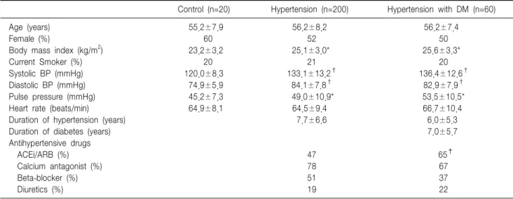
Table 1. Clinical characteristics of the subjects
Control (n=20) Hypertension (n=200) Hypertension with DM (n=60)
Age (years) 55.2±7.9 56.2±8.2 56.2±7.4
Female (%) 60 52 50
Body mass index (kg/m2) 23.2±3.2 25.1±3.0* 25.6±3.3*
Current Smoker (%) 20 21 20
Systolic BP (mmHg) 120.0±8.3 133.1±13.2† 136.4±12.6†
Diastolic BP (mmHg) 74.9±5.9 84.1±7.8† 82.9±7.9†
Pulse pressure (mmHg) 45.2±7.3 49.0±10.9* 53.5±10.5*
Heart rate (beats/min) 64.9±8.1 64.5±9.4 66.7±10.4
Duration of hypertension (years) 7.7±6.6 6.0±5.3
Duration of diabetes (years) 7.0±5.7
Antihypertensive drugs
ACEi/ARB (%) 47 65‡
Calcium antagonist (%) 78 67
Beta-blocker (%) 51 37
Diuretics (%) 19 22
DM, diabetes mellitus; BP, blood pressure; ACEi/ARB, angiotensin-converting enzyme inhibitor/angiotensin receptor blocker. *p<0.05 vs control, †p<0.01 vs control, ‡p<0.05 vs Hypertension.
Pulsed Doppler imaging from the mitral inflow measured early (E) and late (A) velocity and decele- ration time (DT) of E velocity. Mitral annular early diastolic (E’) and systolic (S’) velocities from the me- dial annulus were obtained by using tissue Doppler imaging. The ratio of early mitral inflow (E) to early annular (E’) velocity, as a representative parameter of LV filling pressure, was derived. The Tei index as a well-known parameter of global myocardial function was measured from pulsed Doppler imaging of transmitral inflow and LV outflow.
2. Statistical methods
Data are expressed as mean±SD. Differences between the 3 groups were tested by ANOVA for continuous variables with post-hoc analysis by Scheffe test. Pearson chi-square test was used to test for differences in categorical variables. Univariate analysis by Pearson correlation and multivariate logistic regression analyses were performed to assess the relation of clinical and echocardiographic parameters to LV diastolic function independent of established covariates. Two-tailed p
<0.05 was considered significant. All data were mana- ged by using a commercial statistical program (SPSS for Windows, version 10.0, Chicago, Illinois, USA).
Results
1. Clinical characteristics
As shown in Table 1, hypertensive patients (n=200), hypertensive patients with DM (n=60), and age-, and sex-matched controls (n=20) were enrolled. The hyper- tensive patients with or without DM had higher body mass index, systolic and diastolic BP, and pulse pressure than did the healthy controls. On average, the duration of hypertension was 7.7 years in hypertension group and 6.0 years in the hypertension with DM group. The DM patients had the disease for 7 years on average. Among the antihypertensive drugs, angiotensin-converting enzy- me inhibitor and/or angiotensin-receptor blocker was more commonly used in the hypertension with DM group than in the hypertension group (65% vs 47%).
There were no differences in age, sex, current smoking, or heart rate between the study groups.
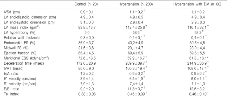
2. Echocardiographic parameters of LV function The hypertensive patients with or without DM had higher IVSd, LV mass index, and relative wall thickness than did the control group, but not LV dimension, FS, or ejection fraction (Table 2). The patients with hyper- tension had lower meridional ESS than did the con- trols. Of the diastolic function parameters, DT, isovolu-
Table 3. Clinical and echocardiographic variables related to left ventricular diastolic function (E/E’ ratio)
Univariate Multivariate
Variables
r P B p
Age 0.304 <0.001 0.069 0.003
LV mass index 0.261 <0.001 0.024 <0.001
Body mass index 0.097 0.11 0.017 0.76
Relative wall thickness 0.178 0.003 −0.721 0.73
Systolic blood pressure 0.136 0.023 0.008 0.54
Diabetes mellitus −0.43 0.27
ACEi/ARB use 0.136 0.71
E/E’, ratio of mitral inflow early (E) velocity to early diastolic mitral annular (E’) velocity; r, Pearson correlation coefficient; B, logistic coefficient;
LV, left ventricular; ACEi/ARB, angiotensin-converting enzyme inhibitor/angiotensin receptor blocker.
Table 2. Echocardiographic parameters of left ventricular function
Control (n=20) Hypertension (n=200) Hypertension with DM (n=60)
IVSd (cm) 0.9±0.1 1.1±0.2† 1.1±0.2†
LV end-diastolic dimension (cm) 4.9±0.4 4.9±0.5 4.9±0.4
LV end-systolic dimension (cm) 3.1±0.3 2.9±0.4 2.9±0.3
LV mass index (g/m2) 83.8±13.7 112.4±25.8† 116.1±32.1†
LV hypertrophy (%) 5.0 58.5† 58.3†
Relative wall thickness 0.3±0.0 0.4±0.1† 0.4±0.1†
Endocardial FS (%) 36.9±3.7 40.2±4.9 39.5±4.5
Midwall FS (%) 21.6±3.6 23.1±4.7 23.0±4.4
Ejection fraction (%) 66.4±4.6 69.4±5.9 69.6±5.5
Meridional ESS (kdyne/cm2) 72.6±18.5 59.9±16.7† 61.8±16.1*
Deceleration time (msec) 172.0±20.9 209.9±39.7† 214.9±36.9†
IVRT (msec) 86.0±9.0 106.3±19.4† 108.0±17.4†
E/A ratio 1.2±0.2 0.9±0.2† 0.9±0.2†
E’ velocity (cm/sec) 8.9±1.4 6.5±1.9† 6.0±1.4†
S’ velocity (cm/sec) 7.9±1.3 7.4±1.4 7.1±1.3
E/E’ ratio 9.2±2.0 11.8±3.7† 12.6±3.2†
Tei index 0.38±0.06 0.45±0.09† 0.46±0.10†
DM, diabetes mellitus; IVSd, end-diastolic interventricular septal thickness; LV, left ventricular; FS, fractional shortening; ESS, end-systolic stress; IVRT, isovolumic relaxation time; E/A, ratio of mitral inflow early (E) velocity to late (A) velocity; E’, early diastolic mitral annular velocity;
S’, systolic mitral annular velocity; E/E’, ratio of mitral inflow early (E) velocity to E’ velocity. *p<0.05 vs control, †p<0.01 vs control.
mic relaxation time, and E/E’ ratio were higher in the hypertensive patients than in the control group. The hypertensive patients had lower E’ velocity, E/A ratio, and Tei index than did the controls.
3. Clinical correlates of echocardiographic parameters of LV diastolic function
We tested univariate analysis with the clinical and echocardiographic variables that can affect the LV dias- tolic function (Table 3). E/E’ ratio, as a marker of LV filling pressure, showed good correlations with age, sys- tolic BP, pulse pressure, relative wall thickness, and LV mass index. On multivariate analysis among the clinical
and echocardiographic parameters, age and LV mass index were independent predictors of LV diastolic dysfunction.
Discussion
In this study, we observed that the hypertensive patients with or without DM had increased LV mass and depressed diastolic, long axis, and global LV func- tion compared with the healthy controls, but no differ- ences were noted between the hypertension and the hypertension with DM groups.
Increased LV mass or LV hypertrophy is a common finding in patients with hypertension and/or DM and is an independent risk factor for developing adverse cardi- ovascular events and progression to heart failure.17-20 High BP and obesity, which often coexist with DM, tend to be associated with preclinical cardiovascular abnormalities and may contribute to the association of diabetes with cardiovascular events. In this study, the patients with hypertension had higher body mass index, BP, and pulse pressure than did the healthy controls.
These characteristics in the hypertensives led to higher LV mass and more depressed LV function, but with preserved LV systolic function. Diastolic dysfunction with preserved LV systolic function is a common find- ing in patients with hypertension and DM even though they do not show any heart failure symptoms. In this study, we did not try to assess heart failure symptoms and excluded the patients with past coronary heart disease, significant arrhythmia, valvular heart disease, or systolic dysfunction. In the real world, however, there may be a much higher prevalence of overt and pre- clinical cardiovascular diseases in patients with hyper- tension and/or DM. Furthermore, those hypertensive patients with subclinical diastolic and long axis dysfun- ction as in this study should be managed and followed up for future adverse cardiovascular events.
In our study, there were no differences in LV func- tion between the hypertension and the hypertension with DM groups, which suggests that DM might not have additive adverse effects on LV function in patients with hypertension. Only age and LV mass are inde- pendent predictive factors of LV diastolic function. But we should take into account multiple complicated vari- ables in claiming a potential link to cardiac structure and function, and it is difficult to conclude such a link in this small observational study.
In conclusion, the hypertensive patients with or with- out DM had higher LV mass and depressed LV dia- stolic, long axis, and global function compared with the healthy controls, but there was no difference between the hypertension and the hypertension with DM groups.
First, this study is cross-sectional and observational and had a relatively small number of participants in a single center. Prospective studies with larger populations will be needed for determining clinical implication. Se- cond, we did not assess more complicated clinical vari- ables that can be associated with depressed LV function.
References
1. Gress TW, Nieto FJ, Shahar E, Wofford MR, Brancati FL. Hyper- tension and antihypertensive therapy as risk factors for type 2 diabetes mellitus. Atherosclerosis Risk in Communities Study. N Engl J Med 2000;342:905-12.
2. Fox CS, Coady S, Sorlie PD, D'Agostino RB Sr, Pencina MJ, Vasan RS, et al. Increasing cardiovascular disease burden due to diabetes mellitus: the Framingham Heart Study. Circulation 2007;115:1544-50.
3. Hunt SA, Abraham WT, Chin MH, Feldman AM, Francis GS, Ganiats TG, et al. ACC/AHA 2005 Guideline Update for the Diagnosis and Management of Chronic Heart Failure in the Adult: a report of the American College of Cardiology/American Heart Association Task Force on Practice Guidelines (Writing Committee to Update the 2001 Guidelines for the Evaluation and Management of Heart Failure): developed in collaboration with the American College of Chest Physicians and the International Society for Heart and Lung Transplantation: endorsed by the Heart Rhythm Society. Circulation 2005;112:e154-235.
4. Devereux RB, Roman MJ, Paranicas M, O'Grady MJ, Lee ET, Welty TK, et al. Impact of diabetes on cardiac structure and function: the strong heart study. Circulation 2000;101:2271-6.
5. Asbun J, Villarreal FJ. The pathogenesis of myocardial fibrosis in the setting of diabetic cardiomyopathy. J Am Coll Cardiol 2006;47:693-700.
6. Fouad FM, Slominski JM, Tarazi RC. Left ventricular diastolic func- tion in hypertension: relation to left ventricular mass and systolic func- tion. J Am Coll Cardiol 1984;3:1500-6.
7. Frohlich ED, Apstein C, Chobanian AV, Devereux RB, Dustan HP, Dzau V, et al. The heart in hypertension. N Engl J Med 1992;327:998- 1008.
8. Levy D, Larson MG, Vasan RS, Kannel WB, Ho KK. The progression from hypertension to congestive heart failure. JAMA 1996;275:1557-62.
9. Di Bonito P, Moio N, Cavuto L, Covino G, Murena E, Scilla C, et al. Early detection of diabetic cardiomyopathy: usefulness of tissue Doppler imaging. Diabet Med 2005;22:1720-5.
10. Wang M, Yip GW, Wang AY, Zhang Y, Ho PY, Tse MK, et al. Tis- sue Doppler imaging provides incremental prognostic value in patients with systemic hypertension and left ventricular hypertrophy. J Hyper- tens 2005;23:183-91.
11. Devereux RB, Alonso DR, Lutas EM, Gottlieb GJ, Campo E, Sachs I, et al. Echocardiographic assessment of left ventricular hypertrophy:
comparison to necropsy findings. Am J Cardiol 1986;57:450-8.
12. de Simone G, Ganau A, Roman MJ, Devereux RB. Relation of left ventricular longitudinal and circumferential shortening to ejection frac- tion in the presence or in the absence of mild hypertension. J Hyper-
tens 1997;15:1011-7.
13. de Simone G, Devereux RB, Roman MJ, Ganau A, Saba PS, Alderman MH, et al. Assessment of left ventricular function by the midwall frac- tional shortening/end-systolic stress relation in human hypertension. J Am Coll Cardiol 1994;23:1444-51.
14. de Simone G, Devereux RB, Koren MJ, Mensah GA, Casale PN, La- ragh JH. Midwall left ventricular mechanics. An independent predictor of cardiovascular risk in arterial hypertension. Circulation 1996;93:259- 65.
15. Quinones MA, Gaasch WH, Cole JS, Alexander JK. Echocardiographic determination of left ventricular stress-velocity relations. Circulation 1975;51:689-700.
16. Reichek N, Wilson J, St John Sutton M, Plappert TA, Goldberg S, Hirshfeld JW. Noninvasive determination of left ventricular end-systolic stress: validation of the method and initial application. Circulation
1982;65:99-108.
17. Casale PN, Devereux RB, Milner M, Zullo G, Harshfield GA, Pick- ering TG, et al. Value of echocardiographic measurement of left ventri- cular mass in predicting cardiovascular morbid events in hypertensive men. Ann Intern Med 1986;105:173-8.
18. Koren MJ, Devereux RB, Casale PN, Savage DD, Laragh JH. Relation of left ventricular mass and geometry to morbidity and mortality in uncomplicated essential hypertension. Ann Intern Med 1991;114:345-52.
19. Devereux RB, de Simone G, Ganau A, Roman MJ. Left ventricular hypertrophy and geometric remodeling in hypertension: stimuli, func- tional consequences and prognostic implications. J Hypertens Suppl 1994;12(Suppl):S117-27.
20. de Simone G, Gottdiener JS, Chinali M, Maurer MS. Left ventricular mass predicts heart failure not related to previous myocardial infarc- tion: the Cardiovascular Health Study. Eur Heart J 2008;29:741-7.