Introduction
Echocardiographic assessment of left ventricular (LV) con- tractility and dimensions is important in the management of patients with congenital heart disease. Conventional two-di- mensional (2D) measures are limited because of volume or pressure-overloaded right ventricles that may distort the septal planes.1) Both M-mode and 2D echocardiography make im- portant geometric assumptions about the LV which leads to inaccuracies in measurements. There is also poor inter- and in-
ORIGINAL ARTICLE J Cardiovasc Ultrasound 2016;24(2):123-127
• Received: December 7, 2015 • Revised: April 26, 2016 • Accepted: May 10, 2016
• Address for Correspondence: Aya M Fattouh, Department of Pediatrics, Cairo University, 1 Aly Basha Ibraheem Street, Elsayedah Zainab, Cairo 11562, Egypt Tel: +20-1001572630, Fax: +20-23646718, E-mail: [email protected]
• This is an Open Access article distributed under the terms of the Creative Commons Attribution Non-Commercial License (http://creativecommons.org/licenses/by-nc/3.0) which permits unrestricted non-commercial use, distribution, and reproduction in any medium, provided the original work is properly cited.
tra-observer variability which limits the use of the technique in follow up of patients and also in scientific studies.2) Introduc- tion of real time three dimensional echocardiographic (RT3DE) imaging techniques is of great interest since this methodology may circumvent many of the above mentioned limitations.3) Validation of the analysis of LV ejection fraction (EF) and vol- umes using RT3DE has been demonstrated in previous stud- ies that used magnetic resonance imaging.4-7) Cardiac magnet- ic resonance (CMR) is not universally available, is not practical
Assessment of Left Ventricular Volume and Function Using Real-Time 3D
Echocardiography versus
Angiocardiography in Children with Tetralogy of Fallot
Faten M Abdel Aziz, MD1, Soha M Abdel Dayem, MD2, Reem I Ismail, MD1, Hebah Hassan, MSc2, and Aya M Fattouh, MD1
1Department of Pediatrics, Cairo University, Cairo, Egypt
2Department of Pediatrics, National Research Centre, Cairo, Egypt
Background: Evaluation of left ventricular (LV) size and function is one of the important reasons for performing echocardiog- raphy. Real time three dimensional echocardiography (RT3DE) is now available for a precise non-invasive ventricular volumetry.
Aim of work was to validate RT3DE as a non-invasive cardiac imaging method for measurement of LV volumes using cardiac an- giography as the reference technique.
Methods: Prospective study on 40 consecutive patients with tetralogy of Fallot (TOF) referred for cardiac catheterization for preoperative assessment. Biplane cineangiography, conventional 2 dimensional echocardiography (2DE) and RT3DE were per- formed for the patients. A control group of 18 age and sex matched children was included and 2DE and RT3DE were performed for them.
Results: The mean LV end diastolic volume (LVEDV) and LVEDV index (LVEDVI) measured by RT3DE of patients were lower than controls (p value = 0.004, 0.01, respectively). There was strong correlation between the mean value of the LVEDV and the LVEDVI measured by RT3DE and angiography (r = 0.97, p < 0.001). The mean value of LV ejection fraction measured by RT3DE was lower than that assessed by 2DE (50 ± 6.2%, 65 ± 4.6%, respectively, p value < 0.001) in the studied TOF cases.
There was good intra- and inter-observer reliability for all measurements.
Conclusion: RT3DE is a noninvasive and feasible tool for measurement of LV volumes that strongly correlates with LV volu- metry done by angiography in very young infants and children, and further studies needed.
KEY WORDS: Tetralogy of Fallot · Echocardiography · Three-dimensional.
in many situations, is expensive, and is a relative contraindica- tion in patients with pacemakers.8) Historically, the determina- tion of the LV volume is obtained by cardiac angiography.9) CMR is very expensive for our patients and not so widely avail- able in our country. So, we aimed to study the validity of RT3DE as a non-invasive cardiac imaging method for measurement of LV volumes using cardiac angiography as the reference tech- nique in group of patients with tetralogy of Fallot (TOF) referred for cardiac catheterization in our institute.
Methods
Study population
The study included 40 children with TOF who were referred to undergo cardiac catheterization with angiography as part of their pre-surgical assessment. In our institute, patients with TOF are referred for cardiac catheterization as part of their pre- operative assessment if there is question about stenosis of the peripheral pulmonary artery, presence, origin, and insertion of major aortopulmonary collateral arteries which should be doc- umented. Those with previous systemic-to-pulmonary artery shunting need cardiac catheterization for visualization of the shunt and the pulmonary artery at the shunt insertion site.
Sometimes assessment of coronary artery anatomy is needed.
Patients with associated pulmonary atresia or aortic arch ab- normalities were excluded. Eighteen healthy, age and sex matched children were included in this study as a control group.
The controls were subjected to 2D and three dimensional (3D) echocardiography to estimate LV volume aiming to compare with that of the studied cases. Informed consent was obtained from the children’s guardians. The study was approved by the Institutional Research Committee.
Methods
All patients were subjected to thorough history taking and detailed clinical examination, 2D echocardiography, and cardi- ac catheterization.
2D echocardiography
Echocardiography was performed for all cases and controls in a supine, left lateral position using a Vivid-7 machine (GE Medical, Horten, Norway) system with probe 3 or 5 MHz (multifrequency transducer) according to the age of patient.
M-mode measurements were done at the level of the tips of the mitral valve leaflets in the parasternal long-axis view of the LV. The parameters measured in systole and in diastole includ- ed: LV dimensions (LV end diastolic dimension, LV end sys- tolic dimension, LV posterior wall thickness), right ventricular diameter, and interventricular septum fractional shortening and EF were also calculated.
RT3DE
It was performed by GE-Vivid 7 ultrasound system using the
technology of the 3000 element matrix probe (3V probe).
This electronic sector probe consisting of X4-2 or X7-2 matrix phased array transducer (center frequency of 2–4 MHz). Ultra- sound images of the ventricle were calculated using Tom Tec 4D Cardio-View RT 1.2 software (GE Medical, Horten, Norway).
RT3DE imaging was performed from the apical window with the patient either in supine or in the left lateral decubitus position. Full LV volume acquisitions were performed over 4 cardiac cycles. Tomtec plug-in was initiated after 3D acquisi- tion. RT3DE data files were analyzed with semi-automatically determined optimal geometry (multiplanar reconstruction to define LV long axis).
After adjustment of the long axis of the ventricle, the initial contour is made on the end systolic and end-diastolic frames of the extracted 4-chamber view from a small set of 6 initial points. LV endocardial contours were traced in every slice semi-automatically frame by frame, with the papillary muscles and trabeculae included in the LV cavity. These contours were corrected manually when necessary and then used to recon- struct for each phase of the cardiac cycle the endocardial sur- face in the 3D space. These reconstructed surfaces were used to calculate LV volume over time throughout the cardiac cycle.
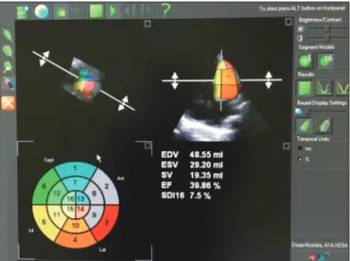
Each volume curve was analyzed to obtain end-diastolic (EDV) and end-systolic (ESV) volumes that were identified as the maximum and minimum values, and EF was computed. The algorithm calculated and displayed automatic contours for the 2-chamber in the long-axis views. When the detected bound- ary is approved, the tool provides both global and segmental information including EDV, ESV, systolic volume, and EF (Fig. 1). The mean analyzing time was calculated.
To determine the reproducibility of LV volume measurements for RT3DE, image analysis was repeated in a random sample of 15 patients by an additional investigator as well as by the same primary reader at least 1 week later. During these repeat-
Fig. 1. Measurements of 3-dimensional left ventricular volumes using 3000 element matrix probe (3V probe) and analyzing the images of the ventricle using Tom Tec 4D Cardio-View RT 1.2 software. EDV: end- diastolic volume, ESV: end-systolic volume, SV: systolic volume, EF:
ejection fraction, SDI: standard deviation index.
ed analyses, the investigators were blinded to the results of all prior measurements. Inter- and intra-observer variability was calculated as the absolute difference of the corresponding pair of repeated measurements in percent of their mean in each pa- tient and then averaged over the 15 patients.
Biplane cineangiography
It was obtained in long axial projections (right anterior oblique 30° in frontal position and left anterior oblique 60°
with 20° cranial angulation in lateral position) at 35 frames/
sec. LV volume was calculated using the following equation assuming that LV geometry approximated with considerable accuracy by an ellipsoid. Correction for image magnification and distortion from non-parallel X-rays was performed ac- cording to the method of Greene et al.10)
LV end diastolic volume (LVEDV) = 4/3 π [(L/2) (M/2) (N/2)] = π/6 (LMN)
Where V is the LV volume, L is the long axis, M is the short axis in one plane and N is the short axis in the other plane.10)
Then, LVEDV index (LVEDVI) was calculated by dividing
LVEDV by body surface area.11) Statistical analysis
All statistical calculations were done using computer pro- grams SPSS (SPSS Inc., Chicago, IL, USA) version 15 for Micro- soft Windows.
Results
The study included 19 males (47.5%) and 21 females (52.5%).
Fourteen patients were offspring of consanguineous marriage.
Their mean age was 3.0 ± 1.8 years; their age ranges between 11 months and 8 years with median 2.35 years, their mean weight was 12.9 ± 3.4 kg, and their mean body surface area was 0.5 ± 0.1 m2. The control group included 18 children with age ranged between 10 months to 6.5 years, median age 2.5 years.
The group included 10 females (56%) and 8 males (44%).
Table 1 shows the demographic, echocardiographic data in- cluding conventional echocardiography and RT3DE and the angiographic data of the included patients. Two cases (5%) only had LVEDVI between 20–30 mL3/m2 and the remaining 38 cases (95%) had a volume > 30 mL3/m2.
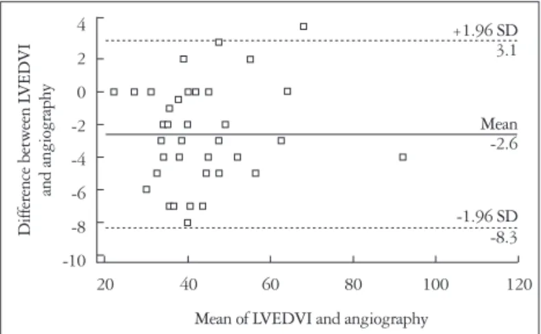
There was a good correlation between RT3DE and cinean- giography for estimation of LVEDVI in all studied patients (r = 0.97, p < 0.001) (Fig. 2). Bland-Altman plot shows the com- parison between the LVEDVI by RT3DE and angiography (Fig. 3). Bias was -2.6, SD was ± 1.96. The upper limit of agree- ment was 3.1 and the lower limit of agreement was -8.3.
We compared the 2D and RT3DE echocardiographic find- ings of both cases and controls as shown in Table 2. LVEDV measured by RT3DE in the studied TOF cases was with a mean value of 22.5 ± 7.4 mL3. The mean LVEDV of the stud- ied TOF cases was less than that of control group, whose mean value was 33.3 ± 19.99 mL3. These results showed clearly that LV volume was less than normal in most of our patients. Statis-
Table 1. Demographic and conventional and RT3DE echocardio- graphic and angiographic data of the studied cases
Variables Mean ± SD
Age (years) (03.0 ± 1.8
Weight (kg) (12.9 ± 3.4
Height (cm) (91.2 ± 12.8
Body surface area (m2) (00.5 ± 0.1
Conventional echocardiography
LVEDD (cm) (02.7 ± 0.30
LVESD (cm) (01.7 ± 0.3
EF (%) (65.3 ± 4.7
FS (%) (39.5 ± 6.3
LPA (cm) (00.8 ± 0.3
RPA (cm) (00.9 ± 0.3
PG across the RVOT (mm Hg) 80.0 (± 14.3)
Macgoon’s ratio (02.5 ± 2.2
RT3DE echocardiography
LVEDV (mL) (22.5 ± 7.4
LVEDVI (mL/m2) (41.5 ± 12.9
ESV (mL) (10.8 ± 3.9
SV (mL) (11.6 ± 4.0
EF (%) (51.5 ± 6.3
Angiographic data
LVEDV (mL) (23.9 ± 7.4
LVEDVI (mL/m2) (44.2 ± 12.6
EF: ejection fraction, ESV: end-systolic volume, FS: fractional shortening, LPA: left pulmonary artery, LVEDD: left ventricular end diastolic dimen- sion, LVEDV: left ventricular end diastolic volume, LVEDVI: left ventricu- lar end diastolic volume index, LVESD: left ventricular end systolic dimen- sion, RPA: right pulmonary artery, RVOT: right ventricular outflow tract, SV: systolic volume, RT3DE: real time three dimensional echocardiography, PG: pressure gradient
Fig. 2. Good correlation between real time three dimensional echocar- diography (RT3DE) and cineangiography for estimation of left ventricular end diastolic volume index (LVEDVI) in all studied patients.
40.00
30.00
20.00
10.00
10.00 15.00 20.00 25.00 30.00 35.00 40.00 r = 0.97
LVEDVI by RT3DE
LVEDVI by angiography
tical analysis showed that there was a significant difference be- tween LVEDV measured by RT3DE in the studied TOF pa- tients and that of control group (p < 0.004) as shown in Table 2.
There was significant under estimation of LVEF by RT3DE echocardiography in comparison to 2D echocardiography (p <
0.001).
Measurement was done without any manual corrections in 5 patients (13%), and with manual corrections in 35 patients (87.5%). The mean analyzing time was 20 min per case.
Intraobserver variability with RT3DE was low (1.9%) while the interobserver variability was acceptable as well (3.7%).
Discussion
We aimed to correlate the RT3DE as a non-invasive tool of
assessment of the LV with the biplane angiography as a refer- ence standard tool.
In our study, the LVEDV and the LVEDVI of our TOF pa- tients were significantly lower than controls. The LV in infants and children with TOF is often smaller than normal but rarely hypoplastic.12) The development of the LV muscle fibers was correlated with the volume of pulmonary blood flow or LV in- flow volume in children with TOF.13)
The LV volume determined with RT3DE was strongly cor- related with that assessed by angiography which was as re- ported by other investigators.9) LVEDV estimated by RT3DE was smaller than that estimated by angiography. This mild underestimation was explained by various reasons. First, the patient’s respiratory movement was difficult to control to ac- quire RT3DE images. Secondly, the transducer was positioned in only an apical acoustic window. Technically, to acquire a RT3DE data set, the whole LV cavity could not always be placed within the image angle.9) Also this is near to the study done by Heusch et al.14) who denoted that LVEDV measured by RT3DE was smaller than those calculated from the angiocardiography but the correlation between the two methods is good (r = 0.93, p = 0.0001).
The above data clearly illustrates that RT3DE can be used as a reliable method for accurate estimation of left heart vol- ume in children.
2D echocardiography alone cannot be used for assessment of the LV volume because 2D echocardiography measures single ventricular dimension rather than a ventricular volume. This dimension can give a good impression of cavity size but less accurate for precise quantification of LV volume.15)
In our study, LVEF measured by RT3DE was significantly underestimated in comparison to that measured by 2D echo- cardiography, which suggested that RT3DE underestimate EF when compared to 2D echocardiography. This was also report- ed by other investigators. The reason for this underestimation may be that RT3DE cannot consistently differentiate between the myocardium and the trabeculae. So, it is recommended to trace the endocardium to exclude trabeculae in the LV cavity for 3D echocardiography. As well, one-beat acquisitions may not successfully capture true end-systole, because of the reduced temporal resolution. This will lead to inaccurate ESV calcula- tions and EF measurements.16)
The study was limited by being performed for patients with only TOF and not other types of congenital heart disease. So, we need future studies in other types of congenital heart dis- ease. In addition, the only available RT3DE transducer had an operating frequency of 2–4 MHz. Finally, the software require manual tracing of the endocardial boundaries so, introduction of automated border detection, if accurate, would make this technique easier, faster and more feasible in the future.
In conclusion, in patients with good acoustic windows, RT3DE using state-of-the-art technology provides accurate and reproducible measurements of global LV volumes.
Table 2. Comparison between 2D and RT3DE echocardiographic findings of the studied cases and controls
Variables Cases,
mean ± SD Controls,
mean ± SD p-value RT3DE
LVEDV (mL) 22.5 ± 7.4 33.3 ± 19.99 0.004 LVEDVI (mL/m2) 41.5 ± 12.9 53.2 ± 20.2 0.01
ESV (mL) 10.8 ± 3.95 15.6 ± 9.2 0.008
SV (mL) 11.6 ± 3.99 17.6 ± 11.1 0.004
EF (%) 51.5 ± 6.3 52.1 ± 4.98 0.8
2D echocardiography
LVEDD (cm) 02.7 ± 0.3 02.9 ± 0.7 0.1
LVESD (cm) 01.7 ± 0.3 02.8 ± 4.3 0.1
LVEDV (mL) 19.4 ± 6.6 28.8 ± 19.6 0.008 LVEDVI (mL/m2) 35.5 ± 9.9 45.3 ± 21.0 0.02
ESV (mL) 06.8 ± 2.7 09.7 ± 6.6 0.02
EF (%) 65.3 ± 4.7 66.8 ± 3.3 0.2
FS (%) 39.5 ± 6.3 37.9 ± 2.8 0.3
LVEDV: left ventricular end diastolic volume, LVEDVI: left ventricular end diastolic volume index, ESV: end-systolic volume, SV: systolic volume, EF:
ejection fraction, LVEDD: left ventricular end diastolic dimension, LVESD:
left ventricular end systolic dimension, FS: fractional shortening, RT3DE:
real time three dimensional echocardiography
Fig. 3. Bland-Altman plot showing comparison between the left ventricular end diastolic volume index (LVEDVI) by real time three dimensional echocardiography and angiography.
4 2 0 -2 -4 -6 -8 -10
20 40 60 80 100 120 Difference between LVEDVI and angiography
Mean of LVEDVI and angiography
+1.96 SD 3.1
Mean -2.6
-1.96 SD -8.3
• Acknowledgements
The study was completely funded by Cairo University.
References
1. Riehle TJ, Mahle WT, Parks WJ, Sallee D 3rd, Fyfe DA. Real-time three-dimensional echocardiographic acquisition and quantification of left ventricular indices in children and young adults with congenital heart disease:
comparison with magnetic resonance imaging. J Am Soc Echocardiogr 2008;
21:78-83.
2. Monaghan MJ. Role of real time 3D echocardiography in evaluating the left ventricle. Heart 2006;92:131-6.
3. Sugeng L, Weinert L, Lang RM. Left ventricular assessment using real time three dimensional echocardiography. Heart 2003;89 Suppl 3:iii29-36.
4. Bu L, Munns S, Zhang H, Disterhoft M, Dixon M, Stolpen A, Son- ka M, Scholz TD, Mahoney LT, Ge S. Rapid full volume data acquisi- tion by real-time 3-dimensional echocardiography for assessment of left ven- tricular indexes in children: a validation study compared with magnetic resonance imaging. J Am Soc Echocardiogr 2005;18:299-305.
5. Mannaerts HF, Van Der Heide JA, Kamp O, Papavassiliu T, Marcus JT, Beek A, Van Rossum AC, Twisk J, Visser CA. Quantification of left ventricular volumes and ejection fraction using freehand transthoracic three- dimensional echocardiography: comparison with magnetic resonance imaging.
J Am Soc Echocardiogr 2003;16:101-9.
6. Jenkins C, Bricknell K, Hanekom L, Marwick TH. Reproducibility and accuracy of echocardiographic measurements of left ventricular parame- ters using real-time three-dimensional echocardiography. J Am Coll Cardiol 2004;44:878-86.
7. Sugeng L, Mor-Avi V, Weinert L, Niel J, Ebner C, Steringer- Mascherbauer R, Schmidt F, Galuschky C, Schummers G, Lang RM, Nesser HJ. Quantitative assessment of left ventricular size and func- tion: side-by-side comparison of real-time three-dimensional echocardiography and computed tomography with magnetic resonance reference. Circulation 2006;114:654-61.
8. Soriano BD, Hoch M, Ithuralde A, Geva T, Powell AJ, Kussman BD,
Graham DA, Tworetzky W, Marx GR. Matrix-array 3-dimensional echo- cardiographic assessment of volumes, mass, and ejection fraction in young pe- diatric patients with a functional single ventricle: a comparison study with cardiac magnetic resonance. Circulation 2008;117:1842-8.
9. Iino M, Shiraishi H, Ichihashi K, Hoshina M, Saitoh M, Hirakubo Y, Morimoto Y, Momoi MY. Volume measurement of the left ventricle in children using real-time three-dimensional echocardiography: comparison with ventriculography. J Cardiol 2007;49:221-9.
10. Greene DG, Carlisle R, Grant C, Bunnell IL. Estimation of left ventric- ular volume by one-plane cineangiography. Circulation 1967;35:61-9.
11. Graham TP Jr, Faulkner S, Bender H Jr, Wender CM. Hypoplasia of the left ventricle: rare cause of postoperative mortality in tetralogy of Fallot.
Am J Cardiol 1977;40:454-7.
12. Matsuda H, Hirose H, Nakano S, Kishimoto H, Kato H, Kobayashi J, Miura T, Kato M, Kawashima Y. Age-related changes in right and left ventricular function in tetralogy of Fallot. Jpn Circ J 1986;50:1040-3.
13. Fukuda J, Izumi T, Matsukawa T, Eguchi S. Development of left ven- tricular muscle in tetralogy of Fallot. Jpn Circ J 1984;48:465-73.
14. Heusch A, Rübo J, Krogmann ON, Bönig H, Bourgeois M. Volume measurement of the left ventricle in children with congenital heart defects:
3-dimensional echocardiography versus angiocardiography. Cardiology 1999;92:45-52.
15. Kaye HH, Tynan M, Hunter S. Validity of echocardiographic estimates of left ventricular size and performance in infants and children. Br Heart J 1975;37:371-5.
16. Lang RM, Badano LP, Tsang W, Adams DH, Agricola E, Buck T, Faletra FF, Franke A, Hung J, de Isla LP, Kamp O, Kasprzak JD, Lancellotti P, Marwick TH, McCulloch ML, Monaghan MJ, Nihoy- annopoulos P, Pandian NG, Pellikka PA, Pepi M, Roberson DA, Shernan SK, Shirali GS, Sugeng L, Ten Cate FJ, Vannan MA, Zamo- rano JL, Zoghbi WA; American Society of Echocardiography; Euro- pean Association of Echocardiography. EAE/ASE recommendations for image acquisition and display using three-dimensional echocardiography.
Eur Heart J Cardiovasc Imaging 2012;13:1-46.