Copyright ⓒ 2011, The Korean Academy of Oral Biology
187
Journal of Oral Biology
Protective Effect of HP08-0106 on Ligature-induced Periodontitis in Rats
Hwa Jung Choi, Hyoung Kwon Cho
1, and Yunjo Soh*
1
Department of Dental Pharmacology, School of Dentistry, and Institute of Oral Bioscience, Brain Korea 21 project, Chonbuk National University, Jeon-Ju 561-756, Korea
2
HanPoong Pharmaceutical Co., LTD., Jeon-Ju 561-841, Korea
(received October 26, 2011 ; revised December 12, 2011 ; accepted December 14, 2011)
Periodontitis is an inflammatory disorder of the periodontium, characterized by destruction of the tooth supporting tissues including alveolar bone and mediated by various pro-inflammatory mediators. Here, we demonstrated that HP08-0106, composed of four crude drugs-Gardenia jasminoides Grandiflora, Angelica gigas Nakai, Rehmannia glutinosa, and Schizonepeta tenuifolia in a weight ratio of 2:2:1:2, perturbs inflammatory responses, osteoclast form- ation in LPS-induced RAW 264.7 cells and alveolar bone resorption in ligature-induced periodontitis. HP08-0106 decreased the protein level of iNOS and COX2 as well as the secreted level of IL-1 β, indicating that HP08-0106 has anti- inflammatory effects. HP08-0106 also inhibited the expression of genes associated with osteoclastogenesis including c-Fos, MMP-9 and TRAP. Moreover, HP08-0106 exhibited a protective effect from alveolar bone loss in ligature-induced periodontitis animal models. Our results strongly suggest that HP08-0106 represent an important therapeutic tool to treat inflammatory disorders associated with bone loss such as periodontitis.
Key words: HP08-0106, periodontitis, anti-inflammatory, osteoclastogenesis
서 론
치주염(periodontitis)은 치주 결합 조직의 파괴, 치주 부 착의 소실, 치조골의 흡수를 야기하는 염증성 질환이다.
세균 산물의 자극하에 치주 염증이 일어나는 동안 다형 핵 백혈구, 림프구, 단핵구, 대식세포는 넓은 범위의 전염 증성 cytokine과 prostaglandin을 합성하며 이를 통한 지 속적인 염증은 일반적으로 치은과 같은 연조직뿐 만 아니 라 치조골을 포함한 주변 경조직 파괴를 동반한다(Park et al., 2010).
세균성 치태를 형성하는 미생물들과 그 부산물들은 치 주조직의 면역-염증반응을 일으켜 치주질환의 근본원인을 제공한다. Lipopolysaccharide (LPS)는 그람 음성의 박테 리아 세포막의 대표적인 구성 성분이며, 염증을 매개하는 macrophage 또는 monocyte를 자극하여 nitoric oxide (NO), tumor necrosis factor-α (TNF-α), interleukins, prostanoids 및 leukotrienes 등의 염증 매개물을 분비하도록 한다(Chen et al., 2005; Hewett et al., 1993; Schumann et al., 1990). 세포 내에서 염증의 전반적인 과정은 COX-2 와 iNOS의 활성화를 통하여 이루어진다(Halliwell et al., 1984). 일반적으로 nitric oxide synthase (NOS) 중에서 iNOS는 염증성 cytokines, interferon-γ 및 LPS의 자극 에 의해서 발현이 증가되는데 이에 의해 생성된 NO는 혈관투과성, 부종 등의 염증반응을 촉진, 심화시킨다(Mu et al., 2001; Ryu et al., 2003). Cyclooxygenase (COX) 는 arachidonic acid를 prostaglandins (PGs)로 전환하는 효소인데 특히 COX-2는 미생물에 의한 감염이나 손상, 여러 가지 요인에 의한 스트레스 반응에 대해 macrophage 에서 발현되어 arachidonic acid로부터 염증 매개 물질인 PGE2를 생성하여 염증 및 면역반응을 촉진시키는 작용을 한다(Kim et al., 2004). 이와 같은 지속적인 염증은 치 주질환에 있어 치조골의 소실로 이어지게 되고 이는 점차 치아의 지지력을 약화시켜 치아의 요동과 상실을 초래할 수 있다(Mundy, 1993; Pitaru et al., 1994). 따라서 최근 치주질환의 치료방법으로 염증 억제와 더불어 궁극적으
*Corresponding author: Yunjo Soh, Department of Dental Pharmacology, School of Dentistry, Chonbuk National Univer- sity, Duck-Jin Dong, Duck-Jin Ku, Jeon-Ju 561-756, Korea Tel: +82-63-270-4038, Fax: +82-63-270-4037
E-mail: [email protected]
로 골 소실을 억제하는 치료법에 대한 연구가 많이 진행 되고 있다(Niikura et al., 2005).
본 연구진은 기존 연구와 전통문헌을 통해 치자(Gardenia Jasminoides), 당귀(Angelica gigas Naka), 건지황(Rehmannia glutinosa), 형개(Schizonepeta tenuifolia var. japonica)가 각각 염증관련 효능이 있음을 확인 하고(Ozaki et al., 2002; Xu et al., 2009; Park et al., 2009; Underwood, 2009), 이들을 적절한 배율로 혼합하여 HP08-0106을 만 들어 치주염에 보호작용을 나타낼 것으로 가정하고 본 연 구를 진행하였다. 염증에 대한 효과를 관찰하기 위해 RAW 264.7 세포를 LPS로 자극하여 TNF-α, IL-1β 분비량의 변화와 COX2 및 iNOS의 단백질량의 차이를 조사하였 고, 파골세포 분화에 대한 효과와 더불어, 흰쥐를 이용한 치주질환 모델동물을 통하여 HP08-0106의 치주질환에 대 한 in vivo 효과를 알아보았다.
재료 및 방법
HP08-0106 제조
HP08-0106의 천연물 원료는 치자, 당귀, 건지황, 형개로 서, 각각 57.0(g), 57.0(g), 28.5(g), 57.0(g)를 준비하여 원 료량의 8배수의 물을 용매로 하여 3시간 1회 추출 후, 200매쉬로 여과하여 60oC 이하에서 감압농축 후, 건조하 였으며, 이는 (유)한풍제약 (전북 전주, 한국)에서 실시하 였다.
실험재료
Dulbecco’s modified Eagles (DMEM), α-minimum essential medium (α-MEM), fetal bovine serum (FBS), trypsin-EDTA 및 세포배양에 필요한 시약은 Gibco labora- tories (Invitrogen Co, Grand Island, USA)로부터 구입하 였다. 마우스 재조합 RANKL은 PeproTech (Rocky Hill, NJ, USA)으로부터, 흰쥐 어금니를 묶는데 쓰인 고무밴드 TRU-FORCE는 TP Orthodontics Inc. (La Porte, IN, USA)에서 구입하였다. Leukocyte Acid Phosphatase Assay Kit와 나머지 실험에 필요한 시약은 문헌에 나온 대로 Sigma (St. Louis, MO, USA)에서 구입하였다(Soh et al., 2000;
Soh et al., 2003).
세포배양과 파골 세포 분화 유도
Murine monocyte/macrophage RAW264.7 세포는 미국 세포주 은행(American Type Culture Collection, Manasas, VA, USA)로부터 구입하여, DMEM 배지에 10% 열처리 된 FBS와 페니실린(100 U/ml)을 첨가하여, 5% CO2, 37oC 와 100% 습도가 유지되는 CO2배양기에서 배양하였다. 파 골 세포 분화를 위해 RAW 264.7 세포를 10% FBS, 2 mM L-glutamate, 100 U/ml 페니실린, 100 µg/ml streptomycin
이 포함된 α- MEM 배지에 잘 섞어주고, well당 3 × 10³ 개의 세포가 되도록 96-well plate에 분주하였다. 50 ng/
ml soluble RANKL 과 HP08-0106을 농도 별로(0, 0.1, 1, 10µg/ml) 처리하여 2~3 일마다 신선한 배지로 교환해 주면서 6일 동안 배양하여 파골 세포 분화를 유도하였다 (Wittrant 등 2004). 배양이 끝난 후, 파골 세포의 분화를 검사하기 위해 세포를 고정하여 TRAP 염색을 시행하였다.
MTT 측정
RAW 264.7 세포를 10% FBS가 포함된 배양배지에 well 당 5 × 10³개의 세포가 되도록 96 well에 분주하고 16시 간 동안 배양하였다. 세포는 다양한 농도의 HP08-0106을 24시간 처리하고, 배양시간이 끝나면 PBS (phosphate- buffered saline)로 3번 씻어주고, 100 µg/ml of MTT [3- (4,5-dimethylthiazol-2-yl)-2,5-diphenyltetrazolium bromide] 용액을 처리하여 37oC에서 2시간 동안 배양하 였다. 다시 한번 PBS로 3번 씻어주고, 200 µl DMSO를 처리하여 그 결과물을 분광 광도계를 이용하여 540 nm에 서 흡광도를 측정하였다.
Tartrate-resistant acid phosphatase (TRAP) staining TRAP 염색법은 이미 실시된 방법(Han et al., 2007)과 Sigma 사의 TRAP 염색용 kit을 구입하여 사용 설명에 따라 파골 세포를 염색하였다. 염색이 끝난 후, 광학 현 미경을 이용하여 핵이 3개 이상인 TRAP-양성 다핵세포 를 계수하여 파골 세포의 생성 지표로 삼았다.
역전사연쇄중합반응(Reverse Transcription -Polymerase Chain Reaction)
총 RNA는 Invitrogen 사의 TRIzol을 이용하여 세포로 부터 분리하였고, SuperScript II reverse transcriptase (Invitrogen)를 이용하여 사용 설명에 따라 cDNA를 합성 하였다. iMAX taq DNA polymerase (iNtRON Biote- chnology, Sungnam, Korea)을 이용하여 PCR을 시행하 였다. PCR primer를 Bioneer (Daejeon, Korea)에서 구입 하였고, primer 서열과 PCR 산물의 크기를 Table 1에 나 열하였다. 94oC 에서 1분 동안 초기 변성을 시키고, PCR 반응은 변성반응을 94oC에서 30초, 결합반응을 30초 그 리고 중합반응을 72oC에서 30초간 30주기를 반복하였고 마지막 중합반응은 72oC에서 10분간 반응시켰다. PCR 산 물을 1.5% agarose gel을 이용하여 전기영동으로 분리하 였고 ethidium bromide로 염색하여, Phosphoimager 와 Quantity One software Version 4.3.1 (Bio-Rad, Hercules, CA, USA)을 이용하여 분석 및 정량 하였다.
Immunoblot analysis
세포를 PBS로 세척하고, lysis buffer [20 mM Tris-HCl (pH 7.5), 137 mM NaCl, 10% glycerol, 1% Triton X-
100, 1 mM phenylmethylsulfonylfluoride (PMSF), 1 mM Na3VO4, 1× protease inhibitor cocktail]를 세포에 첨가 한 후, 원심 분리하여 단백질을 추출해 내었다. 단백질 정 량은 BCA reagent (Pierce, Rockford, IL, USA)를 이용 하였다. 단백질(30~40 µg)을 4 × sample buffer에 섞은 후, 10~12% SDS-PAGE를 시행하고, polyvinylidene difluoride (Bio-Rad) 막에 옮겼다. 5% nonfat skim milk가 포함된 TTBS (0.25% Tween-20 in Tris-buffered saline) 용액에 막을 침투시켜 실온에서 1시간 동안 반응시키고, anti-COX2 (Santa Cruz Biotechnology), anti-iNOS (Santa Cruz Biotechnology), anti-β-actin (Santa Cruz Biotechnology) 항체를 5% nonfat skim milk가 포함된 TTBS에 1:1,000 비율로 희석하여 4oC 에서 16시간 동안 반응시켰다. 그리 고, Horseradish peroxidase-conjugated anti-rabbit or mouse antibodies (Santa Cruz Biotechnology)를 각각 이차 항체 로 이용하여, 5% nonfat skim milk가 포함된 TTBS 에 1:5,000 ~ 1:10,000 비율로 희석하여 실온에서 1시간 동안 반응시켰다. ECL Plus kit (Amersham Biosciences, Piscataway, NJ, USA)를 이용하여 해당 단백질을 관찰하 였다.
치주질환 동물 모델 구축 및 투여
실험에 사용한 동물은 180-230 g의 Sprague-Dawley계 수컷 흰쥐를 (주)다물사이언스 (Daejeon, Korea)에서 구 입하였다. 동물실험은 전북대학교 Institutional Animal Care and Use Committee의 규정에 따라 행하여졌다. 사료와 물을 충분히 공급하면서 1주간 실험환경에 적응시키고, 대 조군과 실험군을 나누어 실험에 사용하였다. 동물 사육실 내의 온도는 20-24oC, 습도는 50-60%로 유지하면서 낮과 밤의 주기는 각각 12시간으로 하였다. 대조군과 HP08- 0106 투여군은 각각 5마리로 하였으며, HP08-0106을 투 여하기 전 날 상악 두 번째 어금니를 염증균(P. gingivalis) 이 처리된 밴드를 이용하여 묶음(ligature)으로 치주염을 유발시켰으며, 대조군과 HP08-0106 투여군(200 및 500 mg/
kg)에 1일 1회 각각 생리 식염수와 시료를 경구투여 하 였다. 동물을 8일 후 희생시키고 상악 어금니부위를 얻 어 탈석회시키고, H&E 염색법을 이용하여 염색하였다.
Niikura 등(2005)의 보고를 참고로 미세 현미경을 통해 치조골의 변화를 해부조직학적으로 비교 분석하였다.
4-hydroxyproline (HYP) assay
HYP 측정은 간성상세포인 HSC-T6 세포를 이용하여 DMEM에서 배양하여 실험을 실시하였다(Choi et al., 2007). 흰쥐 성상 세포주(HSC-T6)에 대조군과 HP08-0106 을 투여하여 배양 후 12N HCl을 첨가하여 105oC에서 18시간 동안 단백질을 가수분해하였다. 0.56%의 완충용 액(chloramine-T)과 Ehrlich’s reagent를 첨가하고 50oC 에서 90분간 반응시키고, 558 nm에서 HYP양을 측정하 였다.
통계처리방법
본 실험의 통계처리는 Student’s t test 방법을 사용하 였으며, 값은 평균과 표준오차로 나타냈다. 대조군과 비 교하여 P < 0.05인 값을 유효성 있는 값으로 표기하였다.
결 과
HP08-0106의 RAW 264.7 세포의 생존능력에 대한 효과 마우스 대식세포인 RAW264.7에 대한 HP08-0106의 생 존능력에 대한 효과를 확인하기 위해서 MTT assay를 실 시하였다. HP08-0106을 농도 별로(0.1, 1, 10 µg/ml) 24 시간 처리한 결과, 각각의 농도에서 HP08-0106가 세포의 생존율에는 영향을 주지 않음을 확인할 수 있었다(Fig.
1). 이는 HP08-0106의 항염증 효과가 단순한 세포의 사 멸에 의한 세포의 염증성 매개물질의 생성 억제가 아니라 HP08-0106 고유의 효과임을 알 수 있었다.
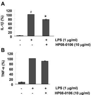
HP08-0106의 TNF-α과 IL-1β의 분비량에 미치는 영향 RAW 264.7 세포에 LPS와 HP08-0106을 처리한 후, TNF-α와 IL-1β의 분비량의 변화를 ELISA kit을 이용하 여 측정하였다. LPS를 처리하지 않은 세포와 비교하였을 때 LPS를 처리한 세포에서 TNF-α와 IL-1β의 분비량이 증가하는 것을 확인하였다. HP08-0106을 10 µg/ml의 농
Table1. Primer sequences for RT-PCR
Target genes (Accession Number)
Primers Forward/Reverse
PCR condition Tm(
oC) size(bp) TRAP
(NM_007388)
5’- ctgctgggcctacaaatcat -3’
3’- ggtagtaagggctggggaag -5’ 55 400
MMP-9 (NM_013599)
5’-cgtcgtgatccccacttact -3’
3’-tcctgggcaagcagtactct -5’ 55 433
c-Fos (NM_010234)
5’-atgggctctcctgtcaacac -3’
3’-tggagtttattttggcagcc -5’ 55 480
GAPDH (NM_008084)
5’- accacagtccatgccatcac-3’
5’-tacagcaacagggtggtgga -3’ 56 452
도로 처리하였을 때 IL-1β의 분비량이 확실히 감소됨을 관 찰하였고, IL-1β의 분비량 감소는 통계학적으로도 유의함 을(P < 0.01) 확인하였다(Fig. 2A). 그러나, TNF-α의 분비 량은 HP08-0106에 의해 큰 영향을 받지 않았다(Fig. 2B).
HP08-0106의 COX2와 iNOS 단백질량에 미치는 영향 HP08-0106이 COX2와 iNOS 단백질량에 영향을 주는
지 알아보기 위해 RAW 264.7 세포에 LPS와 10 µg/ml 의 HP08-0106을 처리하여 COX2와 iNOS의 단백질량을 Western blot의 방법으로 확인하였다. 그 결과, LPS를 처 리한 세포에서 LPS에 의한 COX2의 단백질량이 증가되 는 것을 확인하였고, 또한 10 µg/ml의 HP08-0106에 의해 서 iNOS와 COX2의 단백질량이 모두 현저히 감소하는 것을 확인할 수 있었고, 통계학적으로도 의미 있음(P < 0.01) 을 확인하였다(Fig. 3).
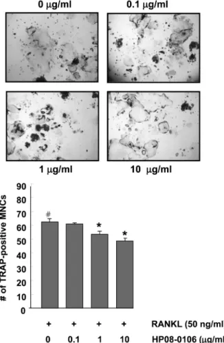
HP08-0106의 파골 세포 분화 유도에 미치는 영향 HP08-0106의 파골 세포 분화에 대한 효능을 확인하기 위해, 마우스 대식 세포인 RAW 264.7 세포에 RANKL (50 ng/ml)을 이용하여 6 일 동안 파골 세포 분화를 유도 하였고, TRAP 염색법을 통해 확인하였다. 그 결과, RANKL 처리된 세포군에서 파골 세포 분화 유도를 확인할 수 있 었고, HP08-0106을 농도 별로(0.1, 1, 10 µg/ml) 함께 처 리한 세포군에서는 HP08-0106의 농도 의존적으로 다핵성 세포인 파골 세포가 감소하는 것을 확인하였다(Fig. 4).
Fig. 1. Effect of HP08-0106 on cell viability in RAW264.7 cells.
RAW 264.7 cells were treated with various concentrations (0, 0.1, 1, 10 µg/ml) of HP08-0106 for 24 h. Cell viability was measured by MTT assay. Data represent mean ± S.D. of three independent experiments triplicate in each run.
Fig. 2. Effect of HP 08-0106 on the LPS stimulated TNF-α and IL- 1β product in RAW 264.7 cells. RAW264.7 cells were serum- starved for 16 h, pretreated with or without 10 µg/ml of HP08-0106 for 30 min, stimulated with LPS (1 µg/ml). The amount of IL-1β (A) and TNF-α (B) secretion was measured by ELISA method.
Data represent mean ± S.D. of three independent experiments trip- licate in each run. Asterisk (*) indicates a significant difference (P < 0.05) compared with the control (
#).
Fig. 3. Effect of HP 08-0106 on the LPS-induced COX2 and iNOS
expression in RAW 264.7 cells. RAW264.7 cells were serum-
starved for 16 h, pretreated with or without 10 µg/ml of HP 08-0106
for 30 min, and stimulated with LPS (1 µg/ml) for 24 h. Cell
extracts were analyzed by Immunoblot analysis with antibodies
against COX2 (A) and iNOS (B). The histograms represent protein
levels compared with loading control, β-actin. Asterisk (*) indicates
a significant difference (P < 0.05) compared with the control (
#).
HP08-0106의 파골 세포 분화 관련 유전자 발현에 미 치는 영향
파골 세포 분화에 따른 유전자 발현의 차이를 알아보기 위해, 파골 세포 분화 지표로 사용되는 c-Fos, MMP-9, TRAP의 유전자 발현량을 RT-PCR을 통해서 확인하였다.
그 결과, HP08-0106을 처리한 세포군에서 c-Fos, MMP- 9, TRAP의 발현이 HP08-0106을 처리하지 않은 세포군에 비해 감소하는 것을 확인하였고, 이들 유전자는 모두 P <
0.01의 값으로 통계학적으로도 의미가 있음을 확인할 수 있었다(Fig. 5).
HP08-0106의 4-hydroxyproline (HYP) 분비량에 대한 영향
HP08-0106의 조직 지지 정도를 관찰하기 위해, 잇몸조 직을 지지하는 콜라겐의 함량을 유추해볼 수 있는 HYP 함량을 측정, 비교하였다. 콜라겐 생성 정도는 HYP에 비
례하고, 간 성상세포가 저산소 상태에서 배양 시 HYP 양 이 유의하게 증가하므로(Choi 등, 2007) 간 성상세포를 이용하였다. 실험 결과 HP08-0106을 처리하지 않은 세포 군과 비교해 10 µg/ml의 HP08-0106을 처리한 세포군에서 HYP 분비량이 증가하는 것을 확인 하였으며, 통계학적으 로 의미가 있음을 확인하였다(Fig. 6). 이 실험 결과는 HP08- 0106이 콜라겐의 합성을 증가시킴을 의미한다.
Fig. 5. Suppression of RANKL-induced osteoclastogenesis-associ- ated gene expression by HP08-0106. RAW264.7 cells were cul- tured with 10 µg/ml HP08-0106 in the presence of RANKL (50 ng/
ml). After 6 days of culture, the mRNA expression level of c-Fos, MMP-9 and TRAP genes was determined by RT-PCR compared with that of GAPDH. The histogram represents the level of the mRNA expression fold compared with that of the control (#). The results are expressed as mean ± S.D. *P < 0.05 versus control (#).
Fig. 6. Effect of HP08-0106 on hydrixyproline (HYP) content. Rat hepatic stellate HSC-T6 cells were cultured in DMEM, and treated with HP08-0106 (10 µg/ml) for 24 h. After incubation, cells were harvested and the content of HYP was measured. Data represent mean ± S.D. of three independent experiments triplicate in each run, *P < 0.05 versus vehicle-treated cells.
Fig. 4. Effect of HP08-0106 on RANKL-induced osteoclastogene-
sis. RAW264.7 cells were cultured in the presence of RANKL (50
ng/ml) for 6 days. HP08-0106 was added into the culture media at a
final concentration of 0, 0.1, 1, 10 µg/ml. The cells were stained for
TRAP. TRAP positive multinucleated cells (# of TRAP
+MNCs)
were counted at magnification 100X and represented at the lower
histogram. Data represent mean ± S.D. of three independent exper-
iments duplicate in each run, *P < 0.05 versus vehicle-treated cells.
치주질환 동물 모델에서의 HP08-0106의 영향 HP08-0106의 동물에서의 효능을 확인하기 위해, 치주염 이 유발된 흰쥐를 이용하여 치조골의 변화를 분석하였다.
그 결과 정상적인 그룹의 치조골과 비교해 ligature와 P.
gingivalis가 처리된 그룹의 치조골에서 해부조직학적 관찰 결과 확실한 골 소실 증가를 확인하였고, HP08-0106을 200 및 500 mg/kg/day 처리한 그룹에서 치조골 소실량이 줄어든 것을 확인할 수 있었다(Fig. 7). 종합적으로 보았 을 때, 이는 HP08-0106이 치주질환으로 인한 치조골 감 소를 억제시키는데 있어 도움이 될 수 있음을 보여준다.
고 찰
치주질환은 세균성 치태에 대한 숙주의 면역, 염증반응 으로 인한 연조직의 부착 소실과 치조골의 파괴를 특징 으로 하는데, 근본적으로는 미생물들과 그 부산물들이 치 주조직의 병적인 변화를 가져오므로 전통적으로 이런 세 균성 요인의 기계적인 제거와 항생제요법이 기본적인 치 주질환의 치료와 예방법이 되어왔다(Niikura et al., 2005).
그러나 최근 염증과 골 소실에 대한 기초 연구가 진행됨 에 따라 치주염에서 나타나는 조직파괴가 주로 숙주의 면역, 염증 방어작용의 활성화에 의한 것이라고 알려진 이후 이러한 면역, 염증반응을 조절하는 새로운 방법들이
개발되고 있다. 현재 이에 대한 대표적인 약제로는 비스 테로이드성 소염제, subantimicrobial dose doxycycline이 나 chemically modified non-antimicrovial tetracyclines, bisphosphonate 등이 있다(Masferrer et al., 1994).
본 연구는 천연 원료로부터의 안전한 치주질환 치료제 개발에 대해 연구하던 중, HP08-0106이라 명명한 치자, 당귀, 건지황, 형개 혼합 추출물이 치주염에 대한 염증 억제 효과와 치조골의 파골 세포 분화 억제에 대해 효능 이 있음을 알게 되었다. 치자(Gardenia Jasminoides)는 열 병, 황달, 토혈, 소갈, 혈뇨, 혈리, 불면증, 결막염 등에 약 재로 사용해 왔으며, 최근에 RAW 264.7 세포에 대해 항 염증 효과가 있다는 보고가 있다(Xu et al., 2009). 당귀 (Angelica gigas Naka)는 최근 신경세포 분화 기능 과 항 산화 효과를 통한 신경세포 보호 효과 등이 알려져 있다 (Jeong et al., 2010; Li et al., 2011). 건지황(Rehmannia glutinosa)은 가공방법에 따라 생지황, 건지황, 숙지황으 로 분류되며, 주요 성분으로는 β-sitosterol, stigmasterol, campesterol 등이 포함된 phytosteol류, 당류, 아미노산, iridoid glycosides, inorganic elements 등이 보고되었으며 (Zhao et al., 2006), 이 중, iridoid glycosides는 free radical 을 억제하는 항산화 작용에 효과적인 것으로 보고되었다 (Kim et al., 2005; Raju et al., 2004). 형개(Schizonepeta tenuifolia var. japonica)는 최근 TNF-α를 억제하여 염증에 효과가 있는 것으로 알려져 있다(Kang et al., 2010). 이와
Fig. 7. Protective effect of HP08-0106 on alveolar bone loss from P.gingivalis and ligature-induced peridodontitis in rats. (A) Macroscopic
appearances and its histological H&E-stained images in the region between the first and second molar roots of rats with ligature-induced peri-
odontitis. (B) Quantitative analysis of the change in alveolar bone length using H&E stained sections. Asterisk (*) indicates a significant dif-
ference (P < 0.05) compared with the control (
#).
같이 이들은 주로 항산화 효능을 공통적으로 가지고 있으 며 현재까지 이들의 골 흡수에 대한 연구는 많이 이루어 지지 않았다.
지속적인 염증과 관련한 골 소실은 파골세포의 활성이 조골세포의 활성에 비해 증가함으로써 골형성량보다 골흡 수량이 증가하여 야기된다. 파골 세포는 조혈세포에서 유 래하는 거대 다핵 세포로, 풍부한 미토콘드리아, 골지체, 리소솜을 갖고 있으며 활성시 세포막이 돌출 함몰되어 형 성된 ruffled border를 통해 탄산탈수효소의 촉매로 이산 화탄소로부터 생성된 수소이온에 의해 골기질의 무기질을 용해시키고 산성상태에서 리소솜의 다른 효소와 더불어 단 백분해효소를 분비하여 유기성분을 분해, 소화시킴으로 골 을 흡수시킨다(Mundy, 1993). 파골세포의 전구세포는 여 러 가지 cytokines과 염증의 병태생리와 관련되어 파골세 포로 분화 및 활성화 된다(Mundy, 1993; Kumar et al., 2001). 특히 치주 질환에서 염증성 사이토카인의 생성은 파골 세포의 주요인자로 알려진 RANKL의 활성을 촉진 한다(Park et al., 2010).
본 연구에서는 HP08-0106의 치주질환에 대한 효능을 연 구하기 위해 TNF-α, IL-1β의 분비량과 COX2 및 iNOS 의 단백질 발현량의 차이를 조사 하였으며, 파골 억제에 대한 수준, 치조골의 길이를 평가하였다. 그 결과 HP08- 0106은 IL-1β와 같은 염증성 사이토카인의 분비량을 감 소시키는 효과를 나타냈으며, 염증관련 단백질인 COX2와 iNOS의 발현에도 큰 억제효과가 있음을 확인하였다. 또 한, HP08-0106은 파골 세포 분화 억제 효과와 c-Fos, MMP-9, TRAP과 같은 파골 세포 활성 관련 유전자 발 현을 감소시켜 파골 세포 분화 억제에도 효과적임을 확인 하였다. c-fos mRNA 발현은 85%정도 억제된 반면 TRAP assay결과 다핵성 파골세포는 의미 있게 감소하였다. 이는 HP08-0106에 함유된 많은 성분 중 일부는 c-fos pathway 를 억제하여 파골세포 분화를 억제하지만 또 다른 HP08- 0106 성분은 분화를 촉진하는 작용을 하여 파골세포 분화 의 정도가 c-fos의 억제 정도에 미치지 못함을 암시한다.
이와 더불어, HP08-0106은 콜라겐의 생성을 증가시켜 조 직 지지에 대한 효능과 치주질환 동물 모델을 통해서 나 타나듯이 치조골의 손실을 감소시키는 효과가 있음을 알 수 있었다. 따라서 본 연구의 결과로부터 HP08-0106의 투 여는 염증 작용의 억제와 파골 세포의 분화 및 활성 억 제를 통하여 치주질환의 진행을 완화시키는데 효과가 있 으며, 치주염 모델 동물을 통하여 생체 내에서 치조골 보 호효과를 가짐으로써 새로운 치주질환 치료제로서의 개발 가능성을 보여주었다.
감사의 글
이 논문은 2008년도 전북지방 중소기업청의 산학 공동
기술개발지원사업의 지원을 받아 연구되었음.
참 고 문 헌