Copyrightⓒ 2010, The Korean Academy of Oral Biology
145
Journal of Oral Biology
Protective Effect of HP08-0111 on Ligature-Induced Periodontitis
Young Ran Park, Hyoung Kwon Cho1, and Yunjo Soh*
Department of Dental Pharmacology, School of Dentistry, and Institute of Oral Bioscience, Brain Korea 21 project, Chonbuk National University, Jeon-Ju 561-756, Korea; 1HanPoong Pharmaceutical Co., LTD., Jeon-Ju 561-841, Korea (received August 23, 2010 ; revised October 6, 2010 ; accepted October 8, 2010)
Periodontitis is an inflammatory disorder of the periodon- tium and is characterized by destruction of the tooth sup- porting tissues, mediated by the upregulation of synthesis and release of a variety of pro-inflammatory factors. Inflam- matory cytokines and prostaglandins upregulate RANKL and its subsequent binding to RANK stimulates osteoclast formation, resorption activity, and survival. In our present study, we investigated the effects of HP08-0111, composed of Coptis japonica (Thunb.) Makino, vitamin C and vitamin E, upon inflammatory responses, osteoclastogenesis and alveo- lar bone loss. HP08-0111 decreased the expression of IL-1β and COX2 on LPS-induced RAW 264.7 cells and inhibited osteoclast-specific genes such as c-Fos, MMP-9, and TRAP.
HP08-0111 also exhibited protective effects against alveolar bone loss in rats with ligature-induced periodontitis. Our results suggest that HP08-0111 is potentially an important therapeutic tool for the treatment of disorders associated with bone loss such as periodontitis.
Key words: HP08-0111, periodontitis, anti-inflammatory, osteoclastogenesis
서 론
경조직과 연조직으로 구성된 치주조직은 치은, 치주인대, 백악질, 치조골로 구분된다. 치주인대는 결합조직으로서 치 근을 둘러싸고 있는 백악질과 치조골을 연결시키는 역할을 하고, 영양공급과 신경 전달 및 치조골의 혈관 통로를 통 하여 골수강과 연결되어 있다. 또한 치조골은 연속적인 골흡수와 골형성을 통해 골 재생이 일어나는 일어나는 기
관으로서, 골흡수와 골형성간 균형이 깨지게 되면 골소실 또는 골화석증이 일어날 수 있다(Mundy, 1993; Pitaru 등, 1994). 치주염의 진행시 염증으로 인해 골형성보다 골 흡 수가 많이 발생하여 치조골의 소실이 야기된다.
최근 여러 가지 cytokines과 염증 질환과의 관련성에 대 한 보고가 발표 되었다(Mundy, 1993; Kumar 등, 2001).
특히 치주 질환에서 염증성 cytokine의 생성은 파골 세포 의 주요인자로 알려진 RANKL의 활성을 촉진한다. 따라 서 골 흡수의 주세포인 파골 세포의 활성이 촉진되는데, 파골 세포는 조혈세포에서 유래하는 거대 다핵 세포로, 최 근 골에서 파골 세포의 형성과 기능에 대한 연구가 많이 진행되고 있다.
Lipopolysaccharide (LPS)는 그람 음성의 박테리아 세포 막의 구성 성분이며, RAW 264.7 세포와 같은 macrophage 또는 monocyte에서 nitoric oxide (NO), tumor necrosis factor-α (TNF-α), interleukins, prostanoids 및 leukotrienes 등의 염증 매개물을 자극하는 역할을 한다(Chen 등, 2005;
Hewett 등, 1993; Schumann 등, 1990). 일반적으로 nitric oxide synthase (NOS) 중에서 iNOS는 염증성 사이토카 인, interferon-γ, 및 LPS의 자극에 의해서 발현이 증가된 다. 정상적인 상태에서 NO는 항균작용과 종양제거의 작 용을 하지만(Weisz 등, 1996), 염증반응 때 발생하는 iNOS 에 의해 생성된 NO는 혈관투과성, 부종 등의 염증반응을 촉진, 심화시킨다고 알려졌다(Mu 등, 2001; Ryu 등, 2003).
Cyclooxygenase (COX)는 arachidonic acid를 prostaglan- dins (PGs)로 전환하는 효소이며, COX-1, COX-2로 구분 된다(Masferrer 등, 1994). COX-1은 항시 발현되는 반면, COX-2는 미생물에 의한 감염이나 손상, 여러 가지 요인 에 의한 스트레스 반응에 대해 macrophage에서 발현되어 arachidonic acid로부터 염증 매개 물질인 PGE2를 생성하 여 염증반응과 면역반응을 촉진시키는 작용을 한다(Kim 등, 2004). 따라서 염증반응이 시작되면 세포는 TNF-α, lymphotoxin, 염증성 사이토카인, mitogens, LPS 등에 의
*Corresponding author: Yunjo Soh, Department of Dental Pharma- cology, School of Dentistry, Chonbuk National University, Duck-Jin Dong, Duck-Jin Ku, Jeon-Ju 561-756, Korea.
Tel : +82-63-270-4038, Fax : +82-63-270-4037, E-mail; [email protected]
해 자극을 받게 되고, 염증반응 매개물인 NF-κB가 활성 화되어 IL-1β, IL-6, IL-8, TNF-α 와 같은 대표적인 염증 관련 물질들을 분비하게 된다. 이로 인해 일부 주요 염증 촉진 단백질의 유전자 발현에 영향을 미치며, COX-2, iNOS 의 활성화를 조절하여 염증반응 전반적인 과정이 이루어 진다(Halliwell 등, 1984).
본 연구진은 치주질환을 치료하기 위한 치료제 개발을 위해 천연 자연물에서 원료를 구하던 중, 기존 연구와 전 통문헌을 통해 황련(Coptis japonica (Thunb.) Makino) 이 항염증, 항균 작용에 우수한 효능이 있음을 문헌을 통 해 확인 하였다(Otsuka 등, 1981; Yoshikawa 등, 1995).
따라서 저자들은 전통적으로 한약재로 많이 쓰이는 황련에 vitamin C 및 vitamine E를 첨가한 혼합물인 HP08-0111 이 치주염에 보호작용을 나타낼 것으로 가정하고 본 연구 를 진행하였다. 항염증에 대한 효과를 관찰하기 위해 RAW 264.7 세포를 LPS로 자극하여 TNF-α, IL-1β 분비량의 변 화와, COX2, iNOS의 발현량의 차이를 조사하였고, 파골 세포에 대한 효과를 검증하기 위해 TRAP 염색법과 RT- PCR 방법을 이용하였으며, 흰쥐를 이용한 치주질환 모델 동물실험을 실시하여 HP08-0111의 치주질환에 대한 효 과를 알아보았다.
재료 및 방법
HP08-0111 제조
HP08-0111의 천연 원료인 황련(Coptis japonica (Thunb.) Makino)을 원료로 하여, 원료량의 8배수의 물을 용매로 하여 3시간 1회 추출 후, 200매쉬로 여과하여 60oC이하의 조건에서 감압농축 후 건조하였으며, 이는 (유)한풍제약(전 북 전주시, 한국)에서 실시하였다.
실험재료
Dulbecco’s modified Eagles (DMEM), α-minimum essential medium (α-MEM), fetal bovine serum (FBS), trypsin-EDTA 및 세포배양에 필요한 시약은 Gibco labora- tories로부터 구입하였다(Gibco, Invitrogen Co, Grand Island, USA). 마우스 재조합 RANKL은 PeproTech로부터(Rocky Hill, NJ, USA), Leukocyte Acid Phosphatase Assay Kit 는 Sigma (St. Louis, MO, USA)로부터 각각 구입하였다.
나머지 실험에 필요한 시약은 Sigma 등에서 구입하였다 (Soh 등, 2000; Soh 등, 2003).
세포배양과 파골 세포 분화 유도
Murine monocyte/macrophage RAW264.7 세포는 미국 세포주 은행(American Type Culture Collection, Manasas, VA, USA)로부터 구입하여, DMEM 배지에 10% 열 억제 처리된 FBS와 페니실린(100 U/ml)이 첨가하여, 5% CO2,
37oC 와 100% 습도가 유지되는 CO2배양기에서 배양하 였다. 파골 세포 분화를 위해, RAW 264.7 세포를 10%
FBS, 2 mM L-glutamate, 100 U/ml 페니실린, 100 µg/ml streptomycin 들이 포함된 α-MEM 배지에 잘 섞어주고, well당 3 × 10³세포가 되도록 96-well plate에 분주하였다.
50 ng/ml soluble RANKL과 HP08-0111을 농도 별로(0, 0.1, 1, 10µg/ml) 처리하여 2~3 일마다 신선한 배지로 교 환해 주면서 6 일 동안 배양하여 파골 세포 분화를 유도 하였다. 배양이 끝난 후, 파골 세포의 분화를 검사하기 위 해 세포를 고정하여 TRAP 염색을 시행하였다.
MTT 측정
RAW 264 세포를 10% FBS가 포함된 배양배지에 5 × 10³ 가 되도록 96 well에 분주를 하고 16 시간 동안 배양하 였다. 세포는 다양한 농도의 HP08-0111을 24 시간 처리 하고, 배양시간이 끝나면 인산 완충 생리식염수(PBS, phos- phate-buffered saline)로 3 번 씻어주고, 100 µg/ml of MTT [3-(4,5-dimethylthiazol-2-yl)-2,5-diphenyltetrazolium bromide] 용액을 처리하여 37oC에서 2 시간 동안 배양하 였다. 다시 한번 PBS로 3 번 씻어주고, 200 µl DMSO를 처리하여 그 결과물을 분광 광도계를 이용하여 540 nm에 서 흡광도를 측정하였다.
Tartrate-resistant acid phosphatase (TRAP) staining TRAP 염색법은 이미 실시된 방법(Han 등, 2007)과 Sigma 사의 TRAP 염색용 kit을 구입하여 사용 설명에 따라 파 골 세포를 염색하였다. 염색이 끝난 후, 광학 현미경을 이 용하여 핵이 3개 이상인 TRAP-양성 다핵세포를 계수하여 파골 세포의 생성 지표로 삼았다.
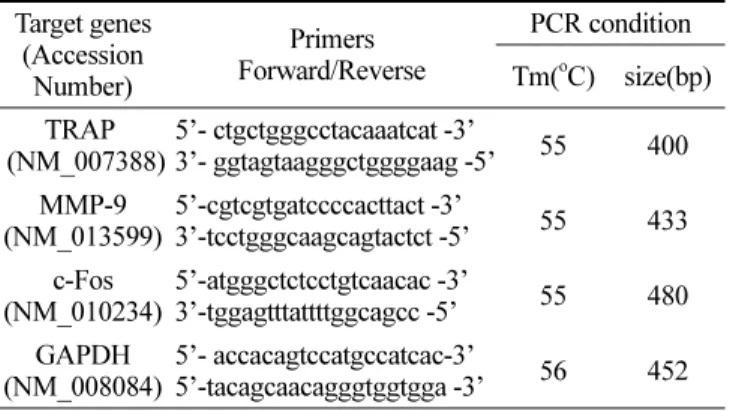
역전사연쇄중합반응(Reverse Transcription-Polymerase Chain Reaction)
총 RNA는 Invitrogen 사의 TRIzol을 이용하여 세포로부터 분리하였고, SuperScript II reverse transcriptase (Invitrogen) 를 이용하여 사용 설명에 따라 cDNA를 합성하였다. iMAX taq DNA polymerase (iNtRON Biotechnology;Sungnam, Korea)을 이용하여 PCR을 시행하였다. PCR primer를 Bioneer (Daejeon, Korea)에서 구입하였고, Primer 서열과 PCR 산물의 크기를 Table 1에 나열하였다. 94oC 에서 1 분 동안 초기 변성을 시키고, PCR 반응은 변성반응을 94oC 에서 30초, 결합반응을 30초 그리고 중합반응을 72oC에서 30초간 30주기를 반복하였고 마지막 중합반응은 72oC에서 10분간 반응시켰다. PCR 산물을 1.5% agarose gel을 이용 하여 전기영동으로 분리하였고 ethidium bromide로 염색 하여, Phosphoimager 와 Quantity One software (Version 4.3.1) (Bio-Rad, Hercules, CA, USA)을 이용하여 분석 및 정량 하였다.
Immunoblot analysis
세포를 PBS로 세척하고, lysis buffer [20 mM Tris-HCl (pH 7.5), 137 mM NaCl, 10% glycerol, 1% Triton X- 100, 1 mM phenylmethylsulfonylfluoride (PMSF), 1 mM Na3VO4, 1× protease inhibitor cocktail]를 세포에 첨가한 후, 원심 분리하여 단백질을 추출해 내었다. 단백질 정량 은 BCA reagent (pierce, Rockford, IL, USA)를 이용하 였다. 단백질(30~40 µg)을 4 ×sample buffer에 섞은 후, 10~12% SDS-PAGE를 시행하고, polyvinylidene difluoride (Bio-Rad) 막에 옮겼다. 5% nonfat skim milk가 포함된 TTBS (0.25% Tween-20 및 Tris-buffered saline) 용액에 막을 침투시켜 실온에서 1시간 동안 반응시키고, anti-COX2 (Santa Cruz Biotechnology), anti-iNOS (Santa Cruz Bio- technology), anti-β-actin (Santa Cruz Biotechnology)항체 를 5% nonfat skim milk가 포함된 TTBS에 1:1,000 비율 로 희석하여 4oC 에서 16시간 동안 반응시켰다. 그리고, Horseradish peroxidase-conjugated anti-rabbit or mouse antibodies (Santa Cruz Biotechnology)를 각각 이차 항체 로 이용하여, 5% nonfat skim milk가 포함된 TTBS 에 1:5,000~1:10,000 비율로 희석하여 실온에서 1시간 동안 반 응시켰다. ECL Plus kit (Amersham Biosciences, Piscat- away, NJ, USA)를 이용하여 해당 단백질을 관찰하였다.
치주질환 동물 모델 구축 및 투여
실험에 사용한 동물은 180-230 g의 Sprague-Dawley계 수 컷 흰쥐를 (주)다물사이언스(한국, 대전)에서 구입하였다.
동물실험은 전북대학교 Institutional Animal Care and Use Committee의 규정에 따라 행하여졌다. 사료와 물을 충분 히 공급하면서 1 주간 실험환경에 적응시키고, 대조군과 실험군을 나누어 실험에 사용하였다. 동물 사육실 내의 온 도는 20-24oC, 습도는 50-60%로 유지하면서 낮과 밤의 주 기는 각각 12 시간으로 하였다. 대조군과 HP08-0111 투여 군 (200 및 500 mg/kg)은 각각 5 마리로 하였으며, HP08- 0111을 투여하기 전 날, 상악 두 번째 어금니를 염증균(P.
gingivalis)이 처리된 밴드를 이용하여 묶음(ligature)으로 치 주염을 유발시켰으며, 대조군과 HP08-0111 투여군 (200
및 500 mg/kg)에 1 일 1 회 각각 생리 식염수와 시료를 경구투여 하였다. 동물을 8일 후 희생시키고 상악 어금니 부위를 얻어 탈석회시키고, H&E 염색법을 이용하여 염색 하였다. Niikura 등(2005)의 보고를 참고로 미세 현미경을 통해 치조골의 변화를 비교 분석하였다.
4-hydroxyproline (HYP) assay
HYP 측정은 간성상세포인 HSC-T6 세포를 이용하여 DMEM에서 배양하여 실험을 실시하였다(Choi 등, 2007).
흰쥐 성상 세포주(HSC-T6)에 대조군과 HP08-0111을 투 여하여 배양 후 12N HCl을 첨가하여 105oC에서 18 시 간 동안 단백질을 가수분해하였다. 0.56%의 완충용액(chlo- ramine-T)과 Ehrlich’s reagent을 첨가하고 50oC에서 90 분간 반응시키고, 558 nm에서 HYP양을 측정하였다.
통계처리방법
본 실험의 통계처리는 Student’s t test 방법을 사용하였 으며, 값은 평균과 표준오차로 나타냈다. 대조군과 비교하 여 P < 0.05 인 값을 유효성 있는 값으로 표기하였다.
결 과
RAW 264.7 세포에 대한 독성
마우스 대식세포인 RAW264.7에 대한 HP08-0111의 독 성을 확인하기 위해서, MTT assay를 실시하였다. HP08- 0111을 농도 별로(0.1, 1, 10 µg/ml) 24 시간 처리한 결과, 각각의 농도에서 HP08-0111에 대한 독성이 나타나지 않아 세포의 생존율에는 영향을 주지 않음을 확인할 수 있었다 (Fig. 1). 이는 HP08-0111의 항염증 효과가 단순한 세포의 사멸에 의한 세포의 염증성 매개물질의 생성 억제가 아니 라 HP08-0111 고유의 효과임을 의미한다.
Table 1. primer sequences for RT-PCR.
Target genes (Accession Number)
Primers Forward/Reverse
PCR condition Tm(oC) size(bp) TRAP
(NM_007388)
5’- ctgctgggcctacaaatcat -3’
3’- ggtagtaagggctggggaag -5’ 55 400 MMP-9
(NM_013599)
5’-cgtcgtgatccccacttact -3’
3’-tcctgggcaagcagtactct -5’ 55 433 c-Fos
(NM_010234)
5’-atgggctctcctgtcaacac -3’
3’-tggagtttattttggcagcc -5’ 55 480 GAPDH
(NM_008084)
5’- accacagtccatgccatcac-3’
5’-tacagcaacagggtggtgga -3’ 56 452
Fig. 1. Effect of HP08-0111 on the cell viability in RAW264.7 cells. RAW 264.7 cells were treated with various concentrations (0, 0.1, 1, 10µg/ml) of HP08-0111 for 24 h. Cell viability was mea- sured by MTT assay. Data represent mean ± S.D. of three indepen- dent experiments triplicate in each run.
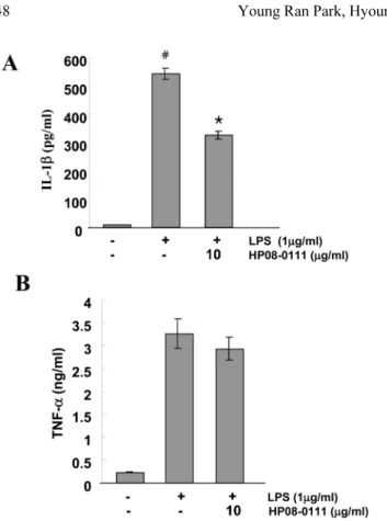
TNF-α, IL-1β의 분비량에 미치는 영향
RAW 264.7 세포에 LPS와 HP08-0111을 처리한 후, TNF- α, IL-1β의 분비량의 변화를 ELISA kit을 이용하여 측정하 였다. LPS를 처리하지 않은 세포와 비교하였을 때 LPS를 처리한 세포에서 IL-1β의 분비량이 증가하는 것을 확인할 수 있으며, HP08-0111을 10 µg/ml의 농도로 처리한 세포와 비교해서 확실한 감소 효과가 있음을 확인 할 수 있었고, 이는 통계학적으로도 의미 있음을(P < 0.01) 확인 할 수 있 었다(Fig. 2A). TNF-α의 분비량은 LPS를 처리한 세포에서 처리하지 않은 세포에 비해 10배 이상 증가하는 것을 확인 할 수 있었으나, HP08-0111은 TNF-α 분비에 큰 영향을 주지 않았다(Fig. 2B). 이를 통해, HP08-0111은 IL-1β 분비 억제 효과가 있음을 확인 할 수 있었다.
COX2, iNOS 발현량에 미치는 영향
HP08-0111이 COX2, iNOS 발현에 영향을 미치는지 알 아보기 위해 RAW 264.7 세포에 LPS와 10 µg/ml의 HP08- 0111을 처리하여 COX2와 iNOS의 발현량을 웨스턴 블롯 으로 확인하였다. 그 결과, LPS를 처리한 세포에서 LPS 에 의한 COX2의 발현이 유도 되는 것을 확인할 수 있었
고, 또한 10 µg/ml의 HP08-0111에 의해서 COX2의 발현 량이 현저히 감소하는 것을 확인할 수 있었고, 통계학적으 로도 의미 있음(P < 0.01)을 확인하였다(Fig. 3A). 그러나 HP08-0111은 iNOS의 발현에 거의 영향을 주지 못했다 (Fig. 3B). 따라서 HP08-0111은 LPS 처리된 RAW 264.7 세포에서 COX2의 발현에 영향을 미친다는 것을 확인 할 수 있었다.
HP08-0111이 파골 세포 분화유도에 미치는 영향 HP08-0111의 파골 세포 분화에 대한 효능을 확인하기 위해, 마우스 대식 세포인 RAW 264.7 세포에 RANKL (50 ng/ml)을 이용하여 6 일 동안 파골 세포 분화를 유도 하였고, TRAP 염색법을 통해 확인하였다. 그 결과, RANKL Fig. 3. Effect of HP 08-0106 on the LPS stimulated COX2 and iNOS expression in RAW 264.7 cells. RAW264.7 cells were serum-starved for 16 h, pretreated with or without 10µg/ml of HP 08-0106 for 30 min, and stimulated with LPS (1µg/ml) for 24 h.
Cell extracts were analyzed by Immunoblot analysis with antibod- ies against COX2 (A) and iNOS (B). The histogram represents the levels of the protein level compared with that of the control, respec- tively. Asterisk (*) indicates a significant difference (P < 0.05) compared with the control (#).
Fig. 2. Effect of HP 08-0106 on the LPS stimulated TNF-α and IL- 1ß product in RAW 264.7. RAW264.7 cells were serum-starved for 16 h, pretreated with or without 10µg/ml of HP08-0111 for 30 min, stimulated with LPS (1µg/ml). The amount of TNF-α (A) and IL-1ß (B) secretion was measured by ELASA method. Data repre- sent mean ± S.D. of three independent experiments triplicate in each run, Asterisk (*) indicates a significant difference (P < 0.05) compared with the control (#).
만 처리된 세포군에서 파골 세포 분화 유도를 확인할 수 있었고, HP08-0111을 농도 별로(0.1, 1, 10 µg/ml) 처리한 세포군에서는 HP08-0111의 농도 의존적으로 다핵성 세포 인 파골 세포가 감소하는 것을 확인하였다(Fig. 4).
HP08-0111이 파골 세포 분화 관련 유전자 발현에 미 치는 영향
파골 세포 분화에 따른 유전자 발현의 차이를 알아보기 위해, 파골 세포 분화 지표로 사용되는 c-Fos, MMP-9, TRAP의 유전자 발현량을 RT-PCR을 통해서 확인하였다.
Fig. 4. Effect of HP08-0111 on RANKL induced osteoclastogenesis. Murine monocytes macrophage RAW264.7 cells were cultured in the presence of RANKL (50 ng/ml) for 6 days. HP08-0111 was added into the culture media at a final concentration of 0, 0.1, 1, 10µg/ml. The cells were stained for TRAP (A). TRAP positive multinucleated cells (# of TRAP+ MNCs) were counted (B). Data represent mean ± S.D. of three independent experiments duplicate in each run, *P < 0.05 versus vehicle-treated cells.
Fig. 5. Suppression of RANKL-induced osteoclastogenesis gene expression by HP08-0111. RAW264.7 cells were cultured with 10 µg/ml HP08-0111 in the presence of RANKL (50 ng/ml). After 6 days of culture, the mRNA expression levels of c-Fos, MMP-9 and TRAP genes were determined by RT-PCR compared with that of GAPDH. The histogram represents the levels of the mRNA fold compared with that of the control (#). The results are expressed as mean ± S.D. *P < 0.05 versus control (#).
그 결과, HP08-0111을 처리한 세포군에서 c-Fos, MMP-9, TRAP의 발현이 HP08-0111을 처리하지 않은 세포군에 비 해 감소하는 것을 확인하였고, 이들 유전자는 각각 P=0.003, P < 0.01, P < 0.01의 값으로 통계학적으로도 의미가 있음 을 확인할 수 있었다(Fig. 5). 그러나 Cathepsin K는 HP08- 0111의 처리에도 변하지 않았다.
HP08-0111의 4-hydroxyproline (HYP) 분비량에 대한 영향 HP08-0111의 조직 지탱 정도를 관찰하기 위해, 잇몸조 직을 지탱, 지지하는 콜라겐의 함량을 유추해볼 수 있는 HYP 함량을 측정, 비교하였다. 콜라겐 생성 정도는 HYP 에 비례하고, 간 성상세포가 저산소 상태에서 배양 시 HYP 양이 유의하게 증가하므로(Choi 등, 2007) 간 성상세포를 이용하였다. 실험 결과 HP08-0111을 처리하지 않은 세포 군과 비교해 HP08-0111을 처리한 세포군에서 HYP 분비 량이 증가하는 것을 확인 하였으며, 통계학적으로 의미가 있음을 확인하였다(Fig. 6).
치주질환 동물 모델에서의 HP08-0111의 영향
HP08-0111의 동물에서의 효능을 확인하기 위해, 치주염 이 유발된 흰쥐를 이용하여 치조골의 변화를 분석하였다.
그 결과 정상적인 그룹의 치조골과 비교해 ligature와 P.
gingivalis가 처리된 그룹의 치조골이 소실되었음을 확인하 였고, HP08-0111을 200 및 500 mg/kg/day 처리한 그룹이 정상적인 그룹과 비슷한 수준임을 확인 할 수 있다(Fig.
7A, B). 이는 HP08-0111이 치주질환으로 인한 치조골 감 소를 억제시키는 효능이 있음을 시사한다.
고 찰
치조골의 파괴는 골 흡수의 주세포인 파골 세포의 활성
에 의해 이루어지는데, 파골세포는 조골세포에서 유래된 거대 다핵세포이다. 최근 골에서 파골 형성과 기능에 대 한 다양한 연구가 진행되어짐에 따라, 본 연구는 치주질 환의 치료제 개발에 대해 연구 하던 중, HP08-0111이라 명명한 황련(Coptis japonica (Thunb.) Makino) 생약 추 출물을 이용하여 치조골에 대한 염증 억제 효과와 파골 세포 분화 억제에 대한 효능에 대해 연구하게 되었다.
황련(Coptis japonica (Thunb.) Makino)은 미나리아제비 과(Ranunculaceae)의 다년생 초본식물로 예로부터 뿌리줄 기를 황련이라 하였으며, 유행성 열병, 장티푸스, 세균성 이 질, 열성 설사 및 복통, 폐결핵, 구토, 비출혈, 하혈, 소갈 증, 회중병, 백일해, 인후의 부종 및 동통, 결막염, 구창, 습 진, 화상 등의 치료에 사용되어 왔다(Otsuka 등, 1981). 황 련은 isoquinoline alkaloids, benzophenan thridine alkalokds, phenolic compounds 등을 포함하고 있으며 항 궤양작용, 간장 개선 작용, 항균작용, 항 염증작용 등의 보고가 있다 (Yoshikawa 등, 1995).
본 연구에서는 HP08-0111의 치주질환에 대한 효능을 연구하기 위해 TNF-α, IL-1β, COX2, iNOS의 분비량 과 발현량의 차이를 조사 하였으며, 파골 억제에 대한 효 능을 확인하기 위해 RAW 264.7 세포를 이용한 파골 세 포 유도, 파골 세포에 특이적인 유전자 발현의 차이를 확 인하였다. 또한 치주질환 모델로부터 HYP 수준, 치조골의 길이를 평가하였다. 그 결과 HP08-0111은 IL-1β와 같은 Fig. 7. Protective effect of HP08-0111 on Alveolar bone loss from P.gingivalis and ligature-induced peridodontitis in rat. Macroscopic appearance of the region between the first and second molar roots in the control and rats with periodontitis (A). Quantitative analysis of the change in alveolar bone length using H&E stained sections (B). Data represent mean ± S.D. of three independent experiments duplicate in each run, Asterisk (*) indicates a significant difference (P < 0.05) compared with the control (#).
Fig. 6. Effect of HP08-0111 on hydrixyproline (HYP) content. Rat hepatic stellate HSC-T6 cells were cultured in DMEM, and treated with HP08-0111 (20µg/ml) for 24 h. After incubation, cells were harvested and the content of HYP was measured (Fig. 6). Data rep- resent mean ± S.D. of three independent experiments triplicate in each run, *P < 0.05 versus vehicle-treated cells.
염증성 사이토카인의 분비량을 감소시키는 효과를 나타냈 으며, 염증관련 단백질인 COX2 발현에도 영향을 끼친다 는 것을 확인하였다. 또한, HP08-0111은 파골 세포 분화 억제 효과와 c-Fos, MMP-9, TRAP과 같은 파골 세포 특 이적인 유전자 발현을 감소시켜 파골 세포 분화 억제에 효과적임을 확인하였다. 또한, HP08-0111은 콜라겐의 생 성을 증가시키는 효능과 치주질환 동물 모델을 통해서 나 타나듯이 치조골의 손실을 감소시키는 효과가 있음을 알 수 있었다. 따라서 본 연구결과는 HP08-0111의 항 염증 작용, 파골 억제 효과 및 치조골 보호효과를 세포실험과 치주염 모델동물에서 밝혀 치주질환 치료제의 개발 가능 성을 제시하였다.
감사의 글
이 논문은 2008년도 전북지방 중소기업청의 산학 공동 기술개발지원사업의 지원을 받아 연구 되었음.
참 고 문 헌
Chen YJ, Hsu KW, Tsai JN, Hung CH, Kuo TC, Chen YL.
Involvement of protein kinase C in the inhibition of lipopolysaccharide-induced nitric oxide production by thapsigargin in RAW 264.7 macrophages. Int J Biochem Cell Biol. 2005;37(12):2574-85.
Choi HJ, Soh Y. An Assay Method for Screening Inhibitors of Prolyl 4-hydroxylase in Immortalized Rat Hepatic Stellate HSC-T6 Cells. J Ap Pharmacol. 2007;15(4):261-65.
Halliwell B, Gutteridge JM. Oxygen toxicity, oxygen radicals, transition metals and disease. Biochem J. 1984;219(1):1-14.
Han KY, Daum Y, Chang EJ, Lee Y, Huang H, Sung SH, Lee ZH, Kim YC, Kim HH. Inhibition of osteoclast differentiation and bone resorption by sauchinone. Biochem Pharmacol.
2007;74(6):911-23.
Hewett JA, Roth RA. Hepatic and extrahepatic pathobiology of bacterial lipopolysaccharides. Pharmacol Rev. 1993;45(4):
382-411.
Kim JY, Jung K, Jeong HG. Suppressive effects of the kahweol and cafestol on cyclooxygenase-2 expression in macrophages.
FEBS Lett. 2004;569(1-3):321-26.
Kim SS, Son YO, Chun JC, Kim SE, Chung GH, Hwang KJ, Lee JC. Antioxidant property of an active component purified from the leaves of paraquat-tolerant Rehmannia glutinosa. Redox Rep. 2005;10(6):311-18.
Kumar S, Bartholomew JV, Rieman DJ, Badger AM, Gowen M, Lee JC. IL-1- and TNF-induced bone resorption is mediated by p38 mitogen activated protein kinase. J Cell Physiol.
2001;187(3):294-303.
Masferrer JL, Zweifel B, Manning PT, Hauser SD, Leahy KM, Smith WG, Isakson PC, Seibert. Selective inhibition of inducible cyclooxygenase 2 in vivo is antiinflammatory and nonulcerogenic. Proc Natl Acad Sci USA. 1994;91(8):3228-32.
Mu MM, Sugiyama T, Koide N, Takahashi K, Mori I, Yoshida T, Yokochi T. The inhibitory action of quercetin on lipopolysaccharide-induced nitric oxide production in RAW 264.7 macrophage cells. J Endotoxin Res. 2001;7(6):431-38.
Mundy GR. Role of cytokines in bone resorption. J Cell Biochem. 1993;53(4):296-300.
Niikura K, Takeshita N, Chida N. A novel inhibitor of vacuolar ATPase, FR202126, prevents alveolar bone destruction in experimental periodontitis in rats. J Toxicol Sci. 2005;30(4):
297-304.
Otsuka H, Sawada T, Goto M. Studies on anti-inflammatory agents. II. Anti-inflammatory constituents from Rhizome of Coptis japonica Makino Yakugaku Zasshi. 1981;101(10):883- 90.
Pitaru S, Narayanan SA. Cellular origins and differentiation control mechanisms during periodontal development and wound healing. J Periodontal Res. 1994;29(2):81-94.
Raju BL, Hou WC, Lai ZY, Liu PC, Hsu FL. Antioxidant iridoid glucosides from Wendlandia formosana. Nat Prod Res. 2004;18(4):357-64.
Ryu JH, Kim JY, Kim YK. Inhibitory activity of plant extracts on nitric oxide synthesis in LPS-activated macrophages.
Phytother Res. 2003;17(5):485-89.
Schumann RR, Flaggs GW, Gray PW, Wright SD, Mathison JC, Tobias PS, Ulevitch RJ. Structure and function of lipopolysaccharide binding protein. Science. 1990;249(4975):
1429-31.
Soh Y, Jeong KS, Lee IJ, Bae MA, Kim YC, Song BJ.
Selective activation of the c-Jun N-terminal protein kinase pathway during 4-hydroxynonenal-induced apoptosis of PC12 cells. Mol Pharmacol. 2000;58(3):535-41.
Soh Y, Shin H, Lee JS, Jang JH, Kim OH, Kang H, Surh YJ.
Oxidative DNA damage and glioma cell death induced by tetrahydropapaveroline. Mutat Res. 2003;544(2-3):129-42.
Weisz A, Cicatiello L, Esumi H. Regulation of the mouse inducible-type nitric oxide synthase gene promoter by interferon-gamma, bacterial lipopolysaccharide and NG- monomethyl-L-arginine. Biochem J. 1996;15(6):209-15.
Xu GL, Li.G, Ma HP, Zhong H, Liu F, Ao GZ. Preventive effect of crocin in inflamed animals and in LPS-challenged RAW 264.7 cells. J Agric Food Chem. 2009;57(18):8325-30.
Yoshikawa K, Kinoshita H, Kan Y, Arihara S. Neolignans and phenylpropanoids from the rhizomes of Coptis japonica var.
dissecta. Chemical and pharmaceutical bulletin. 1995;43(4):
578-81.
Zhao Y, Wen XS, Cui J, Wu WH. Extraction and salting-out purification of alpha-galactosidase and beta-glucosidase from fresh roots of Rehmannia glutinosa. Zhong Yao Cai.
2006;29(2):137-39.