Effect of Fermented Angelica gigas Nakai on Lipid Metabolism in Orotic Acid Model Rats
Hee-Young Ahn, Kyu-Rim Park and Young-Su Cho*
Department of Biotechnology, Dong-A University, Hadan-2-dong Sahagu, Busan 604-714, Korea
Received April 23, 2014 /Revised July 1, 2014 /Accepted July 2, 2014The aim of this study was to identify the effect of fermented Angelica gigas Nakai (A. gigas) on lipid metabolism in orotic acid-induced fatty liver model rats. Sprague-Dawley male rats were randomly divided into four dietary groups (n=6 per group): a normal (N) group fed a standard diet only, OA control, OA acid plus 5% (w/w) A. gigas (OAG), and OA plus 5% (w/w) fermented A. gigas (OFAG).
OA treatment induced enlargement of the liver and accumulation of hepatic triglycerides. The consum ption of fermented A. gigas reduced triglyceride concentrations in the liver and increased the serum lipid concentrations to normal levels. Furthermore, OA treatment significantly decreased serum triglyc eride concentrations without diminishing mRNA expression of microsomal triglyceride transfer protei n (MTP) and protein disulfide isomerase (PDI). Hepatic MTP mRNA expression increased 1.08-fold in response to OA treatment, despite triglyceride accumulation in the liver relative to that of the norm al group. OFAG administration was slightly lower as compared to the OA treatment. This result sugg ests that MTP mRNA expression is not always correlated with hepatic triglyceride accumulation in the OA-induced fatty liver model. However, PDI mRNA expression was significantly increased in the OAG and OFAG groups (1.62-fold and 1.63-fold, respectively) compared with the normal group. The hepatocytes in the OA group contained numerous large fat droplets. These were slightly reduced in the OFAG group.
Key words : Angelica gigas Nakai, fermentation, Microsomal Triglyceride transfer Protein (MTP), orotic acid, Protein Disulfide Isomerase (PDI)
*Corresponding author
*Tel : +82-51-200-7586, Fax : +82-51-200-7505
*E-mail : [email protected]
This is an Open-Access article distributed under the terms of the Creative Commons Attribution Non-Commercial License (http://creativecommons.org/licenses/by-nc/3.0) which permits unrestricted non-commercial use, distribution, and reproduction in any medium, provided the original work is properly cited.
Journal of Life Science 2014 Vol. 24. No. 7. 743~749 DOI : http://dx.doi.org/10.5352/JLS.2014.24.7.743
서 론
참당귀 Angelica gigas Nakai (Umbelliferae)는 예로부터 질 병치료와 건강증진의 목적으로 널리 이용되어온 우리나라 대 표적인 생약재이다 . 당귀는 고혈압, 빈혈, 어혈에 효능이 있으 며 그 밖에도 진정제 , 진통제, 강장제로 사용된다. 특히 약성이 따뜻하고 , 맛은 달고, 무독하여 부작용이 없는 생약재로써 월 경 조절이나 임산부의 출산 전후에는 자궁을 튼튼하게 해주는 작용이 있어 우리나라뿐만 아니라 미국 및 유럽에서도 여성의 건강 관리를 위한 건강보조 식품으로 잘 알려져 있다 [15]. 유 효성분으로는 coumarine계의 decursin, decursinol angelate, nodakenetin, nodakenin, umbelliferone, β-sitosterol, α-pinene, limonene 등이 있으며[13], 그 중 decursin, decursinol angelat e는 주된 성분으로, immunostimulating [11], antibacterial [1 9], antiangiogenic [15], antitumor [18] 등의 약리적인 효과가
있는 것으로 알려져 있다 . Angelica sinensis는 ApoA-IV 전사 를 촉진하여 , 중성지질형성을 감소시킨다고 보고하였고[10], Angelica gigantis의 뿌리에서 분리한 decursin, decursinol ang elate가 mouse의 복막의 대식세포에서 지질 축적을 억제한다 는 것을 확인하였다 [24]. 한편, 저자들의 이전 연구에서 유용 균주들을 이용하여 참당귀 분말을 발효시켰다 . 그 결과 Monas cus purpureus 균주로 발효 시킨 경우 decursin과 decursinol angelate의 함량이 증가함을 확인하였다[5].
최근 들어 , Monascus sp. (홍국균)를 이용한 발효산물은 미 국과 많은 아시아 국가에서 건강식품으로 사용되고 있다 . 홍 국균이 생산하는 홍국색소는 식품첨가물로 이용되고 있으며 , monacolin K, mevinolin, lovastatin, γ-aminobutyric acid (G ABA), acethylcholine과 같은 생리활성물질은 콜레스테롤 생 합성 효소 (HMG-CoA reductase) 활성 억제, 혈압상승 억제, 혈전 용해 작용 및 항산화 활성과 같은 유효성 검증에 관한 연구결과가 보고되고 있다 [27]. 이에, 식품의약품안전처에서 홍국제품을 건강기능식품의 신규 품목으로 등록함으로써 이 들 성분을 이용하여 새로운 건강기능식품개발에 활용하려는 연구가 활발하게 이루어 지고 있다 .
Orotic acid유발 지방간은 간 독성에는 큰 영향을 미치지 않
으면서도 지질 대사에는 큰 영향을 미치는 것으로 알려져 이전
부터 지질 대사에 관련된 지방간 모델로 많이 이용되고 있다
Table 1. Compositions of experimental diets (%)
Component N1) Orotic acid
OA2) OAG3) OFAG4) Casein
Cornstarch Sucrose Cellulose Corn oil Mineral mixture5) Vitamin mixture6) Choline bitartrate DL-Methionine
Angelica gigas Monascus purpureus
Fermented
Angelica gigas
Orotic acid2015 455 10 3.51 0.20.3 00 0
2015 445 10 3.51 0.20.3 00 1
2010 445 10 3.51 0.20.3 50 1
2010 445 10 3.51 0.20.3 05
1
Tatal (%) 100 100 100 100
1)N: Normal.
2)OA: Orotic acid.
3)OAG: Orotic acid+
Angelica gigas
.4)OFAG: Orotic acid+
Monascus purpureus
fermentedAngelica gig as
.5)AIN 93 M-MX mineral mix, MP Biomedicals, Illkirch, France.
6)AIN 93 VX vitamin mix, MP Biomedicals, Illkirch, France.
[12]. Orotic acid를 1% 수준으로 흰쥐에 1주일 이상 섭취시켰 을 때 간장에 중성지방이 이상적으로 축적되어 지방간을 유발 하여 , 비 알코올성 지방간 모델로서도 사용되어 지고 있다[6].
따라서 본 연구에서는 Monascus purpureus균주에 의해 발 효된 참당귀를 기본식이에 첨가하여 orotic acid 유발 지방간 흰쥐의 지질대사 및 조직형태학적 변화를 검토하였다 .
재료 및 방법
실험재료 및 발효조건
참당귀 (Angelica gigas Nakai)는 2013년 3월 산청 산청약초 연구소 (경남, 산청)에서 제공받아 자연건조 후 분쇄하여 실험 에 사용하였다 . Monascus purpureus KCCM1002 (M. purpureu s)는 한국 미생물 보존센터에서 구입하여 발효에 사용하였다.
M. purpureus의 전 배양은 PDA agar로부터 spores를 백금이 를 이용하여 glucose 10%, peptone 5%, KNO
32%, NH
4H
2PO
42%, MgSO
4·7H
2O 0.5% 그리고 CaCl
20.1%가 포함되어 있는 500ml flask에 접종시켜 배양하였다. 배양은 30℃ 에서 72시간 150 rpm의 조건으로 하였다. 전 배양시킨 M. purpureus는 당 귀 뿌리분말에 5% 접종하여, 30℃에서 12일간 발효시켜 실험 재료로 사용하였다 .
실험동물, 식이조성 및 사육조건
실험동물은 6주령의 Sprague-Dawley계 수컷 흰쥐를 ㈜대 한 바이오링크 (충북 음성, 한국)에서 구입하여 온도 22±2℃,
습도 50±5%, 명암주기 12시간(명주기: 07:00~19:00)이 자동 설 정된 동물 사육 실에서 사육하였다 . Table 1과 같이 실험군은 정상군 (N), 오르트산 투여 대조군(OA), 오르트산+당귀 분말 5% (w/w) 투여군(OAG), 오르트산+ M. purpureus 발효 당귀 분말 5% (w/w) 투여군(OFAG) 으로 각 군마다 6마리씩 나누 고 , 식이와 물은 10일간 자유 섭취시켰다. 사육 기간 중 식이 섭취량은 매일 측정하였고 , 체중은 3일에 한번씩 정해진 시간 에 측정하였다 . 본 실험은 동아대학교 동물실험 윤리심의 위 원회의 승인 (승인번호: DIACUC-승인-13-29)을 받아 진행하 였다 .
동물실험, 시료 채취 및 분석시료 조제
동물실험은 10일간 각 군별로 조제사료를 급여하면서 사육 한 후 , 실험 최종일 12시간 이상 절식시킨 후 에테르로 가볍게 마취시켜 해부하였다 . 개복 후 복부 대동맥으로부터 채혈하여 혈액을 채취하고 , 약 30분간 실온에 방치시킨 후 3,000 rpm에 서 20분간 원심분리 하여 혈청을 얻어 혈청 효소 분석에 제공 하였다 . 채혈 후 각 조직을 적출하여 차가운 0.9% 생리식염수 로 세척하고 여과지로 물기를 제거한 후 무게를 측정하고 분 석시료로 제공 하였다 .
혈청 지질농도 및 생화학적 지표분석
혈청 중의 total lipid, triglyceride, total-cholesterol, phosp holipid, nonesterified fatty acid (NEFA)은 의료전문수탁검사 기관인 네오딘의학연구소 (서울, 한국)에 의뢰하여 분석하였 다 . 간 조직의 총 지질은 이전의 실험방법에 준하여 추출하였 으며 [9], 중성지질 농도는 혈청의 lipase-glycerol phosphate oxidase법을 응용한 commercial kit (Sigma-Aldrich, St. Louis MO, USA) 를 사용하였다[21].
Western blot analysis
간장에서 분리된 microsomal triglyceride transfer protein (MTP) 및 protein disulfide isomerase (PDI) 효소 단백질을 8% SDS-polyacryamide gel의 각 well당 각각 20 μg을 전보와 같이 전기영동 하였다 [25]. 분리된 단백질은 300/240 mA/cm
2
조건으로 4℃에서 16시간 동안 nitrocellulose membrane으
로 transfer하였다. 이 membrane을 blocking agent인 10% fat-f
ree milk가 함유된 TBST buffer (10 mM Tris pH 7.5, 100 mM
NaCl, 0.1% Tween 20)로 1시간 동안 실온에서 non-specific
binding protein을 blocking 한 후, TBST buffer로 5분 동안
세척하였다 . 다시 blocking시킨 membrane을 실온에서 1시간
동안 primary 및 secondary antibody로 반응시킨 후 TBST
buffer로 세척하였다. 세척된 membrane에 mouse monoclona
l anti-MTP (97 kDa, BD bioscience), anti-PDI (55 kDa, BD
bioscience)로 4℃에서 overnight하였다. 다시 TBST buffer로
세척시킨 후 goat anti-mouse IgG-HRP (diluted 1:2,000, DB
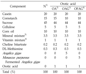
Table 2. Effects of OAG and OFAG on the tissues absolute weight in orotic acid-induced fatty liver model rats (g)
N OA OAG OFAG
Liver Kidney Heart Spleen Testis
Epididymal WAT Perirenal WAT
10.35±0.56c 2.28±0.04a 1.13±0.03a 0.63±0.05a 2.99±0.07a 4.31±0.18a 3.97±0.20a
15.39±0.70a 2.26±0.08a 1.10±0.05a 0.62±0.04a 3.00±0.04a 4.51±0.11a 4.57±0.20a
12.65±0.72b 2.07±0.05b 0.95±0.05b 0.54±0.07a 2.82±0.07ab 2.61±0.09b 1.78±0.32b
12.53±0.62b 2.19±0.05ab 0.97±0.03b 0.51±0.02a 2.78±0.08b 2.51±0.07b 1.80±0.20b Abbreviations are the same as in Table 1.
Values with different letters are significantly different at
p<0.05
. (mean ± S.E., n=6)Fig. 1. Effects of OAG an OFAG on the liver triglyceride content in orotic acid-induced fatty liver model rat. Abbreviatio ns are the same as in Table 1. Values with different letters are significantly different at
p<0.05
(mean ± S.E., n=6).USA)로 incuvation시켰다. Nitrocellulose membrane 상의 단 백질은 Super Signal West Pico Chemiluminescent Substrate 의 image analysis 방법으로 검출하였다. 각각 반응된 MTP 또는 PDI protein band의 상대적 density는 densitometer (Lu mi-Imager F1, Roche, Switzerland)로 정량 하였다.
간 조직의 병리조직학적 관찰
동물해부 직후 적출한 간을 냉각 생리식염수로 관류하여 혈액을 제거시킨 상태에서 조직의 일정한 부위의 일부를 취하 여 10% 중성포르말린 용액에 고정하여 통상적인 조직처리인 파라핀 포매 과정을 거쳐 3~4 μm 두께로 절편 하여 hematox ylin and eosin (H&E) 염색 한 후 광학현미경(Olympus BX41, Olympus Co., Tokyo, Japan)으로 관찰 후 사진촬영을 하였다.
통계처리
실험으로부터 얻어진 결과치는 one-way ANOVA 검정에 의한 평균치와 표준오차 (mean ± SE)로 표시하였으며, 각 실 험군 간의 유의성 검증은 Duncan's multiple range test로 하 였다 [8].
결과 및 고찰
각 장기의 무게
M. purpureus 균주로 발효 시킨 참당귀가 Orotic acid (OA) 유발 지방간 흰쥐의 지질대사에 미치는 영향을 살펴보기 위해 대조군으로 발효 시키지 않은 일반 참당귀와 함께 5% 농도로 10일간 기본식이에 첨가하여 급여하였다. 각 조직의 무게는 Table 2에 나타내었다. 간 조직의 무게는 정상군인 N군과 비 교해 OA 투여 대조군인 OA 군에서 유의적인 증가를 보여 중성지질의 축적에 의한 전형적인 지방간 유발이 확인되었다 . 그러나 OA 투여에 의한 이러한 간 조직의 무게 변화는 OA 투여 실험군인 OAG 군 및 OFAG군에서 유의적으로 감소하 여 이들의 식이첨가로 인해 중성지질을 감소시켜 지방간의 개선효과가 나타난 것으로 사료되나 , N 군보다는 약간 증가 하는 경향을 보였다 . 신장, 심장, 고환의 무게는 N군과 OA
군간 차이는 없었으나 , OA 투여 실험군인 OAG군 및OFAG군 에서 유의적으로 낮았으며 , 부고환 주변 및 신장주변의 지방 조직 무게도 비슷한 경향으로 OAG군과 OFAG군에서 감소하 였다 . 이러한 결과로 보아 참당귀와 발효 참당귀의 식이 첨가 는 체지방 축적 억제 및 지질대사 개선에 어느 정도 영향을 미치는 것으로 사료된다 .
간장의 중성지질 농도
간장은 지질대사에 관련된 중요한 장기로서 혈중 중성지질
합성과 분해로 농도 조절과 밀접한 관련성이 있다 [2]. Orotic
acid 섭취에 의한 지방간 유발 원인은 MTP 활성 저해에 의한
VLDL 분비 저하[2, 26], 중성지질 합성경로의 주요 조절효소
인 phosphatidate phosphohydrolase (PAP) 및 diacylglycerol
acyltransferase (DGAT) 효소 활성의 촉진[2] 및 지방산 산화
억제 [22]가 주요 기작으로 알려져 있다. 본 실험에서 OA 투여
에 의해 간 조직의 중성지질 농도가 정상군 보다 현저히 증가
하여 OA 유발 지방간의 특징을 잘 반영해주는 결과를 보였다
(Fig. 1). OA투여에 의한 간 조직에서 중성지질의 증가는 OA
G군 및 OFAG군에서 유의적으로 낮아지는 경향을 보였고,
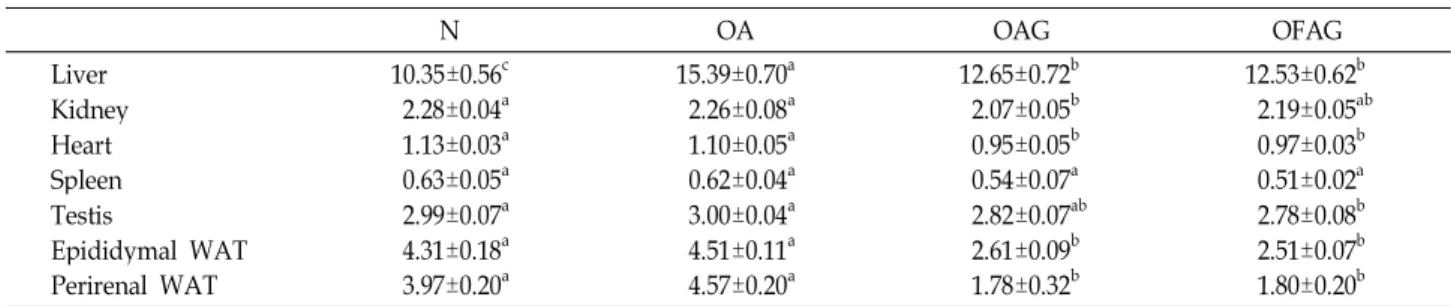
Table 3. Effects of OAG and OFAG on the serum lipid concentrations in orotic acid-induced fatty liver model rats
Plasma lipids (mg/dL) N OA OAG OFAG
Total Lipid Cholesterol Triglyceride Phospholipid
Free fatty acid (mmol/L)
207.1±24.29a 64.50±6.77ab 52.67±10.07a 137.5±10.15a 1.07±0.04a
157.0±14.33b 46.33±4.79b 26.17±8.00b 90.67±8.24b 1.04±0.14a
193.0±10.14a 86.50±4.77a 32.00±3.97b 123.1±8.98a 0.98±0.03a
191.3±22.75a 83.83±9.09a 47.17±2.39ab 119.0±11.08ab
0.86±0.03a Abbreviations are the same as in Table 1.
Values with different letters are significantly different at
p<0.05
. (mean ± S.E., n=6)특히 OFAG 군에서 정상에 가까운 수치를 나타내었다. 간 조 직의 중성지질 농도 감소는 혈중 중성지질 농도의 증가로 나 타나 간으로부터의 VLDL 분비에 긍정적인 영향을 미치는 것 으로 사료된다 . 한편, 알코올을 섭취시킨 동물실험에서 참당 귀 뿌리를 식이 첨가한 군에서 간장 내 총 지질함량과 중성지 질 함량이 감소됨을 확인하였고 [23], 또한 Angelica sinensis 추 출물을 3T3-L1 adipocytes에 의존적으로 배양시킨 결과, 중성 지질 함량이 현저하게 줄어들었다 [10].
혈청지질농도
혈중의 지질 농도는 심혈관계 질환인 동맥경화 , 고혈압, 심 장병 , 고지질혈증 등의 진단지표로 사용되고 있다. 혈중 콜레 스테롤 농도뿐만 아니라 중성지질 농도를 감소시키고 HDL- 콜레스테롤 농도를 증가시키려는 시도가 최근 천연 식물자원 을 대상으로 다방면에서 활발하게 진행되고 있다 [4, 16].
혈청 총 지질 , 중성지질, 총 콜레스테롤 및 인지질 농도는 N군에 비해 OA 군에서 현저히 감소함으로써 간 조직에서 혈중으로 분비되는 지질 운반체 VLDL의 분비 저하가 주요 기작으로 사료된다 (Table 3). OA군에서 감소된 총 지질, 중성 지질 , 콜레스테롤 및 인지질 농도는 OAG군과 OFAG군에서 유의적으로 증가하는 경향이었다 . 특히, OFAG 군은 혈중 중 성지질 함량에서 OA군에 비해 높은 증가수치로 정상 수준으 로 회복되는 경향을 보여 , 간 조직에서 축적되는 중성지질 농 도가 낮아져 어느 정도 지방간이 완화되어 개선되었음을 시사 한다 . 한편, Yu 등은 홍국 첨가 식이를 급여한 흰쥐에서 혈청 중성지방 함량은 2%, 4% Monascus식이군에서 대조군에 비해 모두 유의적으로 감소되었다고 하여 [30] 서로 상이한 결과를 나타내었으나 , Cha 등은 홍국발효홍삼분말을 급여한 흰쥐에 서 혈중 중성지질 농도가 대조군에 비해 홍삼분말 투여군에서 는 감소경향을 보였으나 홍국발효 홍삼분말 투여군에서는 유 의적인 증가를 나타냈다고 [7] 보고하였다. 본 실험에서 발효 당귀 분말의 투여로 높아진 혈중 중성지질 농도의 변화는 홍 국균이 가지는 기능성 물질의 영향과 홍국균에 의해 참당귀가 발효되는 과정에서 증가된 bioconversion에 의한 decursin 과 decursinol angelate 함량의 증가와 관련이 있는 것으로 보이 며 , 이러한 결과는 발효당귀의 식이첨가는 지질대사에 긍정적
인 영향을 주는 것으로 사료된다 . 추후 bioconversion 량에 따른 발효기간 중 변화 양상을 파악할 측정할 필요성도 나타 났다 .
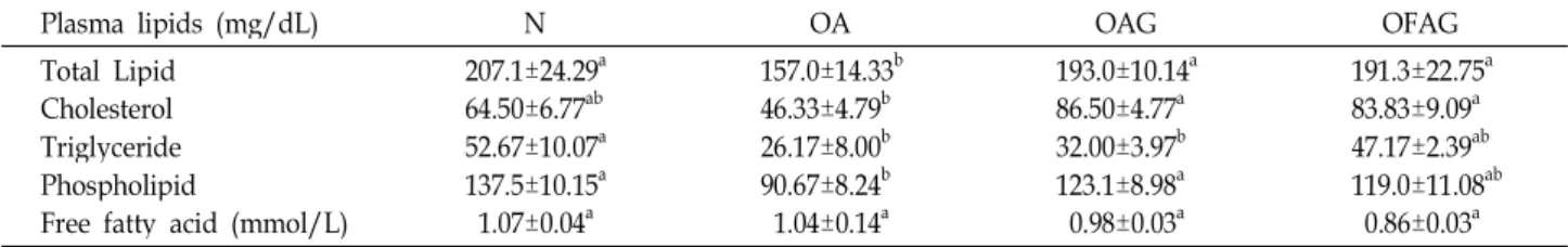
MTP(Microsomal Triglyceride transfer Protein) 및 PDI(Protein Disulfide Isomerase) 활성
간장의 활면소포체에서 합성된 apolipoprotein B
100은 골면 소포체로 이동되면서 극정 지질 성분과 함께 원시형의 초저밀 도리포단백질 (VLDL)을 만들어 Golgi장치에 운반된 후 혈중 으로 분비된다 [17].
최근 간장으로부터 혈중에로의 지질을 운반하는 TG-rich 리포단백의 합성과 분비에 MTP (microsomal triglyceride tra nsfer protein)가 중요한 역할을 담당하는 것으로 시사되어있 다 [1]. 현저한 저지혈증을 나타내는 가족성 무 β-리포단백혈증 (abetalipoproteinemia)의 환자에서 MTP가 유전적으로 결원 되어 있는 것이 해명되어 큰 주목을 받게 되었다 [20].
MTP는 간장 및 소장 상피세포에서 지질 전송 단백질이 microsomal 내강에 존재하는 가용성 단백질로서 97kDa과 58 kDa의 두 subunit로 구성된 heterodimer로 밝혀 졌다. 이중 97kDa인 큰 subunit가 MTP이며, 58kDa의 작은 subunit는 pr otein disulfide isomerase (PDI)로 동정되었다[28].
Orotic acid 유발 흰쥐의 간 조직을 이용하여 western blot 한 결과는 Fig. 2와 같다. MTP활성은 N군에 비하여 OAG군 및 OFAG군에서 1.17배 및 1.07배로 약간 증가하였으며, PDI 활성은 N군과 비교해 볼 때 OAG군 및 OFAG군에서 1.62배 및 1.63배 증가 수치를 나타내었다. 간장의 MTP 활성에는 큰 영향을 미치지 못했지만 PDI 활성과 혈청 중성지질 농도의 사이에는 상관관계가 인정되었으며 , 이러한 결과는 orotic aci d 유발 지방간 흰쥐에 OAG와 OFAG의 급여가 간장 PDI의 활성을 정상적인 수준보다 간장 TG와 apoB와의 결합을 촉진 시켜 혈중에의 VLDL-TG분비가 증가한 것으로 사료된다. 이 에 , OAG와 OFAG는 혈관순환기계 질환의 개선 또는 예방할 수 있는 기능성식품의 후보물질로서 가능성이 제기되었다 .
간장의 morphology
발효당귀가 orotic acid 유발 지방간의 지질대사에 미치는
Fig. 2. Effects of OAG and OFAG on hepatic MTP and PDI western blot analysis in orotic acid-induced fatty liver model rat. Abbreviations are the same as in Table 1. Val ues with different letters are significantly different at
p
<0.05
(mean ± S.E., n=6).Fig. 3. Effects of OAG and OFAG on external look of liver in orotic acid-induced fatty liver model rats. Abbreviations are the same as in
Table 1
.Fig. 4. Hepatic histopathologic changes of central vein in the orotic acid-induced fatty liver model rats. Hepatocyte sta ining was carried out with the hematoxylin and eosin staining method (magnification ×200). Abbreviations are the same as in Table 1.
영향을 형태학적으로 알아 보기 위하여 각 실험군의 동물 처 치 직후 적출한 간장 외관상의 결과는 Fig. 3과 같다. 정상군인 N군의 간 조직은 밝은 선홍색의 정상적인 간 색깔을 띄고 있었으나 OA 군은 황색에 가까운 점들이 분포되어 있고 전체 적으로 선홍색이 희미하게 변화되어 있으며 지방들이 점점 분사 , 침착 되어 있어 과도한 중성지질의 축적으로 비대해진 전형적인 지방간에서의 형태학적 소견과 동일한 결과를 확인 할 수 있었다 . 하지만 OAG 군과 OFAG 군은 N 군과 가까운
선홍빛을 띄고 중성지질이 축적량이 적어 부피도 N 군에 가 장 가까운 형태학적 소견을 보여 OA유발 간 손상에 대한 개선 효과가 있는 것으로 사료된다 . 특히, OFAG 군에서는 붉은 색상을 많이 띄고 있어 지방간이 개선되었음을 나타내고 있는 데 이는 간 조직의 중성 지질 농도와 상관관계를 나타내고 있다 .
간 조직의 병리 조직학적 관찰
H&E 염색을 통한 간 조직을 광학 현미경으로 관찰한 결과 는 Fig. 4와 같다. N 군은 간소엽의 구조가 잘 유지되었으며, 간 세포들은 뚜렷한 둥근핵을 가지고 있으면서 그 간격이 일 정하고 , 세포 간극이 좁은 잘 짜인 소엽 구조를 하고 있었다.
한편 , OA 군은 간세포의 풍선모양 변성, 소포성 지방변성이 소엽 중심대에 주로 나타나 있고 , 간 조직에 전체적으로 지방 세포가 균일하게 배열되어 있을 뿐만 아니라 그 크기도 정상 수준의 세포와 비교하였을 때 상당한 차이가 있는 것으로 보 아 전형적인 지방간 유발이 관찰되었다 [3]. 그러나 OFAG 군 에서는 간소엽을 구성하고 있는 간 세포들이 전반적으로 균일 하게 배열되어 있어 N 군의 간소견과 비슷한 양상을 보였다.
또한 소포성 지방과 지방세포들이 OA군에 비해 OFAG 군에 서 그 크기와 수가 줄어들고 있음을 알 수 있었다 . Wei 등은 M. purpureus로 발효된 쌀이 토끼와 햄스터에서 과도한 지방 섭취에 의해 유발된 지방간을 감소시킨다고 보고했다 [29].
감사의 글
본 연구는 동아대학교 연구비 지원에 의해 이루어졌습니다 .
References
1. Bennett, A. J., Billett, M. A., Salter, A. M. and White, D.
A. 1995. Regulation of hamster hepatic microsomal triglyceri de transfer protein mRNA levels by dietary fats.
Biochem Biophys Res Commun
212, 473-478.2. Cha, J. Y., Cho, Y. S., Kim, I., Anno, T. S., Rahman, M. and Yanagita, T. 2001. Effect of hesperetin, a citrus flavonoid, on the liver triacylglycerol content and phosphatidate phosp hohydrolase activity in orotic acid-fed rats.
Plant Foods Hum Nutr
56, 349-358.3. Cha, J. Y., Jun, B. S. and Cho, Y. S. 2004. Prevention of orotic acid-induced fatty liver in rats by capsaicin.
Food Sci Biotechn ol
13, 597-602.4. Cha, J. Y., Kim, H. J., Jun, B. S. and Cho, Y. S. 2000. Effect of water extract of leaves from
Morus alba
andCudrania tricu spidata
on the lipid concentration of serum and liver in rats.Agric Chem Biotechnol
43, 303-308.5. Cha, J. Y., Kim, H. W., Heo, J. S., Ahn, H. Y., Eom, K. E., Heo, S. J. and Cho, Y. S. 2010. Ingredients Analysis and Biol ogical Activity of Fermented
Angelica gigas
Nakai by Mold.J Life Sci
20, 1385-1393.6. Cha, J. Y., Maeda, Y., Oogami, K., Yamamoto, K. and Yanagi ta, T. 1998. Association between hepatic triacylglycerol accu mulation induced by administering orotic acid and enhence d phospatidate phosphohydrase activity in rats.
Biosci Biotec hnol Biochem
62, 508-513.7. Cha, J. Y., Park, J. C., Ahn, H. Y., Eom, K. E., Park, B. K., Jun, B. S. and Cho, Y. S. 2009. Effect of
Monascus purpureus
fermented Korean red ginseng powder on the serum lipid levels and antioxidative activity in rats.J Korean Soc Food Sci Nutr
38, 1153-1160.8. Duncan, D. B. 1955. Multiple range and multiple F test.
Biom etrics
11, 1-42.9. Folch, J., Lees, M. and Sloan-Stanley, G. H. 1957. A simple method for isolation and purification of total lipids from animal tissues.
J Biol Chem
226, 497-509.10. Guo, A. J., Choi, R. C., Cheung, A. W., Li, J., Chen, I. X., Dong, T. T., Tsim, K. W. and Lau, B. W. 2009. Stimulation of Apolipoprotein A-IV expression in Caco-2/TC7 enterocyt es and reduction of triglyceride formation in 3T3-L1 adipocy tes by potential anti-obesity Chinese herbal medicines.
Chin Med
4, 5-12.11. Han, S. B., Kim, Y. H., Lee, C. W., Park, S. M., Lee, H. Y., Ahn, K. S., Kim, I. H., Kim, H. M. 1998. Characteristic immu nostimulation by angelan isolated from
Angelica gigas
root.Immunopharmacology
40, 39-48.12. Hebbachi, A. M., Seelaender, M. C., Baker, B. W. and Gibbo ns, G. F. 1997. Decreased secretion of very-low-density lipop rotein triglyceride and apolipoprotein B is associated with decreased intercellular triglyceride lipolysis in hepatocytes derived from rats fed orotic acid or n-3 fatty acids.
Biochem J
325, 711-719.13. Heo, J. S., Cha, J. Y., Kim, H. W., Ahn, H. Y., Eom, K. E., Heo, S. J. and Cho, Y. S. 2010. Bioactive meterials and biolog ical activity in the extracts of leaf, stem mixture and root
from
Angelica gigas
Nakai.J Life Sci
20, 750-759.14. Jung, D. J., Porzel, A. and Huneck, S. 1991. Gigasol and other coumarins from
Angelica gigas
.Phytochemistry
30, 710-7 15. Jung, M. H, Lee, S. H., Ahn, E. M. and Lee, Y. M. 2009.12 Decursin and decursinol angelate inhibit VEGF-induced ang iogenesis via suppression of the VEGFR-2-signaling pathwa y.Carcinogenesis
30, 655-661.16. Kim, B. K., Shin, G. K., Jeon, B. S., Bae, D. W. and Cha, J. Y. 2001. Cholesterol-lowering effect of mushroom powder in hyperlipidemic rats.
J Korean Soc Food Sci Nutr
30, 510-515.17. Lamb, R. G. and Fallon, H. J. 1974. Glycerolipid formation from sn-glycerol-3-phosphate by rat liver cell fractions.
Bioch im Biophys Acta
348, 166-178.18. Lee, S., Lee, Y. S., Jung, S. H., Shin, K. H., Kim, B. K. and Kang, S. S. 2003. Antitumor activities of decursinol angelate and decursin from
Angelica gigas. Arch Pharm Res
26, 727- 19. Lee, S., Shin, D. S., Kim, J. S., Oh, K. B. and Kang, S. S.730.2003. Antibacterial coumarins from
Angelica gigas
roots.Arch Pharm Res
26, 449-452.20. Lin, M. C. A., Arbeery, C., Bergquist, K., Kienzle, B., Gordo n, D. A. and Wetterau, J. R. 1994. Cloning and regulation of hamster microsomal triglyceride transfer protein: the reg ulation is independent from that of other hepatic and transp ort of fatty acids and triglycerides.
J Biol Chem
269, 29138-291 21. McGrowan, M. W., Artiss, J. D., Strandbergh, D. R. and Zak,45 B. 1983. A peroxidase-coupled method for the colorimetric determination of serum triglycerides.Clin Chem
29, 538-542.22. Miyazawa, S., Furuta, S. and Hashimoto, T. 1982. Reduction of beta-oxidation capacity of rat liver mitochondria by feedi ng orotic acid.
Biochim Biophys Acta
711, 494-502.23. Oh, S. H., Cha, Y. S. and Choi, D. S. 1999. Effects of
Angelica gigas
Nakai diet on lipid metabolism, alcohol metabolism and liver function of rats administered with chronic ethanol.J Korean Soc Agric Chem Biotehnol
42, 29-33.24. Ohshiro, T., Namatame, I., Lee, E. W., Kawagishi, H. and Tomoda, H. 2006. Molecular target of decursins in the inhibi tion of lipid droplet accumulation in macrophages.
Biol Phar m Bull
29, 981-984.25. Oliveira, C. P., Alves, V. A., Lima, V. M., Stefano, J. T., Debb as, V., Sáa, S. V., Wakamatsu, A., Corrêea-Giannella, M. L., de Mello, E. S., Havaki, S., Tiniakos, D. G., Marinos, E., de Oliveira, M. G., Giannella-Neto, D., Laurindo, F. R., Caldwel l, S. and Carrilho, F. J. 2007. Modulation of hepatic microso mal triglyceride transfer protein (MTP) induced by S-nitroso -N-acetylcysteinein ob/ob mice.
Biochem Pharmacol
74, 290-2 97.26. Pottenger, L. A. and Getz, G. S. 1971. Serum lipoprotein accumulation in the livers of orotic acid-fed rats.
J Lipid Res
12, 450-459.27. Tsukahara, M., Shinzato, N., Tamaki, Y., Namihira, T. and Matsui, T. 2009. Red yeast rice fermentation by selected
Mon
ascus
sp. with deep-red color, lovastatin production but no citrinin, and effect of temperature-shift cultivation on lovast초록:발효당귀가 Orotic acid 유발 흰쥐 지질 대사에 미치는 영향 안희영․박규림․조영수*
(동아대학교 생명공학과)
Orotic acid 1% 수준으로 지방간을 유발시킨 흰쥐에 Monascus purpureus 균주 발효 당귀 5%를 식이 첨가하여 간 조직 및 혈중의 지질대사에 미치는 영향을 검토하였다 . 발효 참당귀 분말을 10일간 식이 급여한 결과, orotic acid 유발 지방간 흰쥐의 간 조직 내 중성지질 농도를 낮추고, 혈청 지질 농도가 정상 수준으로 회복되었음을 확인하였다 . 간 조직 내 MTP mRNA 및 PDI mRNA 발현 정도는 정상군에 비해 발효 당귀군에서 약간 증가하는 경향을 보이거나 , 유의적으로 증가된 수치를 나타내었다. 간 조직의 형태학적 관찰에서는 세포 간극이 좁고 잘 구성된 소엽구조를 하고 있는 정상군에 반해 OA 대조군은 orotic acid 투여로 소포성 지방 변성이 소엽 중심대에 나타나 있고 , 지방세포의 크기와 수가 증가하여 전형적인 지방간 형태의 양상을 보였다. 한편, 발효 당귀군의 간 장은 간세포의 소포성 지방세포들이 현저히 감소함으로써 , 정상 수준의 형태학적 간 성상을 나타내어 지방간 개 선에 긍정적인 영향을 미치는 것으로 판단되며 , 간 조직의 지질 축적 억제로 인한 지질대사 개선에도 효과가 있 는 것으로 사료된다 .
atin production.
Appl Biochem Biotechnol
158, 476-482.28. Watterau, J. R., Aggerbeck, L. P., Laplaud, P. M. and McLean L. R. 1991. Structural properities of the microsomal triglyceri de transfer protein complex.
Biochemistry
30, 4406-4412.29. Wei, W., Li, C., Wang, Y. and Kritchevsky, D. 2006. Effect of
Monascus purpureus
-fermented rice on lipidemia and fattyliver in quail.
Res Commun Mol Pathol Pharmacol
119, 67-75.30. Yu, T. S., Kim, H. H. and Yoon, C. G. 2003. Hepatic Oxygen Free Radical Metabolizing Enzyme Activities and Serum Lip id Profile in Rats Fed Diet Supplemented with