313 ORIGINAL ARTICLE
Korean Circ J 2008;38:313-319
Print ISSN 1738-5520 / On-line ISSN 1738-5555 Copyright ⓒ 2008 The Korean Society of Cardiology
The Protective Effect of Simvastatin on Monocrotaline-Induced Pulmonary Hypertension in Rats
Mi Young Kim, MD1, Young Kyu Kim, PhD2, Yong Wook Jung, MD3, Woo Taek Kim, MD4, Tae Hwan Kwon, MD5 and Dong Seok Lee, MD1
1Department of Pediatrics, 2Physiology and 3Anatomy, Dongguk University College of Medicine, Gyeongju,
4Department of Pediatrics, Daegu Catholic University College of Medicine, Daegu,
5Department of Physiology, Kyungpook National University College of Medicine, Daegu, Korea
ABSTRACT
Background and Objectives: Pulmonary hypertension is characterized by abnormal proliferation of vascular end- othelial cells and smooth muscle cells, and progressive pulmonary microvascular leakage that leads to pulmonary edema. This study was designed to investigate the protective effect of simvastatin on monocrotaline (MCT)- induced pulmonary hypertension and the role of the aquaporin (AQP) water channels. Materials and Methods:
Twenty one 8-week-old rats were randomized to the control, MCT (60 mg/kg, sc) and the MCT plus simvastatin (5 mg/kg/day, po) groups. Four weeks later, the systolic right ventricular pressure, the right ventricular hypertrophy, the medial wall thickness of the peribronchiolar artery and pulmonary arterioles and the renal function were measured to examine the effects of MCT and simvastatin in the rats. Western blotting for lung aquaporin1 (AQP1) and renal aquaporin2 (AQP2) was performed to analyze the effects of MCT and simvastatin on the AQP water channels. Results: Treatment with simvastatin reduced the MCT-induced enhanced right ventricular pres- sure (32.3±2.1 vs. 52.4±3.9 mmHg, respectively; p<0.05), the right ventricular hypertrophy (0.32±0.03 vs.
0.48±0.07, respectively; p<0.05) and the increased medial wall thickness of the peribronchiolar artery (0.14±
0.02 vs. 0.28±0.02, respectively; p<0.05) and pulmonary arterioles (0.15±0.04 vs. 0.29±0.11, respectively; p<0.05).
The decreased expression of lung AQP1 and renal AQP2 protein after MCT treatment was normalized by simva- statin treatment (p<0.05). Additionally, simvastatin treatment significantly reduced the perivascular and inter- stitial edema in the rats’ lungs without major alterations of renal function. Conclusion: These results suggest that simvastatin attenuates the MCT-induced pulmonary hypertension and the pulmonary edema by up-regulation of lung AQP1. Modulation of AQP may be one of the important mechanism of simvastatin. (Korean Circ J 2008;38:313-319)
KEY WORDS: Pulmonary circulation; Pulmonary hypertension; Statins.
Introduction
Pulmonary arterial hypertension (PAH) is a fatal dis- ease that’s caused by progressive narrowing of the pul- monary arterioles and increased pulmonary vascular resistance, and this can result in right ventricular failure and death.1)2) There are several factors that play roles in the pathogenesis of PAH such as genetic mutation of bone morphogenetic protein receptor II (BMPR II)
and activin receptor-like kinase 1 (ALK-1), exposure to drugs, infection and inflammation. These factors even- tually lead to multiple abnormalities, including endo- thelial cell proliferation, matrix production, thrombosis, abnormal vascular vasoconstriction and vascular remo- deling.2)
The currently available treatments for PAH include adjunctive therapy like diuretics, anticoagulant, digoxin and oxygen for right heart failure, and pulmonary va- sodilating agents such as calcium-channel blockers, prostanoids and endothelin receptor antagonists.3) How- ever, these currently available therapies can not reverse the disease process or the considerable morbidity and mortality, although they have a beneficial clinical effect.4) Therefore, new therapies are warranted that are based on a better understanding of the pathophysiology of
Received: November 10, 2007 Revision Received: March 5, 2008 Accepted: April 3, 2008
Correspondence: Dong Seok Lee, MD,Department of Pediatrics, Dongguk University College of Medicine 1090-1 Seokjang-dong, Gyeongju 780- 350, Korea
Tel: 82-54-770-8255, Fax: 82-54-770-8378 E-mail: [email protected]
314·Protective Effects of Simvastatin in PAH
the disease, and especially those therapies that are direc- ted at suppressing inappropriate cellular proliferation in the pulmonary arteries.2)5)
Simvastatin is a well known drug with cardiovascular benefits that exceed their effects on lowering serum cho- lesterol. In addition, simvastatin also exert potent anti- proliferative and proapoptotic effects on vascular smooth muscle cells through the inhibiting the activities of ras and rho guanine triphosphatase (GTPase), which are important for cell proliferation.6) It has recently been demonstrated that simvastatin could reverse established, severe pulmonary hypertension after toxic injury in rats and improve their survival. Other studies also showed that simvastatin treatment potently attenuates chronic hy- poxic pulmonary hypertension in rats and it inhibits vascular remodeling.2)7-10)
The aquaporins (AQPs) are a family of small mem- brane-spanning proteins that are expressed on the plasma membranes of many cell types and they facilitate water transport. In the lung, AQP1 is expressed on the apical and basolateral membranes of the microvascular en- dothelial cells. The abundance of AQP1 in the peribr- onchiolar vessels of perinatal rats may suggest their role in clearance of lung water from the alveolar space.11) Renal AQP2 is present in the vesicles of the collecting duct principal cells that translocate to the apical mem- brane in response to vasopressin.12) AQP2 overexpression has been described in several conditions associated with fluid retention, including congestive heart failure.13) These results suggest that AQP may play a role in the edema formation in PAH. Several animal models of PAH revealed pulmonary microvascular leakage and interstitial inflammation, which resulted in pulmonary edema.14) The aims of this study were examine the pro- tective effect of simvastatin on monocrotaline (MCT)- induced PAH and the role of the AQP water channels.
Materials and Methods
Experimental designs
Twenty one male Sprague Dawley rats (180-200 g) were randomly divided into three groups: group 1 was injec- ted with distillated water (the control group, n=7), group 2 was treated with MCT (the MCT group: MCT was injected at a single dose 60 mg/kg, sc, n=7) and group 3 was MCT rats that were treated with simvastatin (Hanmi Pharmacy, Seoul, Korea) (the MCT+S group, 5 mg/kg/day, po, daily for 28 days from the onset of MCT treatment, n=7). The rats in all three groups were kept in the same room and all of them were sub- jected to the same light-dark cycle. After 28 days, their tissue samples were obtained for morphometric analysis and western blotting. The experimental procedures we used were reviewed and approved by the Animal Care and Use Committee of Dongguk University. The animal
care and use were in accordance with the guidelines of the National Institute of Health.
Measurement of right ventricular pressure
After 28 days, the animals were anaesthetized by an intraperitonial injection of ketamine (100 mg/kg). For measuring the right ventricular pressure, a PE 50 catheter (Becton Dickinson, Franklin Lakes, NJ, USA) was in- serted into the right ventricle via the internal jugular vein, and a fluid-filled PE-50 tube was connected to a pressure transducer (Grass polygraph, Grass instrument CO, Quincy, MA, USA).
Right ventricular hypertrophy and lung morphology After 4 weeks, the rats were euthanized by an anes- thetic overdose and then the right ventricle (RV) free wall was dissected from the left ventricle (LV) and septum (S), and they were weighed separately on the analytic scale. The RV remodeling was assessed by the RV-to- LV plus S weight ratio. For analyzing the vascular re- modeling, the left lung was fixed with a transcardiac infusion of 4% paraformaldehyde. The perfused lung was removed and then paraffin-embedded. Serial coronal sections 5 μm thick were obtained at the lower zone of the lung. Following deparaffinization, the sections were stained with the hematoxylin-eosin (H&E). The medial wall thickness (MWT) of the pulmonary arterioles was measured at the pulmonary arterioles that were 50-100 μm in size and at the peribronchiolar muscular arteries.
The MWT ratio, which is an index of medial wall hy- pertrophy, was determined as the average data of 10 to 15 fields per slice and the MWT ratio was calculated as: [MWT=(external diameter-internal diameter)/exter- nal diameter].
Western blotting of aquaporin1 and aquaporin2 The MCT and/or simvastatin treated lung and kidney tissues were removed and then snap-frozen at -70℃
for Western Blot analysis. The tissue samples were ho- mogenized in ten volumes of homogenizing buffer (0.32 M sucrose, 25 mM imidazole and 1 mM ethylenedia- minetetraacetic acid (EDTA) (pH 7.2) containing 8.5 mM leupeptin and 1 mM phenylmethylsulfonyl fluor- ide), for 10 s with using a polytron. The aliquots were stored at -70℃. Samples of the homogenate were run on 7.5% polyacrylamide mini gels (Bio-rad Mini Prote- an). For each gel, an identical gel was run in parallel and the two gels subjected to Coomassie staining to assure identical loading. After electrophoresis, the protein was transferred to nitrocellulose paper for 2 hours at 400 mA and 120 V in a BioRad transblot system. After trans- fer, the protein bands were identified by Ponceaus S and they were destained with distilled water. The ni- trocelluse sheets were washed in tween phosphate buf- fered saline (PBST) and then incubated with rabbit anti-
Mi Young Kim, et al.·315
AQP1 (Alomone, Jerusalem, Israel) and rabbit anti- AQP2 (Alomone, Jerusalem, Israel) for overnight at 4℃.
The labeling was visualized with horseradish peroxidase- conjugated secondary antibodies (Santa Cruz Biotech- nology, Santa Cruz, CA, USA) and with using an en- hanced chemiluminescence (ECL) system (Amersham Pharmacia Biotech, Little Chalfont, UK). The immu- noblot signal was developed by an ECL system and it was quantified using Scion Image software (version 1.59).
Laboratory findings
On day 26 after treatments, the animals were indi- vidually housed in metabolic cages. The daily dietary intake and urine volume were measured for 2 days. The rats’ blood and urine samples were stored for electro- lyte and renal function testing.
Statistical analysis
All the data is presented as means±SDs. The data was analyzed by one-way analysis of variance (ANOVA) followed by Tukey’s multiple-comparisons test. Multiple- comparisons tests were applied only when a significant difference was determined by ANOVA (p<0.05). P
<0.05 were considered statistically significant.
Results
The effect of simvastatin on the high right ventricular pressure and right ventricular hypertrophy in rats with monocrotaline-induced pulmonary hypertension
The right ventricular pressure was significantly in- creased in the MCT group as compared with that of the controls (52.4±3.9 vs. 24.1±0.26 mmHg, respec- tively, p<0.05), and the right ventricular pressure was markedly suppressed in the simvastatin treatment group (32.3±2.1 mmHg, p<0.05). However, simvastatin tr- eatment did not suppress the right ventricular pressure to normal values (p<0.05) (Fig. 1).
In the MCT group, right ventricular hypertrophy developed and there were significant increases in the RV/ LV+S ratio compared with that of the controls (0.48
±0.07 vs. 0.26±0.02, respectively, p<0.05), and the right ventricular pressure was markedly suppressed in the simvastatin treatment group (0.32±0.03, p<0.05). How- ever, simvastatin treatment did not completely suppress the RV hypertrophy to normal values (p<0.05) (Fig. 2).
The effect of simvastatin on the wall thickness of the pulmonary artery in the rats with
monocrotaline-Induced pulmonary hypertension MCT treatment increased the medial wall thickness (MWT ratio) of the peribronchiolar artery compared with that of the controls (0.28±0.02 vs. 0.10±0.03, respectively, p<0.05), and the medial wall thickness
was significantly suppressed in the simvastatin treated group (0.14±0.02, p<0.05) (Fig. 3A-C). The medial wall thickness of the small pulmonary arterioles (50- 100 μm) was also significantly reduced in the simva- statin treated group as compared with the MCT group (0.15±0.04 vs. 0.29±0.11, respectively, p<0.05) (Fig.
3D-F). This treatment, however, did not completely re- verse the pulmonary arterial wall thickness to a normal value (p<0.05). Perivascular and interstitial edema was clearly seen in the MCT group. However, simvastatin treatment significantly reduced the pulmonary edema (Fig. 3G-I).
The effect of simvastatin on lung aquaporin1 and renal aquaporin2 in the rats with monocrotaline- induced pulmonary edema
To quantitatively evaluate the effect of simvastatin on MCT-induced pulmonary hypertension, the AQP1 and AQP2 expressions were measured in the lung and kidney from the controls and the MCT and/or simvas- tatin treated rats. Western blot analysis demonstrated that the expression of lung AQP1 and renal AQP2 in the MCT group was significantly decreased compared
Right ventricular pressure (mmHg) 60 40
20
0
Control MCT MCT+S
*
*†
Fig. 1. The systolic right ventricle pressure. Simvaststin (S) pre- vented the development of pulmonary arterial hypertension in the monocrotaline (MCT) treated rats. However, simvastatin treat- ment did not suppress the right ventricular pressure to normal values. The results are expressed as means±SDs. *p<0.05 vs.
control, †p<0.05 vs. MCT.
0.6
0.5
0.4
0.3
0.2
0.1
0.0
Control MCT MCT+S
*
*†
RV/(LV+septum) ratio
Fig. 2. The ratio of the right ventricle (RV) to left ventricle (LV) plus septum weight. The monocrotaline (MCT)-induced RV hy- pertrophy was attenuated after simvastatin (S) treatment. However, simvastatin treatment did not completely suppress the RV hy- pertrophy to normal values. Values are expressed as means±
SDs. *p<0.05 vs. control, †p<0.05 vs. MCT.
316·Protective Effects of Simvastatin in PAH
with that of the control group (p<0.05). However, the expression of lung AQP1 and kidney AQP2 was nor- malized after treatment with simvastatin (p<0.05) (Figs.
4 and 5).
Laboratory findings
The serum osmolarity and sodium levels were slightly increased in the MCT group (p<0.05), but they were normalized after simvastatin treatment (p<0.05). Other parameters of renal functions such as the urine output and the Na, K, blood urea nitrogen, creatinine and cre- atinine clearance were not altered (Table 1).
Discussion
PAH is a disease that’s characterized by elevated pul- monary artery pressure, and this can lead to right ven- tricular failure and death.1)2) The median survival of PAH patients was reported to be just a few years with an estimated 5-year survival of 34%.15-17) Yet the newer
medical therapies, besides the adjunctive therapies, have been shown to improve a variety of clinically relevant end-points, including survival, exercise tolerance, the haemodynamics and the quality of life measures.2)5) The introduction of continuous intravenous prostacyclin, a dual endothelin receptor antagonist, for the treatment of PAH has been promising.3) Unfortunately, there is no curative therapy. Therefore, the current therapy for PAH is based on a better understanding of its patho- genesis and an effort to reverse the vascular remod- eling with using phosphodiesterase type 5 inhibitors, elastase inhibitor, platelet derived growth factor (PDGF) receptor antagonist and simvastatin.12)18)19)
Simvastatin improves the cardiovascular outcomes, and this is independent of its effects on cholesterol reduction. In addition to its potent antipoliferative and antiapoptotic effects on the vascular smooth muscle cells,20) simvastatin enhances the production of endo- thelial nitric oxide synthase (NOS)21) and it also has anti- inflammatory effects.22) Simvastatin treatment causes
Control MCT MCT+S
Br Br
Br
Fig. 3. Light microscopic findings of lung. Representative photographs of the peribronchiolar muscular pulmonary artery (A-C, ×200), the pulmonary arteriole (D-F, ×400), and the histological change of the lung (G-I, ×200). The increased medial wall thickness in the monocrotaline (MCT) treatment group was reduced with simvastatin (S) treatment. MCT induced severe perivascular and interstitial ede- ma, but simvastatin treatment markedly reduced the pulmonary edema. Hematoxylin and eosin staining. Bar=50μm. Br: bronchiole.
A B C
D E F
G H I
Br Br
Br
A B C
Mi Young Kim, et al.·317
activation of caspase-3 and pulmonary microvascular
endothelial cell apoptosis in cases of severe pulmonary hypertension.10) It has recently been demonstrated that simvastatin could reverse or attenuate pulmonary hy- pertension.9)23) A small study reported improvements of the 6 min walk tests and the hemodynamics in pa- tients who received simvastatin daily.6) Clinically, we showed that simvastatin decreased the right ventricular pressure, the right ventricular hypertrophy and the me- dial wall thickness of the pulmonary artery in the rats with MCT-induced PAH. These results suggest that simvastatin attenuates the progression of MCT-induced pulmonary hypertension through the reduction of vas- cular resistance. This study was not intended to explore the mechanisms of simvastatin’s effects, but the increased apoptosis of vascular smooth muscle cells and/or the endothelial cells and production of endothelial NOS are might be candidate mechanisms that prevent the in- crease of vascular resistance.
The existence of water-specific membrane channel proteins in selected tissues has been postulated for several decades, and eleven mammalian AQPs have now been identified.12) The abundance of AQP1 in the peribron- chiolar vessels may suggest a role for this protein in clearance of water from the interstitial space.11) In ven- tilator-induced lung injury, AQP water channels have protective effects on pulmonary edema formation.24) However, in a murine model of lipopolysaccharide- induced acute lung injury, depletion of AQP1 did not affect the formation of lung edema, the lung vascular permeability or the lung histology.25) These results sug- gest that any correlation between the decreased expres- sion of AQP1 and lung edema formation is controversial.
In this study, we revealed that the APQ1 expression was decreased in the MCT-induced PAH rat tissues and this was normalized with simvastatin treatment.
The APQ1 expression was also correlated with the
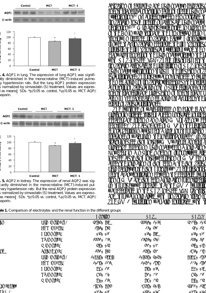
Table 1. Comparison of electrolytes and the renal function in the different groups
Control MCT MCT+S
Serum Osm (mosm/kg) 316.4±6.9 345.5±10.5* 321.6±10.4†
BUN (mg/dL) 16.5±8.3 15±4.1 31±13
Cr (mmol/L) 0.4±0.1 0.5±6.9 0.5±0.2
Na (mmol/L) 144.1±2.5 153.5±4.0* 145±5.1
K (mmol/L) 3.8±0.3 4.2±0.2 4.9±0.8
Urine Volume (mL) 14.1±6.3 13.8±3.2 17.5±2.8
Osm (mosm/kg) 1095.8±489.6 1063.8±303.0 949.7±184.3
BUN (mg/dL) 101.8±11.5 103.2±18.8 125±16.1
Cr (mmol/L) 9.7±1.1 8.8±0.5 9.2±0.9
Na (mmol/L) 7.4±2.0 8.2±2.2 7.3±2.1
K (mmol/L) 9.0±1.5 8.7±2.3 9.9±2.3
Ccr (mL/day) 267.4±94.4 273.8±40.6 331.2±139.6
FENa (%) 0.25±0.1 0.31±0.07 0.28±0.04
Values are expressed as means±SDs. *p<0.05 vs. control, †p<0.05 vs. MCT. Osm: osmolarity, BUN: blood urea nitrogen, Cr: creatinine, Ccr: creatinine clearance ratio, FENa: fractional excretion of Na
Normalized expression of AQP1in the lung (%)
Control MCT MCT+S
120 100 80 60 40 20 0
†
*
Control MCT MCT+S Fig. 4. AQP1 in lung. The expression of lung AQP1 was signifi- cantly diminished in the monocrotaline (MCT)-induced pulmo- nary hypertension rats. But the lung AQP1 protein expression was normalized by simvastatin (S) treatment. Values are expres- sed as means±SDs. *p<0.05 vs. control, †p<0.05 vs. MCT. AQP:
aquaporin.
AQP1 β-actin
AQP2 β-actin
Control MCT MCT+S
0 20 40 60 80 100 120
*
†
Normalized expression of AQP2 in the kidney (%)
Control MCT MCT+S
Fig. 5. AQP2 in kidney. The expression of renal AQP2 was sig- nificantly diminished in the monocrotaline (MCT)-induced pul- monary hypertension rats. But the renal AQP2 protein expression was normalized by simvastatin (S) treatment. Values are express- ed as means±SDs. *p<0.05 vs. control, †p<0.05 vs. MCT. AQP:
aquaporin.
318·Protective Effects of Simvastatin in PAH
resolution of lung edema. Recent papers have reported that AQP depletion would have subclinical effects on water homeostasis, and these effects would become apparent under stressful conditions such as congestive heart failure and pulmonary edema.12)26) Therefore, we suggest that simvastatin might attenuate the formation of pulmonary edema through the decreased expression of AQP1 water channels.
In a previous rat model of congestive heart failure caused by acute myocardial infarction, the renal AQP2 expression was activated in the apical plasma membrane of the renal collecting ducts, which accounted for fluid overload.13) Further, the up-regulation of APQ2 was normalized after fosinopril, valsartan and losartan treatment.27)28) So, we hypothesized that the fluid re- tention with pulmonary edema in PAH would be related with alterations of the renal AQP2 expression.
Our study revealed that the decreased expression of renal APQ2 in MCT-induced PAH rat tissues was not due to functional renal changes such as increased urine output or lowering the sodium concentration in the serum. Therefore, we speculate that the altered expres- sion of renal AQP2 after MCT and/or simvastatin was not associated with the formation and resolution of pulmonary edema. The altered expression of AQP2 may be due to the compromised true glomerular filtra- tion rate and it may be somewhat due to the dehy- dration via MCT-induced renal damage.
In summary, our study demonstrated that simvastatin attenuates the progression of MCT-induced pulmonary hypertension and the pulmonary edema by the incr- eased expression of lung AQP1. Further investigations will be needed to reveal the mechanisms of simvastatin effects and the exact role of APQs in PAH.
Acknowledgments
This work was supported in part by grants from the Dongguk Univ-
ersity Research Fund.
REFERENCES
1) Humbert M, Morrell NW, Archer SL, et al. Cellular and mo- lecular pathobiology of pulmonary arterial hypertension. J Am Coll Cardiol 2004;43(Suppl):13S-24S.
2) Martin KB, Klinger JR, Rounds SI. Pulmonary arterial hyper- tension: new insights and new hope. Respirology 2006;11:6-17.
3) Lee SH, Rubin LJ. Current treatment strategies for pulmonary arterial hypertension. J Intern Med 2005;258:199-215.
4) Kunieda T, Nakanishi N, Satoh T, Kyotani S, Okano Y, Nagaya N. Prognoses of primary pulmonary hypertension and chronic major vessel thromboembolic pulmonary hypertension determined from cumulative survival curves. Intern Med 1999;38:543-6.
5) Puri A, McGoon MD, Kushwaha SS. Pulmonary arterial hyper- tension: current therapeutic strategies. Nat Clin Pract Cardio- vasc Med 2007;4:319-29.
6) Kao PN. Simvastatin treatment of pulmonary hypertension: an observational case series. Chest 2005;127:1446-52.
7) Hu H, Sung A, Zhao G, et al. Simvastatin enhances bone mor-
phogenetic protein receptor type II expression. Biochem Biophys Res Commun 2006;339:59-64.
8) Girgis RE, Li D, Zhan X, et al. Attenuation of chronic hypoxic pulmonary hypertension by simvastatin. Am J Physiol Heart Circ Physiol 2003;285:H938-45.
9) Nishimura T, Vaszar LT, Faul JL, et al. Simvastatin rescues rats from fatal pulmonary hypertension by inducing apoptosis of neointimal smooth muscle cells. Circulation 2003;108:1640-5.
10) Taraseviciene-Stewart L, Scerbavicius R, Choe KH, et al. Simva- statin causes endothelial cell apoptosis and attenuates severe pulmonary hypertension. Am J Physiol Lung Cell Mol Physiol 2006;291:L668-76.
11) King LS, Nielsen S, Agre P. Aquaporin-1 water channel protein in lung: ontogeny, steroid-induced expression, and distribution in rat. J Clin Invest 1996;97:2183-91.
12) King LS, Yasui M. Aquaporins and disease: lessons from mice to humans. Trends Endocrinol Metab 2002;13:355-60.
13) Nielsen S, Terris J, Andersen D, et al. Congestive heart failure in rats is associated with increased expression and targeting of aq- uaporin-2 water channel in collecting duct. Proc Natl Acad Sci U S A 1997;94:5450-5.
14) Reindel JF, Ganey PE, Wagner JG, Slocombe RF, Roth RA.
Development of morphologic, hemodynamic, and biochemical changes in lungs of rats given monocrotaline pyrrole. Toxicol Appl Pharmacol 1990;106:179-200.
15) D’Alonzo GE, Barst RJ, Ayres SM, et al. Survival in patients with primary pulmonary hypertension: results from a national prospective registry. Ann Intern Med 1991;115:343-9.
16) Lee WD, Kim DS, Lee JH, et al. A clinical review of primary pulmonary hypertension. Korean Circ J 2003;33:507-12.
17) Lee WS, Kim KH, Jeong DH, et al. Clinical characteristics and prognostic factors of patients with severe pulmonary hyperten- sion. Korean Circ J 2007;37:265-70.
18) Schermuly RT, Kreisselmeier KP, Ghofrani HA, et al. Chronic sildenafil treatment inhibits monocrotaline-induced pulmonary hypertension in rats. Am J Respir Crit Care Med 2004;169:39-45.
19) Newman JH, Fanburg BL, Archer SL, et al. Pulmonary arterial hypertension: future directions: report of a National Heart, Lung and Blood Institute/Office of Rare Diseases workshop. Circula- tion 2004;109:2947-52.
20) Takemoto M, Liao JK. Pleiotropic effects of 3-hydroxy-3-methyl- glutaryl coenzyme a reductase inhibitors. Arterioscler Thromb Vasc Biol 2001;21:1712-9.
21) Laufs U, Fata VL, Liao JK. Inhibition of 3-hydroxy-3-methyl- glutaryl (HMG)-CoA reductase blocks hypoxia-mediated down- regulation of endothelial nitric oxide synthase. J Biol Chem 1997;
272:31725-9.
22) Son JW, Koh KK, You SM, et al. Effects of simvastatin alone or combined with ramipril on nitric oxide bioactivity and inflam- mation markers in hypercholesterolemic patients. Korean Circ J 2003;33:1053-9.
23) Girgis RE, Mozammel S, Champion HC, et al. Regression of chronic hypoxic pulmonary hypertension by simvastatin. Am J Physiol Lung Cell Mol Physiol 2007;292:L1105-10.
24) Hales CA, Du HK, Volokhov A, Mourfarrej R, Quinn DA.
Aquaporin channels may modulate ventilator-induced lung injury.
Respir Physiol 2001;124:159-66.
25) Su X, Song Y, Jiang J, Bai C. The role of aquaporin-1 (AQP1) expression in a murine model of lipopolysaccharide-induced acute lung injury. Respir Physiol Neurobiol 2004;142:1-11.
26) Agre P, King LS, Yasui M, et al. Aquaporin water channels-from atomic structure to clinical medicine. J Physiol 2002;542:3-16.
Mi Young Kim, et al.·319
27) Staahltoft D, Nielsen S, Janjua NR, et al. Losartan treatment nor- malizes renal sodium and water handling in rats with mild conges- tive heart failure. Am J Physiol Renal Physiol 2002;282:F307-15.
28) Yu CM, Wing-Hon Lai K, Li PS, Lam KY, Leung JC, Lai KN.
Normalization of renal aquaporin-2 water channel expression by fosinopril, valsartan, and combination therapy in congestive heart failure: a new mechanism of action. J Mol Cell Cardiol 2004;36:445-53.