• 검색 결과가 없습니다.

Effect of Garcinia mangostana L. and propolis extracts on the inhibition of inflammation and alveolar bone loss in ligature-induced periodontitis in rats

N/A
N/A
Protected

Academic year: 2021

Share "Effect of Garcinia mangostana L. and propolis extracts on the inhibition of inflammation and alveolar bone loss in ligature-induced periodontitis in rats"

Copied!
7
0
0

로드 중.... (전체 텍스트 보기)

전체 글

(1)

Introduction

Periodontal disease, characterized by alveolar bone resorp- tion and bacterial pathogen-induced inflammation, is a chronic inflammatory disease. It has a significant impact on oral health [1]. Bacteria in subgingival dental plaque (biofilm) are the major causative agent of periodontal disease. Porphyromonas gin- givalis, is one of the key pathogens in periodontal destruction

and is highly associated with periodontal disease development [2]. P. gingivalis induced the secretion of inflammation-induc- ing molecules, including interleukin (IL)-6, IL-8, prostaglandin E 2 (PGE 2 ), inducible nitric oxide synthase (iNOS), cyclooxy- genase (COX)-2, and matrix metalloproteinases (MMPs) and causes osteoclast formation in periodontal tissue [3]. Zhang et al. [4] observed that P. gingivalis invaded alveolar osteoblasts in a periodontitis mouse model. So, it is necessary to study de- Int J Oral Biol 44:55-61, 2019

pISSN: 1226-7155 • eISSN: 2287-6618 https://doi.org/10.11620/IJOB.2019.44.2.55

Effect of Garcinia mangostana L. and propolis extracts on the inhibition of inflammation and alveolar bone loss in ligature-induced periodontitis in rats

Se-Jin Sung 1 , Kyung-Min Kang 1 , Kyung-Hyun Lee 1 , So-Young Yoo 2 , Joong-Ki Kook 3,4 , Dae Sung Lee 2 *, and Sang-Joun Yu 1 *

1 Department of Periodontology, School of Dentistry, Chosun University, Gwangju 61452, Republic of Korea

2 Medi Bio Lab Co., Ltd., Seoul 08389, Republic of Korea

3 Korean Collection for Oral Microbiology, School of Dentistry, Chosun University, Gwangju 61452, Republic of Korea

4 Department of Oral Biochemistry, School of Dentistry, Chosun University, Gwangju 61452, Republic of Korea

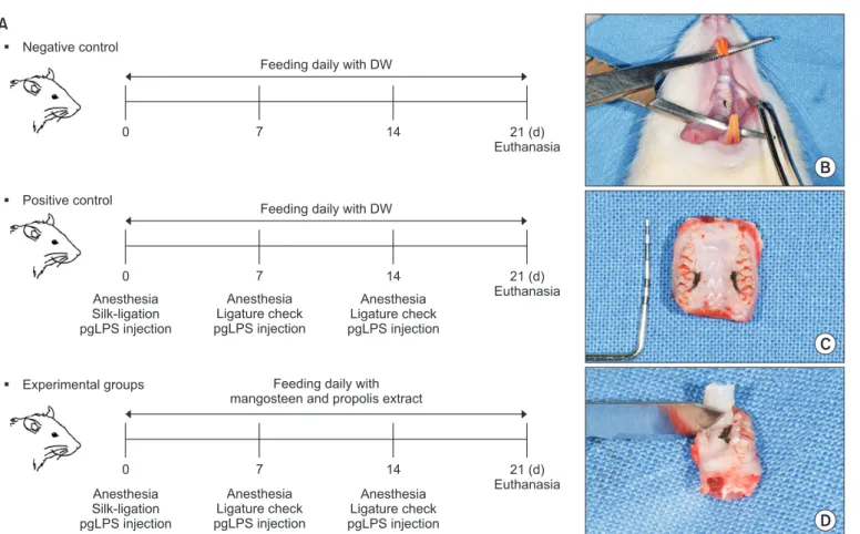
The purpose of this study was to evaluate the effect of mangosteen extract complex (MEC; Garcinia mangostana L.

and propolis extracts) on the inhibition of inflammation and prevention of alveolar bone loss using a ligature-induced periodontitis model. Rat molars were ligatured with silk, and 1 μg/mL of lipopolysaccharide of Porphyromonas gingivalis was injected into the buccal and palatal gingivae of the teeth with or without treatment with the MEC.

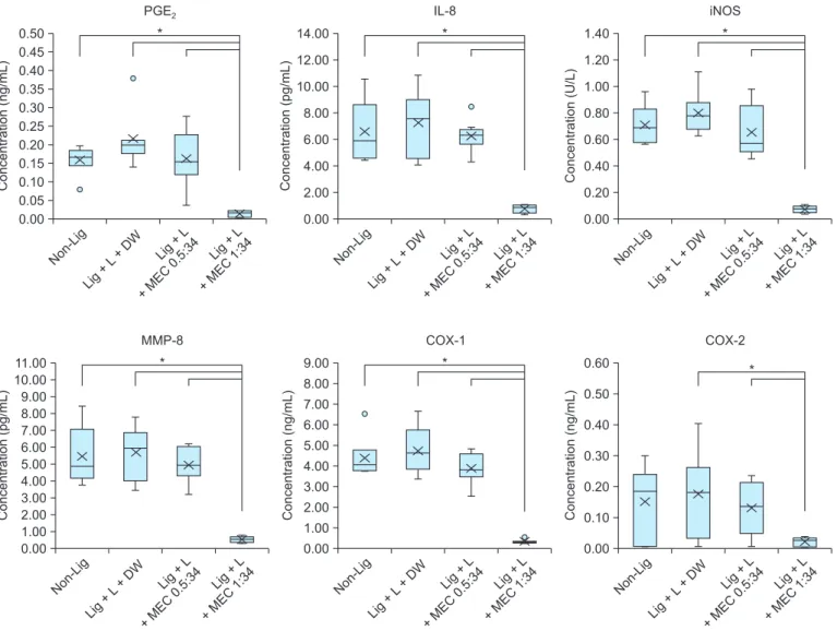
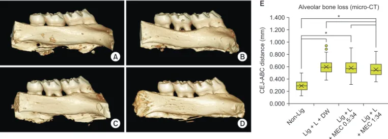
Changes in the expression levels of prostaglandin E 2 (PGE 2 ), interleukin-8 (IL-8), inducible nitric oxide synthase (iNOS), matrix metalloproteinase-8 (MMP-8), cyclooxygenase (COX)-1, and COX-2 in gingival tissues were evaluated using enzyme-linked immunosorbent assays. Alveolar bone loss around the ligated molars was examined using micro-computed tomography. The expression levels of PGE 2 , IL-8, iNOS, MMP-8, COX-1, and COX-2 in gingival tissues were significantly reduced in the group treated with a mixture of 16 µg of mangosteen extract powder and 544 µg of propolis extract powder (ligation [Lig] + lipopolysaccharide extracted from P. gingivalis KCOM 2804 [L] + MEC 1:34). Additionally, alveolar bone loss was significantly reduced in the Lig + L + MEC 1:34 group compared with that in other groups. These results indicate that the MEC could be useful in preventing and treating periodontitis.

Keywords: Mangosteen extract complex, Anti-inflammatory, Alveolar bone loss

Received June 4, 2019; Revised June 9, 2019; Accepted June 11, 2019

*Correspondence to: Sang-Joun Yu, E-mail: [email protected] https://orcid.org/0000-0001-8818-549X

*Correspondence to: Dae Sung Lee, E-mail: [email protected] https://orcid.org/0000-0003-2317-7848 Copyright © The Korean Academy of Oral Biology

CC

This is an open-access article distributed under the terms of the Creative Commons Attribution Non-Commercial License (http://creativecommons.org/licenses/by- nc/4.0/), which permits unrestricted non-commercial use, distribution, and reproduction in any medium, provided the original work is properly cited.

Original Article IJOB

(2)

velopment of materials inhibiting expression of these inflam- matory factors and inhibitory effect of alveolar bone resorption.

Development of the therapy has evolved from use of natural materials. Among these, Garcinia mangostana L. (mangosteen) and propolis have been shown to have anti-inflammatory and osteogenic effects in the wound healing process [5-7].

Propolis is a natural product produced by the honey bee. It possesses several biological properties, such as antioxidant and anti-inflammatory activities [6,8]. Previous research has shown that ethanol extract of propolis on mouse paw edema inhibited production of nitric oxide (NO) [9]. Additionally, bone mineral density was higher in rats that received oral propolis treatment [8]. Mangosteen extract contains abundant xantho- nes such as α, b, and γ-mangosteens [10]. Many biological properties such as anti-inflammatory, anti-oxidative damage and antioxidant activities from xanthones have been reported [11,12]. Previous studies have shown that α-mangosteen sig- nificantly inhibits NO, PGE 2 , tumor necrosis factor (TNF)- α, and iNOS production in lipopolysaccharide (LPS)-stimulated RAW 264.7 cells [3,13].

Animal models have been used to study pathogenesis of periodontal disease and to develop new treatment and pre- vention agents. Ligature-induced periodontitis model in rat has been frequently used in periodontitis-related research [14].

Ligature-induced periodontitis in rats is reliable and extensively used to reproduce periodontitis because periodontal anatomy in the molar is similar to that of humans [15]. The purpose of this study was to evaluate the effect of mangosteen extract complex (MEC; G. mangostana L. and propolis extract) on inhibition of inflammation and alveolar bone loss in ligature- induced periodontitis model in rat.

Materials and Methods

1. Bacteria culture and extraction of lipopolysaccharide

P. gingivalis KCOM 2804 was used in this study, and was obtained from the Korean Collection for Oral Microbiology (KCOM; Gwangju, Korea). The strain was cultured in tryptic soy broth (TSA; BD Difco Laboratories, Franklin Lakes, NJ, USA) supplemented with 0.5% yeast extract, 0.05% cysteine HCl- H 2 O, 0.5 mg/mL hemin, and 2 µg/mL vitamin K 1 at 37℃ in an anaerobic chamber (Bactron I; Sheldon Manufacturing Inc., Cornelius, OR, USA) with 10% H 2 , 5% CO 2 , 85% N 2 gas condi- tion. LPS of P. gingivalis KCOM 2804 was extracted, accord- ing to enclosed protocol using the LPS Extraction Kit (iNtRon,

Seongnam, Korea). Extracted LPS was dissolved in sterilized water filtered with 0.22 µm membrane. Extracted LPS was lyophilized and weighed (P. gingivalis KCOM 2804 strain LPS extracted, was called pgLPS, and pgLPS is denoted by L).

2. Experimental animals

A total of 40 male Wistar rats (weighing 250–400 g) were used in the experiment. In this study, selection and manage- ment of the animals, surgical protocol, and preparation proce- dures, were approved by the Chosun University Institutional Animal Care and Use Committee (CIACUC2017-S0020).

The animals were randomly divided into four groups. The mangosteen complex extracts powder corresponding to each group, were suspended in water and taken into rats. Experi- mental groups were as follows:

1) Non-ligation (Lig) group (negative control, n = 10): non- ligation + distilled water (DW)

2) Lig + L + DW group (positive control, n = 10): silk-ligation + pgLPS injection + DW

3) Lig + L + MEC 0.5:34 group (n = 10): silk-ligation + pgLPS injection + 8 µg mangosteen extract powder and 544 µg prop- olis extract powder (0.5:34)

4) Lig + L + MEC 1:34 group (n = 10): silk-ligation + pgLPS injection + 16 µg mangosteen extract powder and 544 µg propolis extract powder (1:34)

3. Silk-ligature-induced periodontitis rat model

The experiment was conducted for three weeks and all pro- cedures were performed under anesthesia with intramuscular injection of Zoletil ® (20 mg/kg; Virbac, Carros, France) and Rompun ® (7.5 mg/kg; Bayer, Ansan, Korea). A 5-0 silk-ligature (0.5–0.6 mm in diameter) was fixed in the cervical part of the maxillary second molar and all ligatures were positioned sub- gingivally. After ligation, 1 µg/mL pgLPS diluted by phosphate- buffered saline (PBS) was injected into buccal and palatal gingiva of the ligated teeth to induce periodontitis. The pgLPS was reinjected under anesthesia after one and two weeks (Fig.

1).

In experimental groups, mangosteen extract powder and propolis extract powder were suspended in 0.1 mL of sterilized water and administered orally using a feeding needle. In the control groups, the same amount of DW was administered. At three weeks, rats were sacrificed by carbon dioxide inhalation.

After sacrifice, gingival tissue around the ligated maxillary sec-

(3)

ond molar was obtained. It was stored at –80℃, for analysis of expression of specific inflammatory mediators. Remaining tissues were fixed with 10% formaldehyde for taking micro- computed tomography (micro-CT).

4. Expression of inflammatory cytokines in gingival tissues

To measure expression changes of inflammatory mediators (PGE 2 , IL-8, iNOS, MMP-8, COX-1, and COX-2) in gingival tissue, the ELISA kit of the Mybiosource Company (San Diego, CA, USA) was used, according to manufacturer’s instructions.

Stored gingival tissue was homogenized with PBS and centri- fuged to separate the supernatant. The supernatant was used and analyzed according to manufacturer’s instructions. Results are expressed as the mass (ng/mL, pg/mL) or unit (U/L) of each protein present in gingival tissue according to manufac- turer’s instructions.

5. Micro-computed tomography imaging

Collected tissues were scanned with a Quantum GX micro- CT Imaging System (PerkinElmer, Hopkinton, MA, USA) for two minutes at an acceleration potential of 90 kVp, and a beam current of 88 µA. Micro-CT data were reconstructed and formatted using OnDemand3D TM (Cybermed, Seoul, Korea) software. Through this process, three-dimensional structures around the teeth and around the alveolar bone were evaluated in all directions and all widths. Distance between the cemento- enamel junction (CEJ) and coronal plane of alveolar bone crest (ABC) was measured at six sites. It was measured at mesio- buccal, disto-buccal, mid-buccal, mesio-palatal, disto-palatal, and mid-palatal sites of the maxillary left and right second molars.

6. Statistical analysis

Data are presented, as average mean ± standard devia-

Feeding daily with DW

0 7 14 21 (d)

Euthanasia

Feeding daily with DW

Feeding daily with mangosteen and propolis extract 0

0

7

7

14

14

21 (d) Euthanasia

21 (d) Euthanasia Anesthesia

Silk-ligation pgLPS injection

Anesthesia Ligature check pgLPS injection

Anesthesia Ligature check pgLPS injection

Anesthesia Silk-ligation pgLPS injection

Anesthesia Ligature check pgLPS injection

Anesthesia Ligature check pgLPS injection Negative control

A

Positive control

Experimental groups

B

C

D

Fig. 1. Experimental design of silk-ligature induced periodontitis model in rats. (A) Diagram of study. (B) Silk-ligature placement in cervical part of the sec- ond molar. (C) Maxillary tissue of rat. (D) Collection of gingival soft tissue around the second molar area.

DW, distilled water; pgLPS, lipopolysaccharide extracted from P. gingivalis KCOM 2804.

(4)

tion. Statistical analysis was performed, using SPSS (version 12; SPSS Inc., Chicago, IL, USA). To compare the control and experimental groups, the Mann–Whitney U test was used.

Statistical significance was determined by p < 0.05 (95% con- fidence level).

Results

1. Expression of inflammatory cytokines in gingival tissues

Expression level of inflammatory cytokines, such as PGE 2 , IL-8, iNOS, MMP-8, COX-1, and COX-2 in gingiva tissues had a tendency to increase in the group Lig + L + DW than that of

group Non-Lig. In the group Lig + L + MEC 1:34, expression levels of PGE 2 , IL-8, iNOS, MMP-8, COX-1, and COX-2 were significantly decreased compared to the groups Non-Lig, Lig + L + DW, and Lig + L + MEC 0.5:34 (Fig. 2). Compared with the group Lig + L + DW, expression levels of PGE 2 , IL-8, iNOS, MMP-8, COX-1, and COX-2 in the group Lig + L + MEC 1:34 were significantly reduced and in particular, expression of iNOS was downregulated by 91.1% (p < 0.05) (Fig. 2).

Considering these results, the group Lig + L + MEC 1:34 was most effective in inhibiting expression levels of PGE 2 , IL-8, iNOS, MMP-8, COX-1, and COX-2 in gingival tissues of liga- ture-induced periodontitis rats model. The inhibition of inflam- matory cytokines in gingival tissues of rats model suggested that Lig + L + MEC 1:34 may be associated with decreased

Non-Li g 0.50 0.45 0.40 0.35 0.30 0.25 0.20 0.15 Concentration (ng/mL) 0.10

0.00

*

Li g + L + D W Li g + L + MEC

0.5:34 Li g + L + MEC

1:34 PGE 2

Non-Li g 14.00 12.00 10.00 8.00 6.00 4.00 Concentration (pg/mL) 2.00

0.00

*

Li g + L + D W Li g + L + MEC

0.5:34 Li g + L + MEC

1:34 IL-8

Non-Li g 1.40 1.20 1.00 0.80 0.60 0.40 0.20

Concentration (U/L)

0.00

*

Li g + L + D W Li g + L + MEC

0.5:34 Li g + L + MEC

1:34 iNOS

Non-Li g 11.00 10.00 9.00 8.00 7.00 6.00 5.00 4.00 3.00 2.00 1.00

Concentration (pg/mL)

0.00

*

Li g + L + D W Li g + L + MEC

0.5:34 Li g + L + MEC

1:34 MMP-8

Non-Li g 9.00 8.00 7.00 6.00 5.00 4.00 3.00 2.00 1.00

Concentration (ng/mL)

0.00

*

Li g + L + D W Li g + L + MEC

0.5:34 Li g + L + MEC

1:34 COX-1

Non-Li g 0.60 0.50 0.40 0.30 0.20 Concentration (ng/mL) 0.10

0.00

*

Li g + L + D W Li g + L + MEC

0.5:34 Li g + L + MEC

1:34 COX-2

0.05

Fig. 2. Expression of inflammatory cytokines (PGE 2 , IL-8, iNOS, MMP-8, COX-1, and COX-2) in gingival tissues.

Lig, ligation; L, lipopolysaccharide extracted from P. gingivalis KCOM 2804; DW, distilled water; MEC, mangosteen extract complex; PGE 2 , prostaglandin E 2 ; IL-8, interleukin-8; iNOS, inducible nitric oxide synthase; MMP-8, matrix metalloproteinase-8; COX-1, cyclooxygenase-1; COX-2, cyclooxygenase-2.

*p < 0.05; Mann–Whitney U test.

(5)

progression of periodontitis and inhibition of alveolar bone re- sorption.

2. Alveolar bone loss in ligature-induced periodontitis rats model

Alveolar bone loss was significantly increased in the group Lig + L + DW compared to the group Non-Lig. In contrast, it was reduced in the groups Lig + L + MEC 0.5:34 and Lig + L + MEC 1:34 compare to the group Lig + L + DW (Fig. 3).

To evaluate degree of periodontal destruction, the distance between the CEJ and ABC was expressed as the amount of alveolar bone destruction. Distance from the CEJ to ABC in the groups Lig + L + MEC 0.5:34 and Lig + L + MEC 1:34 was sig- nificantly reduced by 4.2% and 13%, respectively, compared to the group Lig + L + DW (p < 0.05) (Fig. 3). These data showed that MEC exhibited an inhibitory effect on alveolar bone loss by marked decrease of CEJ-ABC distances in rats by ligature- induced periodontitis.

Discussion

Periodontitis, one of the most common diseases in adults, is an inflammatory disease caused by periodontal pathogens in plaque on the surface of teeth. These pathogens promote pro- duction of inflammatory cytokines such as PGE 2 , and cytokines IL-1, IL-6, IL-8, TNF-α, and MMPs resulting in bone destruc-

tion [16,17].

Results of this study showed that MEC was significantly inhibited expression levels of inflammatory mediators (PGE 2 , iNOS, MMP-8, IL-8, COX-1, and COX-2) in gingival tissues induced periodontitis by silk-ligature and injection of pgLPS.

Especially, the expression PGE 2 in the group Lig + L + MEC 1:34 markedly decreased than that in the group Non-Lig. In previ- ous study, MEC 1:34 showed anti-inflammatory and in vitro bone formation effects in human gingival fibroblasts and hu- man osteosarcoma cell lines (in submission). This result sug- gests that MEC 1:34 may alleviate the inflammatory state of rat by reducing the expression of PGE 2 at the basic level. PGE 2

is known to be an important inflammatory mediator causing alveolar bone destruction and osteoclast formation [18].

Degree of alveolar bone loss is one of the indicators of se- verity of periodontal disease [19]. Previous studies have shown that 500 µg/mL mangosteen extract did significantly inhibition, in LPS-induced bone destruction by osteoclast [20]. It has been reported that the group treated with 100 mg/kg propo- lis in rats with expanded premaxillary suture reduced alveolar bone loss and a number of osteoclast compared to the con- trol group [21]. Micro-CT study showed significant reduction of alveolar bone loss (13%) in the group Lig + L + MEC 1:34.

This study validates for the first time that MEC (mixture of mangosteen and propolis) reduced alveolar bone loss through inhibition of inflammatory responses in rats with ligature- and pgLPS-induced periodontitis. These results suggest that MEC

Non-Li g 1.400 1.200 1.000 0.800 0.600 0.400 0.200

CEJ-ABC distance (mm)

0.000

*

Li g + L + D W Li g + L + MEC

0.5:34 Li g + L + MEC

1:34 Alveolar bone loss (micro-CT)

A B

C D

E

*

Fig. 3. 3-Dimensional micro-computed tomography (micro-CT) image of alveolar bone loss around the maxillary second molar. Non-Lig group (A), Lig + L + DW group (B), Lig + L + MEC 0.5:34 group (C), Lig + L + MEC 1:34 group (D), and distance between cemento-enamel junction (CEJ) and alveolar bone crest (ABC) (E).

Lig, ligation; L, lipopolysaccharide extracted from P. gingivalis KCOM 2804; DW, distilled water; MEC, mangosteen extract complex.

*p < 0.05; Mann–Whitney U test .

(6)

could be useful in preventing and treating periodontitis.

Acknowledgements

This research was supported by Medi Bio Lab Co., Ltd.

(2017-0716).

Conflicts of Interest

Dae Sung Lee is a co-register on patent (No.: 1018 7939- 80000, Korea) entitled “Pharmaceutical composition for en- hancing osteogenesis comprising Garcinia mangostana extract and propolis extract” and on patent (No.: US 10,022,412 B2 and JP 6387467 B2) entiled “Composition for preventing or alleviating periodontal diseases, containing, as active ingredi- ents, mangosteen extract or α- or γ-mangosteen.”

References

1. Bezerra MM, de Lima V, Alencar VB, Vieira IB, Brito GA, Ribeiro RA, Rocha FA. Selective cyclooxygenase-2 inhibi- tion prevents alveolar bone loss in experimental periodon- titis in rats. J Periodontol 2000;71:1009-14. doi: 10.1902/

jop.2000.71.6.1009.

2. Bumrungpert A, Kalpravidh RW, Chitchumroonchokchai C, Chuang CC, West T, Kennedy A, McIntosh M. Xanthones from mangosteen prevent lipopolysaccharide-mediated in- flammation and insulin resistance in primary cultures of hu- man adipocytes. J Nutr 2009;139:1185-91. doi: 10.3945/

jn.109.106617.

3. Chen Y, Yang L, Lee TJ. Oroxylin A inhibition of lipopoly- saccharide-induced iNOS and COX-2 gene expression via suppression of nuclear factor-kappaB activation. Bio- chem Pharmacol 2000;59:1445-57. doi: 10.1016/S0006- 2952(00)00255-0.

4. Zhang W, Ju J, Rigney T, Tribble G. Porphyromonas gingivalis infection increases osteoclastic bone resorption and osteo- blastic bone formation in a periodontitis mouse model. BMC Oral Health 2014;14:89. doi: 10.1186/1472-6831-14-89.

5. Gutierrez-Orozco F, Chitchumroonchokchai C, Lesinski GB, Suksamrarn S, Failla ML. α-Mangostin: anti-inflammatory activity and metabolism by human cells. J Agric Food Chem 2013;61:3891-900. doi: 10.1021/jf4004434.

6. Ramos AFN, Miranda JL. Propolis: a review of its anti- inflammatory and healing actions. J Venom Anim Tox- ins Incl Trop Dis 2007;13:697-710. doi: 10.1590/S1678- 91992007000400002.

7. Pileggi R, Antony K, Johnson K, Zuo J, Shannon Holliday L. Propolis inhibits osteoclast maturation. Dent Traumatol 2009;25:584-8. doi: 10.1111/j.1600-9657.2009.00821.x.

8. Guney A, Karaman I, Oner M, Yerer MB. Effects of propolis on fracture healing: an experimental study. Phytother Res 2011;25:1648-52. doi: 10.1002/ptr.3470.

9. Tan-No K, Nakajima T, Shoji T, Nakagawasai O, Niijima F, Ishikawa M, Endo Y, Sato T, Satoh S, Tadano T. Anti-inflam- matory effect of propolis through inhibition of nitric oxide production on carrageenin-induced mouse paw edema. Biol Pharm Bull 2006;29:96-9. doi: 10.1248/bpb.29.96.

10. Gutierrez-Orozco F, Failla ML. Biological activities and bio- availability of mangosteen xanthones: a critical review of the current evidence. Nutrients 2013;5:3163-83. doi: 10.3390/

nu5083163.

11. Iikubo K, Ishikawa Y, Ando N, Umezawa K, Nishiyama S. The first direct synthesis of α-mangostin, a potent inhibitor of the acidic sphingomyelinase. Tetrahedron Lett 2002;43:291-3.

doi: 10.1016/S0040-4039(01)02137-2.

12. Martindale J, Bland-Ward PA, Chessell IP. Inhibition of C-fibre mediated sensory transmission in the rat following intraplantar formalin. Neurosci Lett 2001;316:33-6. doi: 10.1016/S0304- 3940(01)02362-X.

13. Tewtrakul S, Wattanapiromsakul C, Mahabusarakam W. Ef- fects of compounds from Garcinia mangostana on inflamma- tory mediators in RAW264.7 macrophage cells. J Ethnophar- macol 2009;121:379-82. doi: 10.1016/j.jep.2008.11.007.

14. Jin Q, Cirelli JA, Park CH, Sugai JV, Taba M Jr, Kostenuik PJ, Giannobile WV. RANKL inhibition through osteoprotegerin blocks bone loss in experimental periodontitis. J Periodontol 2007;78:1300-8. doi: 10.1902/jop.2007.070073.

15. Roeterink CH, van Steenbergen TJ, de Jong WF, de Graaff J.

Histopathological effects in the palate of the rat induced by injection with different black-pigmented Bacteroides strains.

J Periodontal Res 1984;19:292-302. doi: 10.1111/j.1600- 0765.1984.tb00820.x.

16. Fento ğlu Ö, Köroğlu BK, Hiçyılmaz H, Sert T, Özdem M, Sütçü

R, Tamer MN, Orhan H, Ay ZY, Öztürk Tonguç M, Kırzıoğlu

FY. Pro-inflammatory cytokine levels in association between

periodontal disease and hyperlipidaemia. J Clin Periodontol

(7)

2011;38:8-16. doi: 10.1111/j.1600-051X.2010.01644.x.

17. Vernal R, Dezerega A, Dutzan N, Chaparro A, Le ón R, Chandía S, Silva A, Gamonal J. RANKL in human periapical granuloma:

possible involvement in periapical bone destruction. Oral Dis 2006;12:283-9. doi: 10.1111/j.1601-0825.2005.01191.x.

18. Fukushima H, Jimi E, Okamoto F, Motokawa W, Okabe K.

IL-1-induced receptor activator of NF-kappa B ligand in hu- man periodontal ligament cells involves ERK-dependent PGE2 production. Bone 2005;36:267-75. doi: 10.1016/

j.bone.2004.09.011.

19. Tezal M, Wactawski-Wende J, Grossi SG, Ho AW, Dunford

R, Genco RJ. The relationship between bone mineral density and periodontitis in postmenopausal women. J Periodontol 2000;71:1492-8. doi: 10.1902/jop.2000.71.9.1492.

20. Idrus E, Kiswanjaya B. Mangosteen extract inhibits LPS- induced bone resorption by controlling osteoclast. J Int Dent Med Res 2016;9:362-7.

21. Toker H, Ozan F, Ozer H, Ozdemir H, Eren K, Yeler H. A morphometric and histopathologic evaluation of the effects of propolis on alveolar bone loss in experimental periodon- titis in rats. J Periodontol 2008;79:1089-94. doi: 10.1902/

jop.2008.070462.

수치

Fig. 1.  Experimental design of silk-ligature induced periodontitis model in rats. (A) Diagram of study
Fig. 2.   Expression of inflammatory cytokines (PGE 2 , IL-8, iNOS, MMP-8, COX-1, and COX-2) in gingival tissues
Fig. 3.  3-Dimensional micro-computed tomography (micro-CT) image of alveolar bone loss around the maxillary second molar

참조

관련 문서