Introduction of Assessment Indicators of Lung Cancer Management Proposed
by Health Insurance Review and Assessment Service of Korea
1. Organization of lung cancer specialists
This indicator is to assess the composition of the medical staff involved in the lung cancer care in the institutions to be evaluated (Table 1). It aims to improve the coordination and increase opportunities from various perspectives for patients’
care. It is fulfilled when the staffs were consisted of at least one member of the pulmonary medicine, hemato-oncology, tho- racic surgery, pathology, radiation oncology, radiology, and
Indicators and Qualitative Assessment of Lung Cancer Management by Health Insurance Review and Assessment Service (HIRA) of Korea in 2015
Chang Dong Yeo, M.D., Ph.D.
1, Myoung Kyu Lee, M.D.
2, Seung Hyeun Lee, M.D., Ph.D.
3, Eun Young Kim, M.D., Ph.D.
4, Ik Jae Lee, M.D., Ph.D.
5, Heae Surng Park, M.D., Ph.D.
6and Yoon Soo Chang, M.D., Ph.D.
41
Division of Pulmonology, Department of Internal Medicine, Uijeongbu St. Mary's Hospital, College of Medicine, The Catholic University of Korea, Seoul,
2Department of Internal Medicine, Yonsei University Wonju College of Medicine, Wonju,
3Division of Pulmonary and Critical Care Medicine, Department of Internal Medicine, Kyung Hee University School of Medicine, Seoul, Departments of
4Internal Medicine,
5Radiation Oncology, and
6Pathology, Yonsei University College of Medicine, Seoul, Korea
Cancer is the leading cause of death in the Republic of Korea and cancer death accounts for 27.8% of the total deaths, which is not only a social issue but also a concern for the public. Among the cancer death rates, lung cancer mortality account for 34 deaths per 100,000 populations, making it the number one cancer death rate. In a preliminary report on cancer death in 2012, the lung cancer mortality ratio showed the regional variation indicating that there were differences in the qualitative level and the structure among the medical care benefit agency and in the assessment of the treatment process. Therefore, the Health Insurance Review and Assessment Service (HIRA) had begun evaluation of the assessment of lung cancer treatment since 2014 to improve the quality of lung cancer care through evaluation and feeds back the results of lung cancer care process. In this report, authors described the current Indicators for the lung cancer adequacy assessment proposed by HIRA and results of the evaluation reported in 2017.
Keywords: Insurance, Health; Lung Neoplasms; Quality Assurance, Health Care
Address for correspondence: Yoon Soo Chang, M.D., Ph.D.
Research Center for Future Medicine, Yonsei University College of Medicine, 20 Eonju-ro 63-gil, Gangnam-gu, Seoul 06229, Korea Phone: 82-2-2019-3310, Fax: 82-2-3463-3882
E-mail: [email protected] Received: Sep. 30, 2017
Revised: Nov. 5, 2017 Accepted: Nov. 8, 2017 Published online: Dec. 15, 2017
cc It is identical to the Creative Commons Attribution Non-Commercial License (http://creativecommons.org/licenses/by-nc/4.0/).
Copyright © 2018
The Korean Academy of Tuberculosis and Respiratory Diseases.
All rights reserved.
nuclear medicine.
2. Records of smoking history
This indicator is for the physician’s written record on smoking and pack-years (PYs) before treatment. The ratio of smoking record of patients who is treated with lung cancer is calculated as follows: Number of patients with smoking his- tory/Number of patients treated with lung cancer (surgery, chemotherapy, or radiation therapy)×100.
Smoking is not only a major risk factor for lung cancer, but PY is also useful for predicting postoperative complications related to decreased lung function and mucociliary clearance.
In addition, preliminary information on the PY of the patient before surgery is important to plan the treatment modality including surgery, so it is important to identify and record the smoking history and PY before treatment.
3. Records of performance status
This is a measure of the proportion of patients who have an assessment of their performance status (PS) before treatment before surgery, chemotherapy, or radiation therapy for lung cancer. The ratio of performance status record is calculated as follows: Number of patients with a record of performance status before treatment/Number of patients treated with lung cancer (surgery, chemotherapy, or radiation therapy)×100.
This is admitted when the performance status was recorded by the medical staff before the treatment modality, including surgery for pediatric wedge resection, chemotherapy, che- motherapy, radiation therapy, chemotherapy combined with chemotherapy (CCRT) using the Eastern Cooperative Oncol- ogy Group (ECOG), World Health Organization (WHO), or Karnofsky scoring system described in Table 2.
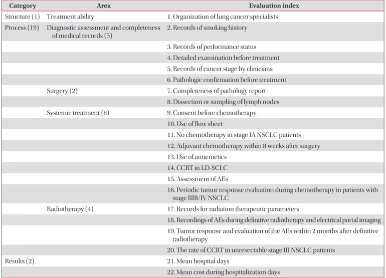
Table 1. Appropriateness indicators of lung cancer management by HIRA of Korea
Category Area Evaluation index
Structure (1) Treatment ability 1. Organization of lung cancer specialists Process (19) Diagnostic assessment and completeness
of medical records (5)
2. Records of smoking history
3. Records of performance status 4. Detailed examination before treatment 5. Records of cancer stage by clinicians 6. Pathologic confirmation before treatment
Surgery (2) 7. Completeness of pathology report
8. Dissection or sampling of lymph nodes Systemic treatment (8) 9. Consent before chemotherapy
10. Use of flow sheet
11. No chemotherapy in stage IA NSCLC patients 12. Adjuvant chemotherapy within 8 weeks after surgery 13. Use of antiemetics
14. CCRT in LD-SCLC 15. Assessment of AEs
16. Periodic tumor response evaluation during chemotherapy in patients with stage IIIB/IV NSCLC
Radiotherapy (4) 17. Records for radiation therapeutic parameters
18. Recordings of AEs during definitive radiotherapy and electrical portal imaging 19. Tumor response and evaluation of the AEs within 2 months after definitive
radiotherapy
20. The rate of CCRT in unresectable stage III NSCLC patients
Results (2) 21. Mean hospital days
22. Mean cost during hospitalization days
HIRA: Health Insurance Review and Assessment Service; NSCLC: non-small cell lung cancer; CCRT: chemotherapy combined with chemo-
therapy; LD: limited disease; SCLC: small cell lung cancer; AE: adverse events.
4. Detailed examination before treatment
This index refers to the percentage of patients who under- went close-up screening examination for lung cancer before treatment including surgery, chemotherapy, or radiation therapy (Table 3). These include (1) chest computed tomog- raphy (CT) that includes upper abdomen and adrenal glands;
(2) positron emission tomography (PET)-CT/PET in the stage IB–III non-small cell lung cancer (NSCLC) patients; (3) brain CT or brain magnetic resonance image (MRI) in the limited disease (LD)–small cell lung cancer (SCLC) and stage II-III NSCLC patients; (4) lung function test in the patients who un- derwent lung cancer surgery and definitive radiation therapy;
(5) mediastinoscopy and endobronchial ultrasound-guided transbronchial needle aspiration or video-assisted thoraco- scopic surgery in N2 NSCLC patients (excluding stage IV); and (6) epidermal growth factor receptor (EGFR) mutation test (monitoring) in the stage IV lung adenocarcinoma patients
1.
Prior to treatment, it is essential to perform highly sensitive tests to determine the status of patients and the lung cancer stage
2,3. (1) Chest CT is an indispensable tool for assessment of tumor size, location, and involvement with surrounding organs, and lymph node
4. (2) PET-CT or PET is useful for determining metastatic disease
2. (3) Brain CT or brain MRI is useful for confirming the presence of brain metastases
5,6. (4) Pulmonary function tests (forced expiratory volume in 1
second [FEV
1], diffusion lung capacity for carbon monoxide) are helpful in assessing the risk associated with surgery or radical radiotherapy
7-9. (5) Mediastinoscopy, endobronchial ultrasound, and mediastinal lymph node dissection should be performed prior to treatment for mediastinal staging if N2 stage is suspected because mediastinal stage has a significant effect on treatment decisions and prognosis
10-12. (6) In NSCLC patients who are not candidate of curative therapy, EGFR mutation test should be performed because EGFR tyrosine kinase inhibitors improve response rates, time to progression, and overall survival compared with systemic chemotherapy in patients harboring activating EGFR mutation
13,14. In case all the relevant tests are been carried out, this Indicator is consid- ered to fulfill the requirement. When the examinations were carried out by other institute(s), including an external agency, it would be recognized when there is corresponding inspec- tion films or result sheets. Regarding the chest CT, it should be taken within 60 days from the starting day of treatment and should include epigastrium and adrenal gland. If there is a written description on the epigastrium and adrenal gland, it is acknowledged and in this case, the reading findings for the upper abdomen should be provided. In case that chest CT scan does not include epigastric and adrenal glands, it would be acknowledged when PET-CT/PET or abdominal CT scan identifies these regions. Regarding pulmonary function tests, FEV
1alone is accepted when FEV
1is equal or more than 80%
Table 2. ECOG/WHO scoring system and Karnofsky PS status scale
ECOG/WHO score system (0–5) Definition Karnofsky score
0 Fully active, able to carry on all pre-disease activities without restriction 90–100 1 Restricted in physically strenuous activity but ambulatory and able to carry out
work of a light or sedentary nature
70–80
2 Ambulatory and capable of all self-care but unable to carry out any work activities;
up and about more than 50% of waking hour
50–60
3 Capable of only limited self-care, confined to bed or chair 50% or more of waking hours
30–40
4 Completely disabled; cannot carry on any self-care; totally confined to bed or chair 10–20
5 Dead
ECOG: Eastern Cooperative Oncology Group; WHO: World Health Organization; PS: performance score.
Table 3. Radiologic tests and their evaluation subjects in patients with lung cancer
Radiologic test Evaluation subject
Chest CT including upper abdomen/adrenal glands (within 60 days from beginning of initial treatment) All patients diagnosed as lung cancer
PET or PET-CT Stage IB–III NSCLC
Brain CT or MRI LD-SCLC
Stage II, III NSCLC
CT: computed tomography; PET: positron emission tomography; NSCLC: non-small cell lung cancer; MRI: magnetic resonance image; LD:
limited disease; SCLC: small cell lung cancer.
of reference value and there are no abnormal symptoms such as dyspnea. EGFR mutation test is recognized if it is confirmed before the start of treatment and in case of radiation therapy on the metastatic lesion, the test after radiotherapy would be accepted.
However, when the patient who does have any lung cancer related diagnosis underwent emergency surgery, the patient refused examination except chest CT, brain CT/brain MRI, and pulmonary function test, and patient status is too poor to take examination, it is excluded from evaluation. In addition, if the EGFR mutation test is not performed due to insufficient specimen, this case shall be excluded from the evaluation by including the reason for exclusion such as the pathologic or the doctor’s medical records.
5. Records of cancer stage by clinicians
Clinical cancer stage is assessed by the ratio of complete records of pretreatment stage by clinicians to all lung cancer patients underwent any treatment modality. For the clinical staging, clinicians should describe the records before treat- ment using TNM staging system according to the American Joint Committee on Cancer (7th edition) for NSCLC and a two-stage classification (LD or extensive disease [ED]) or TNM staging system for SCLC. In patients who underwent nonsurgical treatment, pretreatment staging should be re- ported by one of clinicians including pulmonologist, oncolo- gist, thoracic surgeon, and radiation oncologist. In patients who underwent surgical treatment, thoracic surgeon could document complete records of clinical stage within 28 days after surgery. In cases of neoadjuvant treatment prior to sur- gery, clinicians (pulmonologist, oncologist, thoracic surgeon or radiation oncologist) must document clinical stage before surgery. When the institution shares staging records among clinicians or the trainee documents the clinical cancer stage in a teaching hospital, the final signature by a corresponding specialist is accepted for the evaluation.
6. Pathologic confirmation before treatment
Pretreatment confirmative diagnosis is evaluated by the proportion of patients with pretreatment pathological diag- nosis out of total lung cancer patients who underwent non- surgical treatment. Referral patients who have only pathology reports from outside hospital are also included. Stage IV lung cancer patients with palliative radiation therapy are excluded.
7. Completeness of pathology report
Completeness of surgical pathology report is assessed by the ratio of complete pathology reports to all surgically re- sected lung cancer specimens. Surgical specimens requested to the referral hospitals for pathologic examination are also in-
cluded. Every complete pathology report should document all essential pathological data that will affect patient’s treatment and outcome and pathologists signature. Essential pathologic data include tumor site, tumor size, histologic tumor type ac- cording to the WHO classification, visceral pleural invasion, lymphovascular invasion, perineural invasion, pathologic staging, lymph node status, surgical resection margin status, and nonneoplastic pathologic pulmonary findings such as interstitial pulmonary fibrosis and tuberculosis. For the patho- logic staging, each T and N factor must be clearly defined (e.g., pT2N2 and pT2Nx). Lymph node state should be reported as the number of lymph nodes involved and the total number of lymph nodes submitted. Despite of no residual tumor, lymph node status and nonneoplastic pulmonary findings are re- quired. The reason for missing pathologic parameters should be documented for the complete report.
8. Dissection or sampling of lymph nodes
This index is defined as the proportion of patients under- went lymph nodes dissection or sampling out of all surgically resected lung cancer patients. Systemic mediastinal lymph node dissection or sampling is recommend for complete resection because it discovered pathologic N2 involvement in 24% of patients with clinical stage I and II NSCLC
15. For patients undergoing sublobar resection (segmentectomy or wedge resection), the appropriate N1 and N2 lymph node sta- tion should be sampled unless not technically feasible
16. In pa- tients with N2 disease, more than three ipsilateral mediastinal lymph node stations should be dissected during surgery
15,17. N2 disease is based upon the clinical cancer stage by thoracic surgeon. The patients with (1) postoperative stage IIIB or IV, (2) limited cardiopulmonary function (e.g., chronic obstructive pulmonary disease), (3) inaccessible lymph node less than three stations due to prior surgery, (4) documented reasons for missing lymph node dissection or sampling, (5) pure ad- enocarcinoma in situ or minimally invasive adenocarcinoma, and (6) ground glass opacity on chest CT are excluded.
9. Consent before chemotherapy
This Indicator refers to the percentage of patients who have
provided a written statement on the chemotherapy to the
patient or family member who received chemotherapy and
who have a consent record. The ratio of patient (or family)
agreement for chemotherapy is calculated as follows: Number
of patients with a record of agreement with the description
of chemotherapy/Number of patients treated with chemo-
therapy×100. The patient and physician should discuss the
positive effects and possible risks of the chemotherapy and
the discussion should include the evidence of treatment, treat-
ment related complications and patient’s choice according
to the prognosis. This indicator is admitted when there is a
written consent signed by a doctor who provides sufficient explanation for the chemotherapy and if the nurse is on the job, the doctor’s final signature is required. The purpose of the chemotherapy, the type of chemotherapeutic agents, the time or duration of the chemotherapy, and the main side effects should be included. Finally, a written consent form is issued prior to the initiation of chemotherapy, and a new consent form must be filled out whenever the regimen is changed.
10. Use of flow sheet
This index is the percentage of lung cancer patients who re- ceived chemotherapy using a flow sheet. The ratio of patients using flow sheet among lung cancer patients who received chemotherapy is calculated as follows: Number of patients using flow sheet/Number of patients treated with chemother- apy×100.
By using flow sheet on the chemotherapy, accurate record- ing of chemotherapy and monitoring patient status can fa- cilitate patient status assessment and patient education. This flow sheet should include both oral and parenteral anticancer drugs, excluding oral target drugs, and include five main items such as the purpose of chemotherapy, type of chemotherapy, number of cycles, duration, blood counts such as white blood count or absolute neutrophil count, hemoglobin, and plate- let. However, patients with institutional review board (IRB)- approved clinical trials are excluded from the evaluation.
11. No chemotherapy in stage IA NSCLC patients This index refers to the percent of the stage IA lung cancer patients who had not received adjuvant chemotherapy, and it reflects that the adjuvant chemotherapy is not recommended for the stage IA patients. The ratio of patients who do not receive chemotherapy (stage 1A) is as follows: Number of patients who do not receive adjuvant chemotherapy/Number of patients who underwent resection with lung cancer (stage 1A)×100.
In the cases who did not receive prior therapy, the parame- ter is evaluated for the cancer staging recorded by the thoracic surgeon after surgery, and the cases who have undergone preoperative therapy, it is evaluated based on the clinical can- cer stage recorded by a specialist who is in charge of the pre - treatment. However, patients with IRB-approved clinical trials are excluded from the evaluation.
12. Adjuvant chemotherapy within 8 weeks after surgery This indicator refers to the percentage of patients who un- derwent first adjuvant chemotherapy within 8 weeks of the last therapeutic intervention among patients who underwent surgery for stage II/IIIA of NSCLC (<70 years old, ECOG 0–1).
The ratio of adjuvant chemotherapy within 8 weeks after sur-
gery is calculated as follows: Number of patients who under- went adjuvant chemotherapy within 8 weeks after surgery/
Number of patients who underwent surgery for stage II/IIIA of NSCLC (<70 years old, ECOG 0–1)×100.
Stage II/IIIA patients with NSCLC are required to adjuvant chemotherapy after surgery and it is recommended to per- form adjuvant chemotherapy within 8 weeks considering the period of recovery from surgery and surgical complications.
Exclusion criteria of this indicator (the ratio of adjuvant che- motherapy within 8 weeks after surgery) are as follows: (1) patients who were transferred to other institutions within 8 weeks after surgery or died, (2) patients who underwent pre- operative therapy, (3) patients who underwent postoperative chemoradiotherapy or only palliative therapy, (4) patients who scheduled postoperative radiation therapy for adjuvant therapy, (5) patients with IRB-approved clinical trials.
Regardless of dosage regimen, all administered (oral or par- enteral) chemotherapeutic agents are included in this Indica- tor. Cancer staging is based on the following criteria: (1) clini- cal cancer stage recorded by physician before neoadjuvant chemotherapy, (2) cancer stage recorded by thoracic surgeon after surgery when the patients did not receive neoadjuvant chemotherapy. PS is based on the records that were evaluated before the start of adjuvant chemotherapy.
13. Use of antiemetics
This index is the proportion of patients provided with a serotonin antagonist among those who received moderate- to-moderate risk emetogenic chemotherapeutic agents. The patients who underwent emetogenic chemotherapy of mod- erate to high risk may suffer nausea and vomiting at least 4 days and should be protected during periods of risk. It will be evaluated the percentage of the patients who provided anti- emetic agents before administration of chemotherapeutic agents with moderate to severe nausea and vomiting. How- ever, patients with IRB-approved clinical trials are excluded.
14. CCRT for patients with LD-SCLC
This Indicator is the rate of patients who underwent concur- rent CCRT in LD-SCLC. The PS is based on the records that were evaluated before the start of CCRT and the score of the PS should be 0–2
18. Radiation therapy should be given when chemotherapy begins one to three cycles, and the combined use of radiotherapy and chemotherapy should be performed within 1 day
19,20.
15. Assessment of adverse effects
This is an indicator for the percentage of cases who received
chemotherapy for lung cancer and had an assessment of the
side effects of chemotherapy before each treatment cycle. As-
sessment of adverse reactions to chemotherapeutic agents such as non-hematologic toxicity (nausea/vomiting, diarrhea, systemic asthenia, skin rash, peripheral neuropathy, etc.), nephrotoxicity, hepatotoxicity and bone marrow suppression are recommended before administration of chemotherapy.
However, cases that do complete one cycle after the onset of chemotherapy and IRB-approved clinical trials are excluded.
Regardless of oral or parenteral regimens, all assessments of the adverse effect should be included and approved by the physician if the anticancer drug side-effects are listed on the medical record, including flow sheet, before each cycle. When evaluating side effects, one or more side effects should be in- dicated and if there are no side effects, it should be indicated as “None.” If oral anticancer drugs are administered alone, side effects should be evaluated every cycle within the first 3 months of administration, and every two to three cycles or every 2 to 3 months after 3 months of administration.
16. Periodic tumor response evaluation during chemotherapy in patients with stage IIIB/IV NSCLC Physician should record periodic tumor response evalua- tion during chemotherapy in patients with NSCLC of stage IIIB/IV. They should describe tumor response as complete remission (CR), partial response (PR), stable disease (SD), or progressive disease (PD) according to WHO or Response Evaluation Criteria In Solid Tumor (RECIST) criteria. Tumor response assessment should be done every two to three cycles or every 2 to 3 months. The lung cancer stage is based on the clinical cancer stage recorded by the specialist before the initiation of chemotherapy. Tumor response assessment methods are used by imaging modalities such as chest X-ray, CT, MRI, and PET-CT, etc.
17. Records for radiation therapeutic parameters There should be a record of the therapeutic radiation pa- rameters as follows: total radiation dose, radiation dose per fraction (or a number of the fraction), and treatment site.
18. Recordings of adverse events during definitive radiotherapy and electrical portal imaging
The radiation oncologists usually interview patients who receive radiotherapy once a week in the course of treatment to evaluate the side effects of radiotherapy. This Indicator is used to evaluate whether the side effects and the accuracy of the radiation field placement using electronic portal imaging devices (EPID). The radiation oncologist must record once a week, and a delay of 1–2 days due to holidays or other reasons is possible. If there is EPID checklist in the picture archiving and communication system, the documents accepted as chart records.
19. Tumor response and evaluation of the adverse events within 2 months after definitive radiotherapy Radiation oncologists must record and should describe tumor response as CR, PR, SD, or PD according to WHO or RECIST criteria. If there is no side effect, it should be recorded as “None.”
20. The rate of CCRT in unresectable stage III NSCLC patients
This indicator is the rate of patients who underwent CCRT in unresectable stage III NSCLC. Radiotherapy should be performed on the thoracic lesion and should be performed on the same day as chemotherapy
21,22. The PS is based on the records that were evaluated before the start of CCRT and the score of the PS should be 0–1
23. The age of the patient should be less than 70 years.
21. Mean hospital days
This index refers to the average length of hospital stay for patients who underwent surgery for lung cancer. The cases where the number of hospitalization day exceed the upper value or fall below the lower value are excluded and the for- mula is as follows.
Upper limit=X > [Q3+2.5|Q3-Q1|], Lower limit=X < [Q1- 2.5|Q3-Q1|]
X: total medical expenses or length of hospitalization days, Q1: 1st quartile, Q3: 3rd quartile
22. Mean cost during hospitalization days
This indicator is the average cost of patients who are hospi- talized for lung cancer surgery. This index also excludes the cases whose hospitalization days are extremely high or low and therefore exceed the upper limit or fall below the lower limit. The definitions for the upper and lower values are the same as those described in the indicator 21.
Results for Assessment in Lung Cancer in Korea
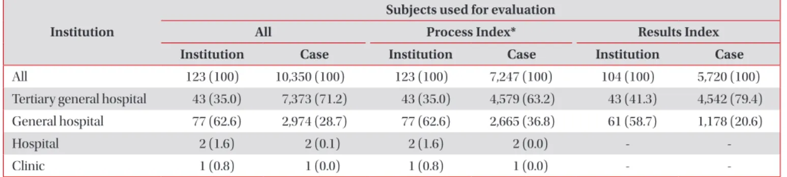
241. Subjects used for evaluation
Final subjects used for evaluation were 10,350 cases from
123 institutions. A total of 7,247 cases from 123 institutions
were subjects used to evaluate “Process Index.” Of these in-
stitutions, tertiary general hospitals and general hospitals ac-
counted for 97.6%. A total of 5,720 cases from 104 institutions
were subjects used to evaluate “Results Index,” including 4,542
cases from 43 tertiary general hospitals and 1,178 cases from 61 general hospitals. Detailed proportions of subjects used for evaluation in terms of institution and index are summarized in Table 4.
In terms of treatment modalities, 50.4%, 34.1%, and 15.5% of cases underwent surgery, chemotherapy, and radiotherapy, respectively. Male cases accounted for 69.7%. Those who were 60 years old or more accounted for 74.1% of cases. NSCLC cases accounted for 82.5% while SCLC cases accounted for 17.5%. Of 5,981 NSCLC cases, 1,777 (29.7%), 505 (8.4%), 1,087 (18.2%), and 2,612 (43%) cases were at stages 1, 2, 3, and 4, respectively. Among 1,243 SCLC cases, 369 (29.7%) cases had LD-SCLC while 874 (70.3%) had ED-SCLC.
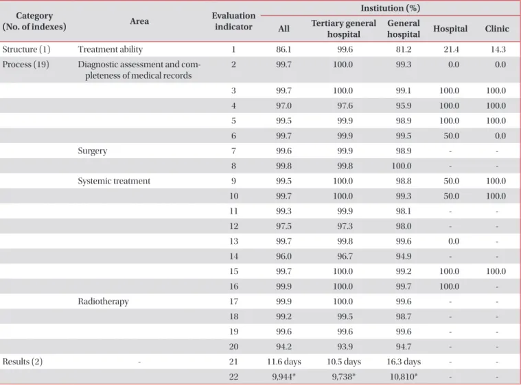
2. Results of evaluation 1) Structural category
This evaluation category consisted of one index: “constitu- tion of specialists.” It indicated whether an institution had all required specialists for lung cancer diagnosis and treatment, including pulmonologist, oncologist, thoracic surgeon, pa- thologist, radiation oncologist, radiologist, and nuclear medi- cine specialist. Fulfillment rates of this index in tertiary general hospitals, general hospitals, and clinics were 99.6%, 81.2%, and 14.3%, respectively. Radiologist showed the highest fulfillment rate (100%) while radiation oncologist showed the lowest ful- fillment rate (69.8%). Distribution of specialists according to different types of institutions is shown in Figure 1.
2) Process category
Process category consisted of four areas and 19 indexes.
“Area of diagnostic assessment and completeness of medical records” consisted of five indexes: “Record of smoking history,”
“Record of performance status,” “Detailed examination before treatment,” “Record of cancer stage by clinicians,” and “Patho- logic confirmation before treatment.” Fulfillment rates for these five indexes were 99.7%, 99.7%, 97.0%, 99.5%, and 99.7%,
respectively. “Area of surgery” consisted of two indexes: “Com- pleteness of pathology report” and “Dissection or sampling of lymph nodes.” The two indexes also showed high fulfillment rates (99.6% and 99.8%, respectively).
“Area of systemic treatment” included eight indexes: “Con- sent before chemotherapy,” “Use of flow sheet,” “No chemo- therapy in stage IA,” “Adjuvant chemotherapy within 8 weeks after surgery,” “Use of antiemetics,” “CCRT in LD-SCLC,”
“adverse events (AEs),” and “Regular response evaluation for stage IIIB/IV NSCLC.” These eight indexes also showed high fulfillment rates (99.5%, 99.7%, 99.3%, 97.5%, 99.7%, 96.0%, 99.7%, and 99.9%, respectively). “Area of radiotherapy” con- sisted of four indexes: “Records for radiotherapy,” “Assessment of AEs during curative radiotherapy and regular image cali- Table 4. Distribution of subjects used for evaluation
Institution
Subjects used for evaluation
All Process Index* Results Index
Institution Case Institution Case Institution Case
All 123 (100) 10,350 (100) 123 (100) 7,247 (100) 104 (100) 5,720 (100)
Tertiary general hospital 43 (35.0) 7,373 (71.2) 43 (35.0) 4,579 (63.2) 43 (41.3) 4,542 (79.4) General hospital 77 (62.6) 2,974 (28.7) 77 (62.6) 2,665 (36.8) 61 (58.7) 1,178 (20.6)
Hospital 2 (1.6) 2 (0.1) 2 (1.6) 2 (0.0) - -
Clinic 1 (0.8) 1 (0.0) 1 (0.8) 1 (0.0) - -
Values are presented as number (%).
*If an institution had less than 150 cases, the evaluation was made for all subject cases. If an institution had more than 150 cases, the evalua- tion was made for those sampled from subject cases.
Figure 1. Distribution of specialists according to different types of institutions. Radiologist showed the highest fulfilment rate (100%) while radiation oncologist showed the lowest (69.8%).
Upper general hospital 100
80
60
40
20
Clinic
Workforce(%)
0
Hospital General
hospital
000000100
0100050.0000
60.899.756.995.591.773.090.8
99.610097.7100100100100
Pulmonologist Medical oncologist Thoracic surgeon Pathologist
Radiation oncologist Radiologist
Nuclear medicine specialist