Hidradenitis suppurativa (HS, also known as acne in- versa, acne tetrad and pyoderma fistulans) is an uncom- mon chronic suppurative inflammatory disease that af- fects the apocrine gland-bearing follicular epithelium (1- 3). Clinically, it is characterized by recurrent chronic skin infections and the formation of sinus tracts and considerable scars (1). Its most common locations are the axilla, perineum and groin, and these areas have the highest density of follicular structures (2). The diagnosis of hidradenitis suppurativa is primarily clinical, so there are few reports about its magnetic resonance imaging (MRI) findings. We present here the MRI findings of a patient with hidradenitis suppurativa that caused a pos- terior neck mass.
Case Report
A 37-year-old man presented with a posterior neck mass. He had skin lesions on the posterior neck since puberty and he underwent medical treatment. But the lesion recurrently waxed and waned, so the extent of the lesion became wider. Physical examination revealed a hard, elevated, non-tender mass (longest dimension:
20 cm) with multiple sinuses that produced yellowish pus. He had another lesion of same nature on the left buttock. Clinically, the dermatologist had suspected the disorder to be hidadenitis suppurativa and referred the patient to a plastic surgeon for surgical removal. MRI was performed for planning of the operation. MRI demonstrated an ill-defined soft tissue mass with multi- ple variable sized cystic lesions. The soft tissue mass measured 12×10×4 cm in the subcutaneous fat layer, it showed low signal intensity with patchy increased sig- nal intensity on both the T1- and T2-weighted images and it was not gadolinum-enhanced. The cystic lesions within the low signal mass showed increased signal in-
J Korean Radiol Soc 2007;56:537-540
─ 537 ─
Hidradenitis Suppurativa Presenting with a Posterior Neck Mass: A Case Report1
Seung Young Lee, M.D., Min Hee Jeon, M.D., Il Heon Bae, M.D., Gi Seok Han, M.D., Sang Hoon Cha, M.D., Sung Jin Kim, M.D., Kil Sun Park, M.D.
1Department of Diagnostic Radiology, Chungbuk National University Hospital
Received January 18, 2007 ; Accepted April 10, 2007
Address reprint requests to : Seung Young Lee, M.D., Department of Radiology, Chungbuk National University Hospital, 62 Gaeshin-dong, Heungduk-gu, Cheongju, Chungbuk 361-763, Korea
Tel. 82-43-269-6472 Fax. 82-43-269-6479 E-mail: [email protected]
Hidradenitis suppurativa is a rare disorder that is characterized by recurrent chronic skin infections and the formation of sinus tracts and considerable scaring. A 37-year- old man presented with a hard posterior neck mass. Multiple pus-producing sinuses were detected in the skin covering the mass. MRI demonstrated an ill-defined, soft tis- sue mass with multiple variable sized cystic lesions. The soft tissue mass measured 12
×10×4 cm in the subcutaneous fat layer, it contained multifocal cystic lesions that re- vealed higher signal intensity on both the T1- and T2-weighted images, as compared with the adjacent neck muscles. The mass was not enhanced on the post-contrast T1 weighted images. Some of the cystic lesions extended to the skin. The mass was re- moved surgically and confirmed to be hidradenitis suppurativa.
Index words :Hidradenitis suppurativa
Seung Young Lee, et al: Hidradenitis Suppurativa Presenting With a Posterior Neck Mass
─ 538 ─
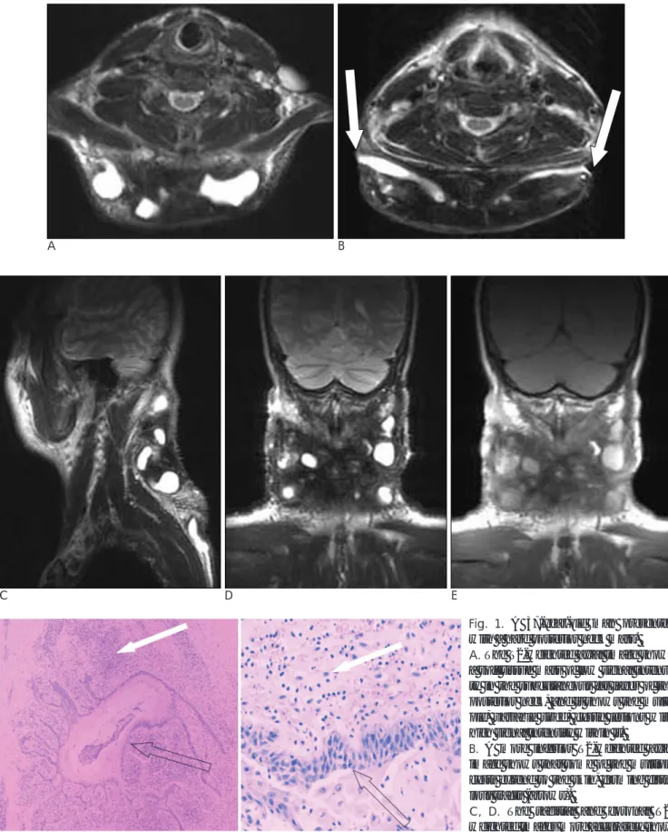
A B
C D E
F
Fig. 1. A 37-year-old man presented with a hard posterior neck mass.
A. The T2-weighted axial image shows a soft tissue mass of low signal intensi- ty in the subcutaneous fat layer of the posterior neck, and it shows the multi- ple, variable sized, cystic lesions with high signal intensity within it.
B. A more inferior T2-weighted axial image shows that some of the multiple cysts extend to the skin, forming fistu- lous tracts (arrows).
C, D. The sagittal and coronal T2- weighted images more accurately show the extent of disease. The mass in- volves the skin and subcutaneous layer and it does not extend to the muscle or bone.
E. The non-contrast enhanced, T1-weighted coronal image shows the heterogenous low signal intensity of the mass and the multi- ple cystic lesions of high signal intensity within it.
F. The gross specimen and photomicrography reveal chronic inflammation and fibrosis (solid arrows) with multiple epidermal cysts (open arrows) and some of them are ruptured.
tensity in comparison with the neck muscle on the T1- weighted image and bright signal intensity was noted on the T2-weighted image, and some of them extended to the skin. Surgical removal and skin grafts were per- formed. Pathologically, the lesion revealed chronic in- flammation and fibrosis with multiple epidermal cysts (some ruptured) without malignant cells.
Discussion
Hidradenitis suppurativa is a misnomer because the process does not arise from the sweat glands, but rather, it arrises from infected hair follicles. Hidradenitis sup- purativa is a response to follicular occlusion that’s prob- ably caused by frictional trauma (1, 4). Follicular hyper- keratosis is the initial event, leading to occlusion, occa- sional secondary apocrine involvement and follicular rupture with the resultant inflammation and possibly secondary infection (2). Sinus tract formation arises from rupture of the abscess within the follicular epitheli- um. These sinus tracts may dissect into the deeper struc- tures such as muscles, fascia and even bowels (1, 5).
Hidradenitis suppurativa is a recurrent disease with a highly variable clinical course. The clinical diagnostic criterias are wide and they include recurrent disease, scarring and a multifocal location (2). In this case, the patient had long standing, recurrent skin lesions in the posterior neck and left buttock, so clinically, the derma- tologist had a high suspicion of hidradenitis suppurativa.
For patients with hidradenitis suppurativa, tender papules or deep-seeded nodules develop, and the nodule may slowly resolve; however, it often expands and coa- lesces with surrounding nodules to form a large, painful inflammatory abscess that may rupture spontaneously.
The lesion then heals with fibrosis, dermal contractures and rope-like elevation of the skin; sinus tracts may also develop. On the MR imaging of our patient, this ap- peared as a soft tissue mass with low signal intensity on the T1- & T2-weighted images in the subcutaneous fat layer, which corresponds to chronic inflammation and fibrosis. The multiple cystic lesions within the mass cor- respond to epidermal cysts or abscessed cysts.
Microscopically, some of them rupture or extend to the skin, and so they formed sinus tracts. We think the lo- calized patchy increased signal intensity may corre- spond with ruptured cyst and spilled cystic material.
Systemic antibiotics, hormonal therapy and corticos-
teroids may be effective, but the common experience is one of initial response followed by subsequent relapse when such treatment is withdrawn. Intra-lesional cor- ticosteroids are a good option for isolated inflamed le- sions. But surgery is essential in many cases, especially for localized, recurrent, refractory and advanced dis- ease. Incision and drainage shows a recurrence rate of 100% while wide excision and surgical reconstruction has a recurrence rate of 25% at a median interval of 20 months (2, 6). More aggressive and definitive surgical re- moval of all the involved tissue, beyond the clinically in- volved margin, is the most effective treatment modality.
The incidence of complication from hidradenitis suppu- rativa such as septicemia, anemia, fistula and squamous cell carcinoma may be reduced by making the early di- agnosis and adequate treatment (2).
The diagnosis of hidradenitis suppurativa is primarily clinical, but at the time of surgical excision, surgeons must consider the size or extent of the lesion, and whether or not they involve the surrounding structures such as muscle or bone so they can plan reconstruction.
MRI gives sufficient and objective findings for this. In our case, the disease involved the skin and subcuta- neous tissue without involvement of muscle and bone, so the resected area could be reconstructed by skin grafting.
Hidradenitis suppurativa is an uncommon disorder in dermatology, and there are few reports about its radio- logical findings. In our experience, the MRI findings of HS correspond very well to the surgical and pathologic findings.
References
1. Russ E, Castillo M. Lumbosacral epidural abscess due to hidradenitis suppurativa. AJR Am J Roentgenol 2002;178:770-771 2. Wiseman MC. Hidradenitis suppurativa: a review. Dermatol Ther
2004;17:50-54
3. Nadqir R, Rubesin SE, Levine MS. Perirectal sinus tracks and fistu- las caused by hidradenitis suppurativa. AJR Am J Roentgenol 2001;
177:476-477
4. Jansen T, Plewig G. What’s new in hidradenitis inversa (alias hidradenitis suppurativa). J Eur Acad Dermatol Venereol 2000;14:
342-343
5. Mortimer PS, Lunniss PJ. Hidradenitis suppurativa. J R Soc Med 2000;93:420-422
6. Loo WJ, Rytina E, Todd PM. Hidradenitis suppurativa, Dowling- Degos and multiple epidermal cysts: a new follicular occlusion tri- ad. Clin Exp Dermatol 2004;29:622-624
J Korean Radiol Soc 2007;56:537-540
─ 539 ─
Seung Young Lee, et al: Hidradenitis Suppurativa Presenting With a Posterior Neck Mass
─ 540 ─
대한영상의학회지 2007;56:537-540
경부종괴로 발현한 화농성 한관염: 1예 보고1
1충북대학교병원 영상의학과
이승영・전민희・배일헌・한기석・차상훈・김성진・박길선
화농성 땀샘염은 드문 질환으로 만성 재발성 피부감염으로 피부로 누출관을 만들며, 상당한 반흔조직 형성을 특 징으로 한다. 37세 남자 환자가 후경부 종괴를 주소로 내원했다. 종괴를 덮은 피부에는 농을 포함하고 있는 다수의 구멍이 관찰되었다. 자기공명영상에서 종괴는 후경부의 피하지방층에 위치하는 12×10×4 cm 가량의 종괴로, 내 부에 다수의 낭종을 포함하고 있는 경계가 잘 그려지지 않는 고형 종괴였다. 종괴는 T1과 T2에서 모두 저 신호강 도 종괴내부에 부분적으로 증가한 신호강도를 가지는 양상이며 조영증강은 거의 되지 않았다. 종괴 내부의 낭성부 분은 T2에서 물과 비슷하고 T1에서는 물보다 높은 신호강도를 보이며, 그들 중 일부는 피부로 연결된 누출관을 형 성하고 있었다. 종괴는 수술로 제거되었으며, 임상적으로 그리고 병리학적으로 화농성 땀샘염으로 확진되었다.