- 211 - 대한족부족관절학회지: 제13권 제2호 2009
J Korean Foot Ankle Soc. Vol. 13. No. 2. pp.211-213, 2009
족관절에 발생한 미만성 색소 융모 결절성 활액막염의 개방적 절제술(1예 보고)
청주성모병원 정형외과, 가톨릭대학교 의과대학 정형외과학교실*
김보현⋅권순억⋅강신택⋅박세욱*
Open Synovectomy in Diffuse Pigmented Villonodular Synovitis of Ankle Joint (A Case Report)
Bo-Hyeon Kim, M.D., Soon-Eok Kwon, M.D., Shin-Taek Kang, M.D., Se-Wook Park, M.D.*
Department of Orthopedic Surgery, Cheongju St. Mary’s Hospital, Cheongju, Korea Department of Orthopedic Surgery, Catholic University College of Medicine, Seoul, Korea*
=Abstract=
Pigmented villonodular synovitis (PVNS) is a rare proliferative disorder that affects synovium, tendon sheath and bursa.
Although the condition can present in any joint, knee joint is the most commonly affected site and only 2.5% of cases occur in foot and ankle joint. PVNS occurs in two types: localized and diffuse. Localized type is characterized by focal involvement of the synovium with either nodular or pedunculated masses, Diffuse type affects virtually the entire synovium. Diffuse type has reported more recurrence rate. We have experienced a patient who has diffuse type PVNS of ankle joint and report an optimal method of surgical treatment.
Key Words: Ankle, Pigmented villonodular synovitis, Diffuse type, Excision
∙Address for correspondence Soon-Eok Kwon, M.D.
Department of Orthopedic Surgery, Cheongju St. Mary’s Hospital, 589-5 Jujung-dong Sangdang-gu, Cheongju-si Chungcheongbuk-do, 360-568, Korea
Tel: +82-43-219-8142 Fax: +82-43-219-8447 E-mail: [email protected]
색소 융모 결절성 활액막염(pigmented villonodular synovitis: PVNS)은 관절, 건막 및 점액낭에 존재하는 활 액막에서 기시하여 증식을 일으키는 특발성 질환으로 유병 율이 매우 낮은 것으로 알려져 있다6,7). 신체의 어느 관절에 나 발생할 수 있는 것으로 보고되나 슬관절에 가장 호발하 고 족관절에는 매우 드물게 발생한다6,11). 조직학적으로는
양성 종양으로 분류되나 연부 조직 및 골을 침범하고 연골 을 파괴하는 등의 성질을 띠며 관절 및 사지에 기능적, 형태 적 문제를 초래하기도 한다. 개방적 절제술, 관절경 절제술 등의 수술적 방법, 방사선 조사 또는 화학요법 등을 통하여 치료가 이루어지고 있으나 미만형 색소 융모 결절성 활액막 염의 경우, 개방성 절제술을 통한 활액막 전 절제술이 가장 효과적인 치료법으로 받아들여지고 있다6,7). 저자들은 족관 절에 발생한 미만형 색소 융모 결절성 활액막염의 증례에 대하여 개방적 전 절제술을 시행하고 효과적인 치료법에 대 하여 문헌 고찰과 함께 보고하고자 한다.
증례 보고
31세 여자 환자로 내원 13개월 전부터 특별한 외상력 없
김보현⋅권순억⋅강신택⋅박세욱
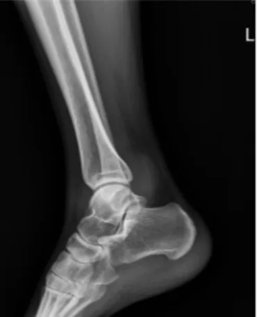
- 212 - Figure 1. Plain lateral radiograph of left ankle shows soft tissue shadow around ankle but no bony abnormality.
A B C
Figure 2. Sagittal MRI of Lt ankle. (A) T1 weighted image shows low signal intensity mass in anterior and posterior aspect of ankle joint.
(B) T2 weighted image shows high signal intensity of anterior and posterior mass. (C) T1 weighted with Gd enhanced image shows slightly high signal intensity.
이 발생한 좌측 족관절의 통증 및 종물을 주소로 내원하였 다. 종물이 촉지되기 전 타병원에서 보존적 치료를 받아왔 으나 증세의 호전이 없었으며 최근 증상이 악화되는 양상을 보였다. 내원 당시 시행한 이학적 검사상 좌측 족관절의 전 방, 내측, 후외방에 걸쳐 경계가 불명확한 종물이 촉지되었 으며 관절면의 압통이 있었다. 피부 발적 및 국소 열감 등 감염의 증후는 없었으며 통증으로 인한 관절 운동의 제한이 있었다. 시행한 단순 방사선 소견상 족관절 전후면 연부 조 직의 음영 증가 소견이 관찰되었으나 골 파괴 소견은 없었 다(Fig. 1). 자기 공명 영상 검사를 시행하였으며 T1 강조 시상면 영상에서 족관절의 전방과 후방 관절강을 넓게 채우 고 있는 이질성의 저신호 강도를 보이는 종물이 관찰되었 다. T2 강조 시상면 영상에서도 균일하지 않은 고신호 강도 의 종물이 관찰되었으며 관절액이 증가한 소견을 보였다.
종물은 조영 증강되었으며, 주변 골조직의 저명한 변화소견 은 없었다(Fig. 2).
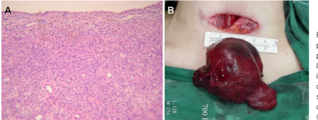
좌측 족관절의 전방 도달법 및 후방 도달법을 이용한 개 방적 절제술로 5×2×2.5 cm 크기의 경계가 명확한 종물 2 개를 제거하였으며, 비후된 활액막에 대하여 전 절제술을 시행하였다. 절제된 종물은 노란색을 띠는 미만성의 활액막 증식 소견을 보였으며 혈색소의 침착으로 보이는 적갈색 부 분이 전체적으로 산개되어 있었다. 병리 조직 검사상 포말 세포와 혈철소를 포함한 포식세포 등으로 구성된 유두상 돌 기, 조직구와 다핵성 거대 세포의 침윤 및 활액막 조직내의 거대세포와 유사 조직구 세포 등 미만성 색소 융모 결절성 활액막염과 일치하는 소견을 보였다(Fig. 3). 수술 직후 감 염 등의 합병증 소견은 없었으며 24개월 추시 관찰에서 환 자는 재발의 징후 없이 통증과 관절 운동 제한도 사라졌다.
고 찰
색소 융모 결절성 활액막염은 1852년 Chassaignac1)이 중지의 굴곡건에 발생한 것을 처음 기술한 이후 Jaffe 등3) 이 1941년에 비종양성 염증 반응으로 규정하였으나 현재는 양성 종양성 병변으로 받아들여지고 있다. 연간 백만 명 당 1.8명 정도에서 발생하는 드문 질환으로 슬관절 등의 대형 관절에서 많이 발생하나 족부 및 족관절에는 2.5%정도의 낮은 유병률을 보인다4,6,7). 임상적 증상으로는 이환된 관절 의 동통, 종물 촉지 및 부종 등을 주로 호소하며, 때로는 증 식된 활액막에 의한 골파괴나 관절 연골의 침식으로 관절의 동통성 강직을 보이기도 한다9). 종종 골관절염, 류머티스 관절염의 초기로 오인할 수 있으며 일반적으로 초기 단순 방사선 검사상 정상소견을 보여 임상적으로 진단이 어렵거 나 늦어지는 경우가 있다. 색소 융모 결절성 활액막이 발생 하는 원인은 아직 정립되지 않았으나 외상으로 인한 반복적
족관절에 발생한 미만성 색소 융모 결절성 활액막염의 개방적 절제술(1예 보고)
- 213 -
A B
Figure 3. Gross and microscopic photos of the mass. (A) Gross photo shows well marginated large mass. (B) Microscopic find- ings shows villous projections composed of foamy cells, hemo- siderin-containing phagocytes, giant cells and chronic inflammatory cells (H&E, ×40).
인 국소 출혈에 의하거나 원인이 밝혀지지 않는 자극에 의 해서 활액막에 염증이 발생한다는 가설8,10) 및 지방대사의 이상에 의해 발생한다는 설4) 등이 제기되고 있으며 본 증례 에서도 정확한 외상 및 대사이상 등의 과거력은 없었다.
색소 융모 결절성 활액막염은 활액막의 일부에 결절이 나 자루형 종물 형태로 국한되어 나타나는 국소형과, 이환 된 관절의 전 활액막을 침범하며 점진적으로 골 및 관절조 직의 파괴를 동반하는 미만형으로 구분된다2,8). 국소형 색 소 융모 결절성 활액막염에 비해 미만형에서 재발율이 높은 것으로 보고되고 있다7). 미만형의 경우 부적절한 활액막의 제거에 의해 병변의 완전한 제거가 이루어지지 않아 재발이 흔한 것으로 알려져 있다5,7). 족관절의 경우 슬관절 등과 같 이 큰 관절과는 다르게 관절경을 통한 충분한 활액막을 제 거하기가 용이하지 않아 절개하여 개방적으로 활액막을 제 거하는 것이 더 유용하다7,8). 개방적 절제술을 실시할 경우 에도 미만형은 족관절의 해부학적 구조상 기술적으로 완전 한 절제 및 노출이 어렵기 때문에 광범위한 절개를 통한 전 절제술을 실시하여 활액막을 모두 제거해야 한다. 본 증례 에서도 자기 공명 영상 소견상 종물의 크기가 크고 미만형 으로 전 관절을 침범하고 있어 전방 도달법 및 후방 도달법 에 의한 개방적 절제술을 시행하여 관절내 활액막을 모두 제거하였다. 수술 후 재발을 방지하기 위하여 방사선 조사, 화학요법 등의 방법이 제시되고 있으나 방사선 조사로 인한 방사선 노출의 부작용, 관절 강직, 젊은 환자에서는 종양의 발생 등의 부작용이 보고되고 있다5). 색소 융모 결절성 활 액막염의 정확한 발생 원인이 증명되지 않은 상태이므로 수 술 후 방사선 조사 및 화학 요법 등을 시행하는 것은 주의가 필요하다. 그러므로 족관절에 발생한 미만형 색소 융모 결 절성 활액막염에서 치료 및 재발 방지를 위해 관절의 적절 한 노출 및 개방적 활액막 제거술을 시행하여 이환된 관절 의 활액막을 완전히 제거하는 것이 중요하다고 생각된다.
REFERENCES
1. Chassaignac M: Cancer de la gaine des tendons. Gaz Hop Civ Milit, 47: 185-186, 1852.
2. Granowitz SP, D’ Antonio J and Mankin HL: Pathogenesis and long-term end results of pigmented villonodular synovitis.
Clin Orthop, 114:335-351, 1976.
3. Jaffe HL, Lichtenstein L and Sutro CJ: Pigmented villonodular synovitis, bursitis and tenosynovitis. Arch Pathol, 31: 731- 765, 1941.
4. Klompmaker J, Veth RP, Robinson PH, Molenaar WM and Nielsen HK: Pigmented villonodular synovitis. Arch Orthop Trauma Surg, 109: 205-210, 1990.
5. Lee M, Mahroof S, Pringle J, Short SC, Briggs TW and Cannon SR: Diffus pigmented villonodular synovitis of the foot and ankle treated with surgery and radiotherapy. Int Orthop, 29: 403-405, 2005
6. Mayers BW and Masi AT: Pigmented villonodular synovitis and tenosynovitis: a clinical epidemiologic study of 166 cases and literature review. Medicine(Baltimore), 59: 223-238, 1980.
7. Mendenhall WM, Mendenhall CM, Reith JD, Scarborough MT, Gibbs CP and Mendenhall NP: Pigmented villonodular synovitis: review. Am J Clin Oncol, 29: 548-550, 2006 8. Sakkers RJ, de Jong D and van der Heul RO: X-chromosome
inactivation in patients who have pigmented villonodular synovitis. J Bone Joint Surg Am, 73: 1532-1536, 1991.
9. Saxena A and Perez H: Pigmented villonodular synovitis about the ankle: a review of the literature and presentation in 10 athletic patients. Foot Ankle Int, 25: 819-826, 2004.
10. Singh R, Grewal DS and Chakravarti RN: Experimental production of pigmented villonodular synovitis in the knee and ankle joints of rhesus monkeys. J Pathol, 98: 137-142, 1969.
11. Sung JH, Kim WY, Han CW, Yoon JK and Kim JY:
Simultaneous pigmented villonodular synovitis and synovial chondromatosis in the ankle joint. J Korean Orthop Assoc, 33:
477-483, 1998.