A Rare Case of Limited Muscle
Involvement in Polyarteritis Nodosa
INTRODUCTION
Polyarteritis nodosa (PAN) is a systemic vasculitis that can affect multiple organs in its diffuse form and is associated with high rates of morbidity and mortality. From the standpoint of pathology, PAN is necrotizing arteritis predominantly involving medium and small muscular arteries, without glomerulonephritis or vasculitis in the arterioles, capillaries, or venules (1, 2). PAN has protean manifestations, and it may present as a systemic disease or in its limited form, be confined to a single organ (2). The limited form of PAN is rare and may pose a diagnostic challenge; it can mimic other common diseases, and its nonspecific presentation may lead to confusion or delays in diagnosis.
Musculoskeletal system involvement in PAN is known to present as myalgia, arthralgia, polymyositis-like syndrome, asymmetric nondeforming polyarthritis, or rarely, as intermittent claudication, acute leg ischemia, or myopathy (2). Muscular involvement in PAN may occur as a component of systemic PAN or as a limited form. Patients with the limited form of PAN are known to present with pain, swelling, and tenderness of the lower leg (3). Magnetic resonance imaging (MRI) findings of muscular involvement in PAN have been reported in other studies, all in cases with the limited form of PAN (4). Most of the previous studies have reported nonspecific edematous signal changes in the affected muscles with hyper-signal intensities on T2-weighted or fat-saturated sequences (5), which may be found in all clinical mimics such as inflammatory or infectious myositis. Ultrasound is particularly useful in the diagnosis of large- and medium-sized vessel vasculitis, as the characteristic wall thickening of the affected arteries allows for the confirmation of a suspected diagnosis. However, only a few studies have reported ultrasonography findings for the limited form of PAN. Herein, we describe a rare case of PAN involving skeletal muscle as indicated on
This is an Open Access article distributed under the terms of the Creative Commons Attribution Non-Commercial License (http://creativecommons.org/licenses/ by-nc/4.0/) which permits unrestricted non-commercial use, distribution, and reproduction in any medium, provided the original work is properly cited. Received: January 16, 2021 Revised: February 22, 2021 Accepted: February 23, 2021
Correspondence to:
Ro Woon Lee, M.D.
Department of Radiology, Inha University College of Medicine, 27 Inhang-ro, Jung-gu, Incheon 22332, Korea.
Tel. +82-32-890-2762 Fax. +82-32-890-2743 E-mail: [email protected]
Copyright © 2021 Korean Society of Magnetic Resonance in Medicine (KSMRM)
Case Report
Polyarteritis nodosa (PAN) is a systemic vasculitis involving small- and medium-sized arteries, which presents with necrotizing inflammation. PAN occurs as a systemic disease or as a limited form confined to a single organ. Few cases have been reported with single organ involvement, and even fewer have been reported with skeletal muscle involvement. Herein, we report the ultrasonography and magnetic resonance imaging findings in a rare case of PAN with limited muscle involvement in a 66-year-old man.
Keywords: Polyarteritis nodosa (PAN); Vasculitis; Myositis; Magnetic resonance imaging (MRI)
Sung Oh Song1, Ro Woon Lee1, Mie Jin Lim2, Seong Ryul Kwon2, Won Park2
1Department of Radiology, Inha University College of Medicine, Incheon, Korea 2
MRI and ultrasonography. The laboratory features and the pathologic findings in the present case were consistent with the limited form of PAN.
CASE REPORT
A 66-year-old man was admitted to the hospital with a 1-week history of myalgia and muscle weakness in both thighs. The patient had abrupt lower leg weakness in both legs and was unable to walk or rise from a chair. His initial temperature was 37.9°C, heart rate was 80 beats/min, blood pressure was 128/80 mmHg, and respiratory rate was 26 breaths/min. All peripheral pulses were palpable and cardiac (cardiac sound assessment and echocardiogram), chest, and abdominal examinations (inspection, palpation, percussion, and auscultation) were normal. There were no focal neurologic deficits, and the results of the mental status examination were normal. He had no demonstrable erythema or rash.
The initial laboratory test results were as follows: leukocyte count, 14,560/μL with 85% neutrophils; hemoglobin level, 14.1 g/dL; erythrocyte sedimentation rate (ESR), 117 mm/h (reference range, 0-30); C-reactive protein (CRP) level, 19.32 mg/dL (reference range, 0-0.4); blood urea nitrogen (BUN), 23.3 mg/dL; creatinine, 1.01 mg/dL; creatinine phosphokinase (CPK), 56 U/L (reference range, 30-188); aldolase, 9.7 U/L (reference range, < 7.6); and angiotensin-converting enzyme, 52 U/L (reference range, 20-70). Liver function tests and urine sediment were normal. The patient’s plasma D-dimer level was 1.5 μg/mL (reference range, < 0.5). Hepatitis B and C serology, cold agglutinin, anti-nuclear antibody (ANA), and anti-neutrophil cytoplasmic antibody (ANCA) were all negative.
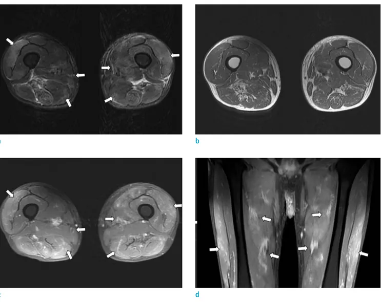
The MRI of both thighs revealed multifocal ill-defined intramuscular T2 signal alteration around both sides of the vastus and adductor muscle groups, rectus femoris, and semitendinosus muscle of the thigh on T2-weighted images, more predominantly distributed around the intramuscular vascular branches (Fig. 1). These areas were isointense on pre-contrast T1-weighted images but were seen as variably sized small multifocal patchy enhancing nodules or tubular, branching enhancing areas with fuzzy margins following intravenous gadolinium administration. There was no increased uptake on the bone scan.
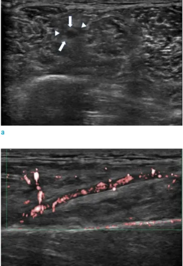
Additionally, ultrasonography was used to evaluate both thighs. Ultrasonography with a 5-12 MHz linear probe showed prominent segmental circumferential wall
thickening of the patent intramuscular artery in the right vastus lateralis muscle (Fig. 2). Doppler ultrasound was also performed for the corresponding lesion. However, Doppler and color Doppler studies showed no significantly increased vascular flow within the thickened vascular wall and perivascular area.
Muscle biopsy was performed intraoperatively, with specimens obtained from the right vastus lateralis muscle. Multifocal fibrinoid necrosis was observed in the muscle and adipose tissue and leukocytoclastic and mononuclear inflammatory cell infiltrations were noted in the medium- and small-sized arteries of the perimysium (Fig. 3), indicating PAN. The patient was diagnosed with PAN confined to both thigh muscles. The thigh pain of the patient gradually improved after treatment with high-dose prednisolone (1 mg/kg/d). The ESR, CRP, and CPK levels were restored to normal after two weeks of treatment.
DISCUSSION
PAN is a necrotizing vasculitis involving medium and small arteries but sparing the venous system (1, 2). The characteristic pathological findings of PAN are fibrinoid necrotizing inflammatory foci in the walls of small- and medium-sized arteries, with multiple small aneurysms. During the acute phase, the elastic lamina is gradually destroyed, and the media undergoes fibrinoid necrosis. Patients with PAN typically experience constitutional symptoms such as fever, malaise, weight loss, and diffuse aching along with polyarthritis, peripheral neuropathy, and various manifestations in the kidneys and gut (6). Less frequently, patients may experience musculoskeletal symptoms such as myalgia, arthralgia, and polymyositis-like syndrome. As in the present case, the extremities, especially the lower limbs, can be the sole or first-affected body parts. Several similar cases have been described in the literature under different names such as calf muscle vasculitis, limb restricted vasculitis, and muscular PAN (7, 8). In one previous case report, although the calf was the main muscle involved, other lower limb areas, particularly the quadriceps, were also affected (7).
The main feature of muscular PAN is the noticeable paucity of systemic organ manifestations, with the exception of cutaneous or testicular involvement (7). The symptoms of muscular PAN are often confused with myopathy, as in the present case. Therefore, different kinds of inflammatory myopathies should be considered in the
diagnosis of muscular PAN. In our differential diagnosis, we also considered myopathies, including polymyositis (PM) and dermatomyositis (DM). The report of myopathy in PAN is uncommon because the involvement of other organs may lead to a diagnosis before significant muscle involvement becomes apparent (7). The progression from a limited form to systemic PAN is rare (less than 10%) and extra-musculoskeletal major organ involvement has not been observed after follow-up in other case series of the limited form of PAN with musculoskeletal involvement (8). In cases of muscular PAN, serum CPK levels are normal or increased,
and electromyography inconsistently shows a myopathy pattern. As indicated by the uncertainty of these findings, histopathologic confirmation of necrotizing arteritis is important in the diagnosis of muscular PAN.
Systemic PAN commonly results in the formation of an abdominal aneurysm and frequently involves the renal artery. Conventional angiography and magnetic resonance angiography (MRA) have been used to detect PAN. In several individual cases of the limited form of PAN, MRI findings of skeletal muscle involvement have been reported (4). Most previous studies have reported nonspecific
Fig. 1. Axial image of T2-weighted fat-saturated magnetic resonance imaging (a) shows multifocal patchy T2 high-signal intensity foci in the intramuscular area of both thighs involving the vastus and adductor muscle groups, rectus femoris, and semitendinosus muscle (arrows). (b) No demonstrable intramuscular signal abnormality is seen on the pre-contrast T1-weighted axial image of either thigh at the same level. (c, d) Post-contrast fat-suppressed T1-weighted axial and coronal images show multifocal contrast-enhancement anterior and posterior compartments in both thigh muscles, which had previously shown intramuscular T2 signal alteration (arrows) more predominantly distributed around the intramuscular vascular branches.
a b
Fig. 2. Transverse ultrasound scan image with a 5-12 MHz linear probe (a) of the intramuscular vessel branch of the vastus lateralis muscle in the right thigh shows irregular perivascular increased echogenicity in the arterial wall (arrows) and perivascular low echogenic hollow (arrowheads). (b) The longitudinal scan image in the corresponding area shows thick and linear vascular wall thickening (arrows) in the intramuscular arterial branch of the vastus lateralis muscle. (c) No remarkable increase in the vascular flow of the thickened vessel wall or the perivascular area of the intramuscular arterial branch is seen on color Doppler ultrasound.
a
c
b
Fig. 3. Incisional biopsy was performed on the left gastrocnemius muscle. Microscopic features (a) show small- and medium-sized arteries in the intramuscular tissue (left image, hematoxylin and eosin stain, × 100 magnification). (b) The arterial wall contains fibrinoid necrotizing inflammation characteristic of polyarteritis nodosa (right image, hematoxylin and eosin stain, × 200 magnification).
intramuscular patchy high-signal intensities in the affected muscles on T2-weighted or short-T1 inversion recovery (STIR) sequences, which may also be found in all of the aforementioned clinical mimics, including inflammatory myopathies such as PM or DM. Kang et al. (4) reported that muscle involvement in PAN may show fluffy, nodular, enhancing lesions centered on vessels on contrast-enhanced images and may be accompanied by fascial or periosteal enhancement. In the present case, MR images showed multifocal patchy T2 high-signal intensity foci with enhancement in the muscle groups of both thighs, predominantly distributed around the deep intramuscular vascular branch. The increased signal intensity foci in the intramuscular area on T2-weighted images is thought to be edema, which is an increase in intracellular or extracellular free water and suggestive of the acute phase of muscular PAN (4). However, accompanying fascial and periosteal enhancement, indicating that PAN may manifest as fasciitis and periostitis, indicative of the subacute-to-chronic phase, were not observed in the present case.
We found multiple enhancing foci around the intramuscular branching vessels on contrast-enhanced MR images. The pathophysiology of skeletal muscle involvement of PAN has been explained in several ways, including ischemia resulting from the occlusion of blood vessels, the involvement of muscle fibers by inflammation around the blood vessels, and muscle atrophy due to extensive damage of the peripheral nerves (9, 10). Biopsy specimens of the involved muscle in PAN have shown areas of atrophy, the degeneration of muscle fibers, and myonecrosis, which is indicative of ischemic injury (9, 11). Deteriorated blood flow due to the occlusion or narrowing of blood vessels can lead to fuzzy diffusion of the contrast media into the extravascular space, which exhibits multiple enhancing foci centered on blood vessels on contrast-enhanced images.
Hyper-signal intensity appearing predominantly or exclusively in T2-weighted or STIR sequences indicates increased intracellular or extracellular free water in muscle. In our patient, the MR image findings were most likely consistent with muscle edema, which is the principal feature of muscle inflammation. However, because myopathies of traumatic, metabolic, and degenerative origin and peripheral neuropathies exhibit these same changes, some authors prefer the term edema-like abnormalities rather than edema (12). It is, therefore, unlikely that a diagnosis of the limited form of vasculitis may be made based solely on MRI findings. In particular, the patchy, asymmetric, and distal distribution of the lesions resembles that found in
focal myositis (12).
Despite an apparently low specificity, MRI of skeletal muscle could constitute a useful complementary examination for limited PAN (13). Nonetheless, further study needs to be undertaken to evaluate the precise diagnostic sensitivity of MRI. Another application of MRI could be to select the optimal site for a muscle biopsy to ensure that the sampled tissue comes from an affected area, whereas serial MRI might be helpful for monitoring the course of the disease and response to treatment (13).
Additionally, in some cases, ultrasonography has been proposed as a valuable tool for the evaluation of vasculitis. Three typical ultrasonographic findings are related to inflamed arteries, including intimal edema forming a hypoechoic ring at the periphery of the vessel lumen (halo sign), lumen stenosis, and occlusion. In our case, prominent thickening in the vessel wall of the intermuscular vascular branch and a perivascular low echogenic halo was noted. This is the first case report of ultrasonography findings of PAN with limited muscle involvement.
In summary, we present a rare case of the limited form of PAN that initially involved skeletal muscles. Also, we believe that the recognition of intramuscular vessel wall thickening as seen on ultrasonography or MRI may help suggest a diagnosis of limited PAN involving skeletal muscle, among the numerous diseases showing hyper-signal intensities in the affected muscles on T2-weighted or fat-saturated MR sequences, even if there are no clinical features of systemic PAN.
Ethical Approval
This study was approved by the Institutional Review Board, which waived the requirement for informed patient consent.
REFERENCES
1. Jennette JC, Falk RJ, Bacon PA, et al. 2012 revised International Chapel Hill Consensus Conference Nomenclature of Vasculitides. Arthritis Rheum 2013;65:1-11
2. Colmegna I, Maldonado-Cocco JA. Polyarteritis nodosa revisited. Curr Rheumatol Rep 2005;7:288-296
3. Hernandez-Rodriguez J, Alba MA, Prieto-Gonzalez S, Cid MC. Diagnosis and classification of polyarteritis nodosa. J Autoimmun 2014;48-49:84-89
in Polyarteritis Nodosa: Report of Eight Cases With Characteristic Contrast Enhancement Pattern on MRI. AJR Am J Roentgenol 2016;206:378-384
5. Revelon G, Rahmouni A, Jazaerli N, et al. Acute swelling of the limbs: magnetic resonance pictorial review of fascial and muscle signal changes. Eur J Radiol 1999;30:11-21 6. Guillevin L, Pagnoux C, Teixeira L. Polyarteritis nodosa
and microscopic polyangiitis. In Ball GV, Bridges SL, eds. Vasculitis. 2nd ed. Oxford: Oxford University Press, 2008:335-364
7. Plumley SG, Rubio R, Alasfar S, Jasin HE. Polyarteritis nodosa presenting as polymyositis. Semin Arthritis Rheum 2002;31:377-383
8. Khellaf M, Hamidou M, Pagnoux C, et al. Vasculitis r e s t r i c t e d t o t h e l o w e r l i m b s : a c l i n i c a l a n d histopathological study. Ann Rheum Dis 2007;66:554-556 9. Matsubara S, Mair WG. Ultrastructural changes of skeletal
muscles in polyarteritis nodosa and in arteritis assoicated with rheumatoid arthritis. Acta Neuropathol 1980;50:169-174
10. Pearson CM, Currie S. Polymyositis and related disorders. In Walton JN, ed. Disorders of voluntary muscle. Edinburgh, UK: Churchill Livingstone, 1974:614-652
11. Plumley SG, Rubio R, Alasfar S, Jasin HE. Polyarteritis nodosa presenting as polymyositis. Semin Arthritis Rheum 2002;31:377-383
12. Reimers CD, Schedel H, Fleckenstein JL, et al. Magnetic resonance imaging of skeletal muscles in idiopathic inflammatory myopathies of adults. J Neurol 1994;241:306-314
13. Gallien S, Mahr A, Rety F, et al. Magnetic resonance imaging of skeletal muscle involvement in limb restricted vasculitis. Ann Rheum Dis 2002;61:1107-1109