INTRODUCTION
Reactive lymphoid hyperplasia (RLH), known as pseudolym- phoma, is a rare condition that has been observed in the liver.
It is characterized by a marked proliferation of polyclonal and non-neoplastic lymphoid cells with the formation of abundant follicles that have active germinal centers (1, 2). It is usually localized and well demarcated from the surrounding tissue.
RLH is thought to represent an immune-mediated reactive phenomenon, and may arise in association with a malignant tumor (2, 3). We report a case of RLH of the liver that mim- icked a metastatic carcinoma, based on radiological findings in a patient with renal cell carcinoma. In particular, the his- tiocyte-rich RLH pattern observed in this case, which seems to be an uncommon feature of RLH, is discussed.
CASE REPORT
A 46-yr-old woman underwent a radical right nephrectomy for stage I renal cell carcinoma of the clear cell type. A follow- up computed tomography (CT) scan was done 14 months later
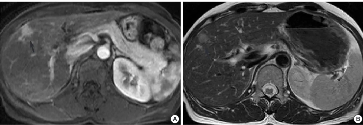
and revealed a new mass in segment 5 of the liver. It was 1.0 cm in diameter and well-defined round-shaped mass showing high attenuation at arterial phase imaging and slight low atten- uation at portal and equilibrium phase imaging. For further evaluation of this mass, a magnetic resonance (MR) examina- tion was performed. On T2-weighted MR imaging, this mass showed an intermediate hyperintensity-like liver malignancy (Fig. 1A). On gadolinium-enhanced MR imaging, this mass showed bright nodular enhancement at arterial phase imag- ing (Fig. 1B) and peripheral rim-like enhancement at delayed phase imaging, which was interpreted as a metastatic renal cell carcinoma or hypervascular hepatocellular carcinoma. A physical examination and chest roentgenogram were unremark- able. Laboratory data were all within the normal range and the results of liver function tests were normal (aspartate aminotrans- ferase[AST]21 U/L, alanine aminotransferase [ALT]33 U/L, total bilirubin 0.66 mg/dL, alkaline phosphatase 94 U/L, and lactate dehydrogenase [LDH]154 U/L). A test for the hepatitis B antibody was positive. The level of carcinoembryonic anti- gen was 3.7 ng/mL (normal -5), and CA19-9 was slightly ele- vated (41.98 U/mL, normal -36). Alpha-fetoprotein levels and anti-mitochondrial antibodies were not available. A diagno-
156
Ho Sung Park, Kyu Yun Jang, Young Kon Kim*, Baik Hwan Cho�, Woo Sung Moon
Departments of Pathology, Radiology*, and Surgery�, Chonbuk National University, Medical School, Institute for Medical Sciences and the Center for Healthcare Technology Development, Jeonju, Korea
Address for correspondence Woo Sung Moon, M.D.
Department of Pathology, Chonbuk National University, Medical School, San 2-20 Geumam-dong, Deokjin-gu, Jeonju 561-180, Korea
Tel : +82.63-270-3086, Fax : +82.63-270-3135 E-mail : [email protected]
*“This work was supported by the Korea Research Foundation Grant funded by the Korean Government (MOEHRD)” (The Regional Research Universities Program/Center for Healthcare Technology Development).
Unusual Morphologic Features
Reactive lymphoid hyperplasia (RLH) of the liver is a rare entity and has also been termed nodular lymphoid lesion or pseudolymphoma of the liver. We report a case of hepatic RLH exhibiting unusual histiocyte-rich histologic features in a 47-yr-old woman in conjunction with a renal cell carcinoma. A follow-up computed tomog- raphy scan was done 14 months after a right radical nephrectomy for renal cell car- cinoma revealed a nodular lesion in segment 5 of the liver. The lesion was interpret- ed as metastatic renal cell carcinoma or hepatocellular carcinoma based on the history of the patient and radiologic findings. Wedge resection of segment 5 was done with sufficient distance from the mass. Microscopically, the lesion was com- posed predominantly of peculiar histiocytic proliferation and was characterized by lymphoid aggregates forming a lymphoid follicle with germinal centers. The present case and prior cases reported in the literature suggest that RLH of the liver appear to be a heterogenous group of reactive inflammatory lesions that are often associ- ated with autoimmune disease or malignant tumors.
Key Words : Pseudolymphoma; Liver Neoplasms; Lymphoid Hyperplasia, Reactive
Received : 26 March 2007 Accepted : 13 June 2007
sis of metastatic renal cell carcinoma from the previous renal cell carcinoma was presumed, based on the prior history of the patient and radiological findings, and wedge resection of
segment 5 including the mass was performed ensuring ade- quate distance from the mass. Grossly, the resected liver seg- ment contained a well-circumscribed, yellowish-white, soli-
Fig. 1. (A) The respiratory-triggered T2-weighted turbo spin-echo MR imaging shows a small mass with intermediate high signal intensity (arrow) in liver segment 5. (B) The gadolinium-enhanced arterial phase MR imaging shows small bright nodular enhancement (arrow) in the same location as in A.
A B
Fig. 2. (A) A cut section of the resected liver shows a well-circumscribed, yellowish-white mass in segment 5. (B) The lesion is composed predominantly of peculiar histiocytic proliferation and lymphoid aggregate forming lymphoid follicles on the edge of the nodule. Note the dense infiltrate of lymphocytes in several portal tracts immediately around the nodule (arrows, ×20, H&E). (C, D) The lesion is mainly composed of aggregates of epithelioid histiocytes simulating non-caseating granuloma (C×200, D ×400, H&E). (E) Hyalinized structures in the nod- ule (arrow, ×200, H&E). (F) CD68 immunostaining highlights the large number of histiocytes (×200, CD68).
B C
D E F
A
forming a lymphoid follicle with germinal centers (Fig. 2B, C). Most of the follicles were observed on the edge of the nod- ule. There was also marked hyalinization in part of the mass, and several bile ductules were observed on the edge of the nodule. In the surrounding liver tissue, a marked periduc- tular fibrosis with prominent lymphocytic infiltration was also observed (Fig. 2B). However, the hepatic parenchyma distant from this nodule was normal and lymphoid infiltra- tion was not detected in the portal tracts. The bile duct sys- tem contained no stones. Lymphoid cells positive for L-26 (B cell marker, DAKO, Glostrup, Denmark) were distribut- ed mainly in the germinal centers, while those positive for UCHL-1 (T cell marker, DAKO) were present around the germinal centers. There were no cytokeratin-positive malig- nant epithelial cells in the nodule. CD68 (histiocyte mark- er, DAKO) immunostaining highlighted the large number of histiocytes with epithelioid cell features (Fig. 2D). HMB45 (marker for angiomyolipoma, DAKO)-positive cells were not detected. Use of special stains, such as periodic acid-Schiff, Grocott, and Ziehl-Neelsen stains failed to demonstrate any microorganisms in the lesion. A polymerase chain reaction analysis for mycobacteria was negative.
DISCUSSION
In the liver, RLH has also been variously termed as nodular lymphoid lesion (2, 4) and pseudolymphoma (5-9). A review of the literature has revealed that 17 cases of RLH of the liver have been reported to date (Table 1). Including the present case, the age of patients ranged from 36 to 85 yr (average, 60.5 yr). There was a female predilection with a female-to-male ratio of 8:1. In terms of underlying disease, chronic hepati- tis (6, 8), autoimmune disease such as Sjogren s disease (9), chronic thyroiditis (10), primary biliary cirrhosis (2), CREST syndrome (2), and diabetes mellitus (1, 7) have been report- ed. Hence, autoimmune mechanisms seem to be most like- ly involved in the RLH occurrence.
By definition, the diagnosis of RLH is based on a polymor- phous lymphocytic infiltration, and often numerous germinal centers, without cytologic atypia; however, on the basis of the published pathologic descriptions, this seemed to be variable both in extent and in cellular composition. In nine cases, lym- phoid infiltration was seen in the portal areas around the nod- ule. However, including the current case, lymphoid infiltra- tion was confined to the portal tracts immediately around the nodule; the rest of the hepatic parenchyma was normal, sug- gesting that this lesion was local rather than diffuse (1-3, 7, 10-13). We can speculate that several lymphoid follicles in these portal areas conglomerated and formed an RLH lesion.
Tanizawa and colleagues reported a RLH of the liver charac- terized by an angiofollicular pattern with interfollicular hyalin- ization mimicking Castlemans desease (14). In five cases includ- ing the case reported here, hyalinized trabecular structures in the nodule were present (2, 11, 13, 14). The peculiar histio- cytic proliferation in the present case consisted of a granulo- matous arrangement of epithelioid histiocytes, which is sim- ilar to the characteristic features of a non-caseating granuloma.
However, periodic acid-Schiff, Grocott, and Ziehl-Neelsen stains failed to demonstrate any microorganisms in the lesion.
Furthermore, the histologic findings of lymphoid follicles with active germinal centers in our case are not seen in other gran- ulomatous lesions, such as sarcoidosis. Among the RLH of the liver cases we reviewed, only one case exhibited aggregates of epithelioid histiocytes in the nodule as seen in the case present- ed here. These findings suggest that RLH of the liver appear to represent a heterogenous group of reactive inflammatory lesions that share a varying degree of inflammation, rather than a specific entity.
Including the present patient, seven cases of hepatic RLH accompanying malignant tumors have been reported (3, 5, 8, 11, 13): in two each cases of colon cancer (3) and gastric can- cer (5, 8). One patient with multiple carcinomas (gastric, cecal, and colon cancer) has presented with hepatic RHL (13). Pan- tanowitz and colleagues have described an RLH of the liver in a patient with a renal cell carcinoma (11). Surprisingly, the histopathologic findings including the presence of lymphoid follicles with germinal centers, hyalinized trabecular structures, and lymphocytic infiltration in the portal tracts around the nodular lesion, are very similar to those seen in the present case. These findings suggest a possible correlation between hepatic RLH and renal cell carcinoma. However, there have been very few cases, so it is not clear whether the renal cell carcinoma was involved in the onset of the hepatic RLH. We can speculate that the etiology of RLH of the liver may be related to an immunologic abnormality that is caused by the malignant tumor itself or previous surgery for the tumors. The prognosis of the RLH associated with malignancies is good, and most patients treated by resection of this lesion have shown no recurrence or progression to lymphoma (3, 11).
Although our case showed some features of an inflammatory pseudotumor, it has other histologic findings that are not seen in an inflammatory pseudotumor, including the absence of fibroblastic proliferation, a lack of prominent fibrosis, collec- tion of foamy histiocytes, or occlusive endophlebitis. Further- more, the histologic features of inflammatory pseudotumors do not necessarily contain lymphoid follicles that are always found in RLH (7). Thus, our lesion is histologically different from an inflammatory pseudotumor.
Radiologically, RLH should be differentiated from other
. .
solid focal hepatic lesions as both conditions show interme- diate hyper-intensity on T2-weighted MR imaging. In par- ticular, the RLH in our case showed bright nodular enhance- ment on arterial phase MR imaging, which may be misinter- preted as hypervascular metastasis from a renal cell carcinoma or a hypervascular hepatocelluar carcinoma. The imaging find- ings of our case might be similar to those of previous reports
demonstrating variable enhancement of RLH using contrast- enhanced CT, CT during angiography, and direct hepatic an- giography (3, 6, 8, 10, 12). However, since the imaging modali- ties used in published reports were variable, a further evalu- ation with a large number of cases will be required in order to define clearly the radiological findings of this entity.
Rt., right; Lt., left; Seg, segment.
Special histologic features in addition to lymphoid Cases (reference) Age/Sex Size (cm)
follicles with germinal center Associated disease location
Grouls (5) 85/F 1.4×0.8 Gastric cancer
0.8×0.7 Rt. lobe
Isobe et al. (1) 59/F 0.9 Lymphocytic infiltration in the portal tracts around the Diabete mellitus Lt. lobe nodular lesion
Ohtsu et al. (6) 42/F 1.5×1.3 Chronic hepatitis B
Seg 6
Katayanagi et al. (7) 66/F 1.5×1.0 Lymphocytic infiltration in the portal tracts around the Diabete mellitus Rt. lobe nodular lesion
Tanizawa et al. (14) 67/F 2.0 Angiofollicular structures mimicking Castleman s disease Lt. lobe Hyalinized interfollicular spaces
Kim et al. (8) 72/M 1.7×1.5 Chronic active hepatitis of the non-tumorous liver Chronic hepatitis C
Seg 3 Gastric cancer
Sharifi et al. (2) 52/F 0.4 Lymphoepithelial lesions Primary biliary cirrhosis
Rt. lobe Strands of amorphous, dense hyalinized materials Thick-walled arterioles
56/F 1.5 Rare lymphoid follicles CREST syndrome
Lt. lobe Aggregates of epithelioid histiocytes
Lymphocytic infiltration in the portal tracts around the nodular lesion
56/M 0.7 Lymphoepithelial lesions Chronic diverticular
? Scattered giant cells and epithelioid histiocytic aggregates disease
Drug-induced hepatitis
Nagano et al. (10) 47/F 1.0 Chronic thyroiditis
Seg 7 Hemangioma of the liver
Pantanowitz et al. (11) 69/F 1.7×1.0 Numerous hyalinized trabeculated structures Renal cell carcinoma Rt. lobe Lymphocytic infiltration in the portal tracts around the
nodular lesion
Okubo et al. (9) 49/F 2.0 Sjogren s syndrome
Rt. lobe
Takahashi et al. (3) 77/F 1.5 Lymphocytic infiltration in the portal tracts around the Colon cancer
Seg 3 nodular lesion
64/F 0.9×0.7 Lymphocytic infiltration in the portal tracts around the Colon cancer
Seg 2 nodular lesion
Maehara et al. (12) 72/F 1.3×1.0 Lymphocytic infiltration in the portal tracts around the
Seg 3 nodular lesion
Willenbrock et al. (4) 36/F 1.8 Epithelioid cells, and giant cells in the interfollicular area Focal nodular hyperplasia
Seg 8 Lymphoepithelial lesions Hemangioma
Sato et al. (13) 75/F 1.4 Hyalinized fibrous tissues Gastric, cecal, and
Rt. lobe Lymphocytic infiltration in the portal tracts colon cancer around the nodular lesion
Present case 46/F 1.0×1.0 Florid aggregates of epithelioid histiocytes Renal cell carcinoma Seg 5 Rare lymphoid follicles
Lymphocytic infiltration in the portal tracts around the nodular lesion
Hyalinized materials
Table 1. Cases of reactive lymphoid hyperplasia of the liver in the literature and the present case
. .
thology.
REFERENCES
1. Isobe H, Sakamoto S, Sakai H, Masumoto A, Sonoda T, Adachi E, Nawata H. Reactive lymphoid hyperplasia of the liver. J Clin Gastroen- terol 1993; 16: 240-4.
2. Sharifi S, Murphy M, Loda M, Pinkus GS, Khettry U. Nodular lym- phoid lesion of the liver: an immune-mediated disorder mimicking low- grade malignant lymphoma. Am J Surg Pathol 1999; 23: 302-8.
3. Takahashi H, Sawai H, Matsuo Y, Funahashi H, Satoh M, Okada Y, Inagaki H, Takeyama H, Manabe T. Reactive lymphoid hyperplasia of the liver in a patient with colon cancer: report of two cases. BMC Ga- stroenterol 2006; 6: 25.
4. Willenbrock K, Kriener S, Oeschger S, Hansmann ML. Nodular lym- phoid lesion of the liver with simultaneous focal nodular hyperplasia and hemangioma: discrimination from primary hepatic MALT-type non-Hodgkin s lymphoma. Virchows Arch 2006; 448: 223-7.
5. Grouls V. Pseudolymphoma (inflammatory pseudotumor) of the liver.
Zentralbl Allg Pathol 1987; 133: 565-8.
6. Ohtsu T, Sasaki Y, Tanizaki H, Kawano N, Ryu M, Satake M, Hasebe T, Mukai K, Fujikura M, Tamai M, Abe K. Development of pseudolym- phoma of liver following interferon-alpha therapy for chronic hep-
A case of pseudolymphoma of the liver with chronic hepatitis C. J Hep- atol 1997; 26: 209-14.
9. Okubo H, Maekawa H, Ogawa K, Wada R, Sekigawa I, Iida N, Mae- kawa T, Hashimoto H, Sato N. Pseudolymphoma of the liver associ- ated with Sjogren s syndrome. Scand J Rheumatol 2001; 30: 117-9.
10. Nagano K, Fukuda Y, Nakano I, Katano Y, Toyoda H, Nonami T, Nagasaka T, Hayakawa T. Reactive lymphoid hyperplasia of liver coex- isting with chronic thyroiditis: radiographical characteristics of the disorder. J Gastroenterol Hepatol 1999; 14: 163-7.
11. Pantanowitz L, Saldinger PF, Kadin ME. Pathologic quiz case: Hep- atic mass in a patient with renal cell carcinoma. Arch Pathol Lab Med 2001; 125: 577-8.
12. Maehara N, Chijiiwa K, Makino I, Ohuchida J, Kai M, Kondo K, Mor- iguchi S, Marutsuka K, Asada Y. Segmentectomy for reactive lymphoid hyperplasia of the liver: report of a case. Surg Today 2006; 36: 1019- 23.
13. Sato K, Ueda Y, Yokoi M, Hayashi K, Kosaka T, Katsuda S. Reactive lymphoid hyperplasia of the liver in a patient with multiple carcino- mas: a case report and brief review. J Clin Pathol 2006; 59: 990-2.
14. Tanizawa T, Eishi Y, Kamiyama R, Nakahara M, Abo Y, Sumita T, Kawano N. Reactive lymphoid hyperplasia of the liver characterized by an angiofollicular pattern mimicking Castleman s disease. Pathol Int 1996; 46: 782-6.
. .