Korean J Gastroenterol Vol. 75 No. 1, 56-59 https://doi.org/10.4166/kjg.2020.75.1.56 pISSN 1598-9992 eISSN 2233-6869
IMAGE OF THE MONTH
Korean J Gastroenterol, Vol. 75 No. 1, January 2020 www.kjg.or.kr
담낭암과 동반된 담낭 천공에 의한 간농양
장영환, 이세환, 황정아
1, 안혜인
2순천향대학교 천안병원 소화기내과, 영상의학과1, 병리과2
Liver Abscess Arising from Gallbladder Perforation with Gallbladder Cancer
Younghwan Jang, Sae Hwan Lee, Jeong Ah Hwang1 and Hyein Ahn2
Division of Gastroenterology, Departments of Radiology1 and Pathology2, Soonchunhyang University Cheonan Hospital, Cheonan, Korea
CC This is an open access article distributed under the terms of the Creative Commons Attribution Non-Commercial License (http://creativecommons.org/licenses/
by-nc/4.0) which permits unrestricted non-commercial use, distribution, and reproduction in any medium, provided the original work is properly cited.
Copyright © 2020. Korean Society of Gastroenterology.
교신저자: 이세환, 31151, 천안시 동남구 순천향6길 31, 순천향대학교 천안병원 소화기내과
Correspondence to: Sae Hwan Lee, Division of Gastroenterology, Soonchunhyang University Cheonan Hospital, 31 Suncheonhyang 6-gil, Dongnam-gu, Cheonan 31151, Korea. Tel: +82-41-570-3692, Fax: +82-41-574-5762, E-mail: [email protected], ORCID: https://orcid.org/0000-0001-8320-5914
Financial support: None. Conflict of interest: None.
증례: 64세 남자 환자가 내원 10일 전부터 시작된 발열, 오한, 우상복부 통증을 주소로 내원하였다. 환자는 당뇨병과 고지혈증으로 약물 치료 중이었으며, 6개월간 해외 여행력은 없었다. 내원 당시 활력징후는 혈압 130/70 mmHg, 맥박수 138회/분, 체온 39.6℃로 측정되었다. 촉진 시 우상복부 압통 을 호소하였다. 말초혈액 검사에서 백혈구 9,920/mm3, 혈색 소 10.5 g/dL, 혈소판 148,000/mm3로 측정되었고, 생화학 검사에서 AST 116 U/L, ALT 105 U/L, 총 빌리루빈 1.3 mg/dL, ALP 225 U/L, GGT 113 U/L로 측정되었다. 종양표지자는 AFP 0.61 ng/mL로 정상 범위였고, CA 19-9 35.61 U/mL로 약간 증가되어 있었다. 응급실에서 시행한 복부 CT에서 간 좌엽 제4분절에 약 9.3 cm 크기의 테두리 조영증강을 보이는 낭성 병변이 관찰되었다. 담낭은 내강이 약간 작고 벽의 비후 와 조영증강 소견을 보였으며, 담낭 주변부의 침윤 및 담낭 주변 액체 저류가 있었다(Fig. 1A, B). 간농양과 병발된 담낭 염으로 진단하고 경험적 항생제를 투여하였으며, 간농양에 대 한 경피적 농양 배액술을 시행하였다. 환자의 임상증상과 혈 액학적 검사는 호전되었으며 배액량도 감소하여 배액술 시행 11일 경과 후 복부 CT를 추적하였고 간농양의 크기는 현저하 게 감소하였으나 담낭벽의 불규칙한 비후는 호전이 없었다 (Fig. 1C, D). 담낭 병변에 대한 정확한 평가를 위하여 자기공 명영상을 촬영하였고, 담낭 체부에 담낭벽의 비후와 조영증강
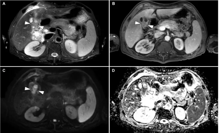
이 보이며 해당 부위의 내강이 좁고, 확산강조영상에서 제한 확산 소견을 보여 담낭암의 가능성을 배제할 수 없었다(Fig. 2).
이후 외과와 상의 후 복강경하 담낭 절제술을 시행하였고, 중 등도 분화 샘암종으로 진단되어 인근 림프절 곽청술 및 간실 질 절제술을 추가로 시행하였다(Fig. 3).
진단: 담낭암과 동반된 담낭 천공에 의한 간농양 자발성 담낭 천공은 흔하지 않은 담낭 질환이다. 급성 담낭 염에서 합병증으로 발생하는 담낭 천공의 발생 비율은 0.8-3.8%로 보고되어 있다.1만성 담낭염에서 합병되는 담낭 천공은 그보다 더 낮으며, 담낭암과 연관하여 천공이 발생하 는 경우는 매우 드물다. 담낭 천공을 일으키는 원인으로는 담 석, 감염, 악성 종양, 외상, 스테로이드 치료, 당뇨, 동맥경화 등 혈류 공급의 이상이 있을 수 있다.2,3병인은 담낭관의 폐색 으로 인하여 담즙 정체와 담낭 팽만이 일어나게 되고, 정맥과 림프액 흐름이 막히면서 혈관 손상이 담낭의 괴사를 초래하여 발생하는 것으로 알려져 있다.4천공이 호발하는 위치는 혈관 공급이 가장 적은 담낭 저부이다.5
담낭 천공은 1934년 Niemeier6에 의하여 처음 분류되었 다. 1형은 급성으로 발생하며 복막염을 동반하는 형태이며, 2형은 아급성으로 천공 주변부에 국한되어 발생하고 담낭 주 변 농양이나 국소적 복막염 소견을 보인다. 3형의 경우 만성
Jang Y, et al. Liver Abscess Arising from Gallbladder Perforation with Gallbladder Cancer
57
Vol. 75 No. 1, January 2020
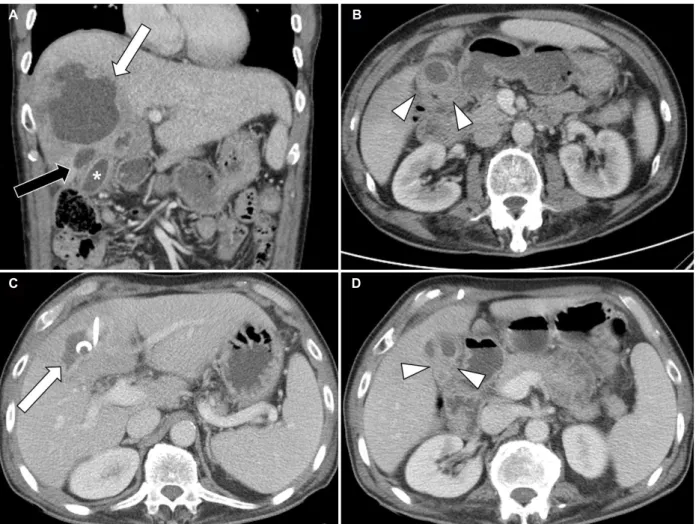
Fig. 1. Initial abdomen pelvic computed tomography. (A) About a 9.3 cm irregular rim enhancing lesion (arrows) was seen in segment 4 of the liver. A small pericholecystic abscess (black arrow) was also noted and the lumen of the gallbladder (asterisk) was mildly shrunken. (B) Irregular enhancing wall thickening (arrowheads) was observed in the gallbladder. Follow-up computed tomography. (C) The size of liver abscess (arrow) was markedly decreased with percutaneous drainage. (D) Enhancing wall thickening (arrowheads) persisted in the gallbladder.
적으로 진행하여 주변으로 누공을 형성하는 형태이다.6 담낭 천공으로 인하여 간농양이 발생한 경우는 비교적 드물며 보고 된 논문에 따라 Niemeier 분류법에 적용되지 않는다고 기술 하는 경우도 있고 2형으로 분류하는 경우도 있다.7,8
담낭 천공의 증상은 비특이적이다. 우상복부 통증, 고열, 우 상복부 종물과 압통 등의 증상과 혈액 검사상 간 효소와 ALP 수준의 상승을 보일 수 있다.9이는 일반적인 급성 담낭염과의 소견과 비슷하여 감별 진단이 쉽지 않다. 복부 초음파는 수술 전에 담낭 천공을 진단할 수 있는 영상 검사이다. 담낭벽의 비후(>3 mm), 담낭 확장(>3.5-4.0 cm) 등의 소견을 관찰할 수 있으며, 담낭벽의 결손 부위(hole sign)를 확인할 수 있다 면 진단에 매우 특이적이다.10 CT는 진단과 수술 전 계획을 세울 때 도움을 줄 수 있으며 천공을 진단하는 데 있어서 초음 파보다 민감도가 높은 검사이다. 또한 담낭 주변의 변화, 농양 의 확인에도 유용하다.11,12담낭벽의 결손을 평가하는 데 있어
MRI의 경우 높은 연부조직 해상력과 다면촬영능을 보여 진단 기법 중 하나로 이용할 수 있다.13또한 본 증례와 같이 담낭암 의 진단에 있어서는 매우 특이적인 검사이다.
담낭 천공의 치료는 Niemeier 분류법에 따라 달라지게 되 는데 1형의 경우 담낭 절제술을 기본으로 주변의 농양이 존재 할 경우 수술적 배액을 하게 된다. 2형의 경우 수술 전에 초음 파 유도하 경피적 배액술을 먼저 시행하여 감염이 호전된 후 복강경하 담낭 절제술을 시행하며, 3형의 경우는 담낭 절제술 과 함께 누공의 수술적 복구를 같이 시행하는 것을 추천한 다.10 본 증례에서는 담낭암과 이로 인한 담낭 천공으로 이차 성 간농양이 발생한 것으로 최종 진단하였으며, 드물지만 간 농양과 동반된 담낭염 혹은 담낭 천공에서 담낭암 병발 여부 도 감별 질환으로 고려해볼 수 있겠다.
A B
C D
58
장영환 등. 담낭암과 동반된 담낭 천공에 의한 간농양The Korean Journal of Gastroenterology Fig. 3. Pathologic findings of the gallbladder. (A) Gross findings of the
gallbladder. (B) Microscopic finding shows adenocarcinoma of the gallbladder invading through the wall into the adjacent soft tissue (H&E, ×12.5).
Fig. 2. Gallbladder magnetic resonance imaging. (A) Wall thickening (arrowheads) with annular narrowing was seen in the body on the T2-weighted image. (B) Contrast-enhanced wall thickening (arrowheads) on the portal phase. Hyper-intensity (arrowheads) on the diffusion-weighted imaging (C) and hypo-intensity (arrowheads) on the apparent diffusion coefficient map (D) were considered to be a malignant wall thickening.
REFERENCES
1. Stefanidis D, Sirinek KR, Bingener J. Gallbladder perforation: risk factors and outcome. J Surg Res 2006;131:204-208.
2. Isch JH, Finneran JC, Nahrwold DL. Perforation of the gallbladder.
Am J Gastroenterol 1971;55:451-458.
3. Madrazo BL, Francis I, Hricak H, Sandler MA, Hudak S, Gitschlag K. Sonographic findings in perforation of the gallbladder. AJR Am J Roentgenol 1982;139:491-496.
4. Taneja S, Sharma A, Duseja AK, Kalra N, Chawla Y. Spontaneous perforation of gallbladder with intrahepatic bilioma. J Clin Exp Hepatol 2011;1:210-211.
5. Abu-Dalu J, Urca I. Acute cholecystitis with perforation into the peritoneal cavity. Arch Surg 1971;102:108-110.
6. Niemeier OW. Acute free perforation of the gall-bladder. Ann Surg 1934;99:922-924.
7. Hussain T, Adams M, Ahmed M, Arshad N, Solkar M. Intrahepatic perforation of the gallbladder causing liver abscesses: case studies and literature review of a rare complication. Ann R Coll Surg Engl 2016;98:e88-e91.
8. Singh K, Singh A, Vidyarthi SH, Jindal S, Thounaojam CK.
Spontaneous intrahepatic type II gallbladder perforation: a rare cause of liver abscess - case report. J Clin Diagn Res 2013;7:
2012-2014.
9. Peer A, Witz E, Manor H, Strauss S. Intrahepatic abscess due to gallbladder perforation. Abdom Imaging 1995;20:452-455.
10. Derici H, Kara C, Bozdag AD, Nazli O, Tansug T, Akca E. Diagnosis and treatment of gallbladder perforation. World J Gastroenterol 2006;12:7832-7836.
11. Morris BS, Balpande PR, Morani AC, Chaudhary RK, Maheshwari M, Raut AA. The CT appearances of gallbladder perforation. Br J Radiol 2007;80:898-901.
A B
C D
A B
Jang Y, et al. Liver Abscess Arising from Gallbladder Perforation with Gallbladder Cancer
59
Vol. 75 No. 1, January 2020 12. Cristian D, Grama F, Burcoş T. Laparoscopic treatment of a hep-
atic subcapsular abscess secondary to gallbladder perforation:
case report. Chirurgia (Bucur) 2014;109:132-135.
13. Masood M, Ali M, Burgaul R, Smith A. Hepatic abscess secondary to gallbladder perforation: case report and literature review.
Scott Med J 2008;53:1-6.