Correlation between Subscapularis Tears and the Outcomes of Physical Tests and Isokinetic Muscle Strength Tests
Ho-Su Jang1, Doo-Hwan Kong2,3, Suk-Hwan Jang1,3
1Department of Orthopedic Surgery, 2Sports Medical Center, 3Sports Medical Research Institute, Inje University Seoul Paik Hospital, Inje University College of Medicine, Seoul, Korea
Background: The aim of this study was to investigate the correlation between the type of subscapularis tendon tears diagnosed during arthroscopy and the outcomes of physical tests and of isokinetic muscle strength tests.
Methods: We preoperatively evaluated physical outcomes and isokinetic muscle strength of 60 consecutive patients who underwent an arthroscopic rotator cuff repair and/or subacromial decompression. We divided the patients into five groups according to the type of subscapularis tear, which we classified using Lafosse classification system during diagnostic arthroscopic surgery.
Results: When we performed a trend analysis between the outcomes of the physical tests and the severity of subscapularis tendon tear, we found that both the incidence of positive sign of the collective physical tests and that of individual physical tests increased significantly as the severity of the subscapularis tear increased (p<0.001). Similarly, the deficit in isokinetic muscle strength showed a tendency to increase as the severity of subscapularis tear increased, but this positive correlation was statistically significant in only the deficit between those with Lafosse type II tears and those with Lafosse type III tears.
Conclusions: Although no single diagnostic test surpasses above others in predicting the severity of a subscapularis tear, our study im- plies that, as a collective unit of tests, the total incidence of the positive rate of the physical tests and the extent of isokinetic strength defi- cit may correlate with severity of subscapularis tears.
(Clin Shoulder Elbow 2016;19(2):90-95)
Key Words: Shoulder; Rotator cuff; Arthroscopy; Physical examination; Muscle strength Clinics in Shoulder and Elbow Vol. 19, No. 2, June, 2016
http://dx.doi.org/10.5397/cise.2016.19.2.90
Received September 13, 2015. Revised December 8, 2015. Accepted January 10, 2016.
Correspondence to: Suk-Hwan Jang
Department of Orthopedic Surgery, Inje University Seoul Paik Hospital, 9 Mareunnae-ro, Jung-gu, Seoul 04551, Korea Tel: +82-2-2270-0028, Fax: +82-2-2270-0023, E-mail: [email protected]
Financial support: This study was supported by the 2012 Inje University research grant. Conflict of interests: None.
Introduction
The prevalence of rotator cuff lesions increases with age, and cuff lesions are known to be the most common cause of chronic shoulder pain in patients over middle-aged.1) The subscapu- laris muscle, as the strongest and the largest of the rotator cuff muscles, contributes to the passive and active stability of many structures of the shoulder and plays a prominent role in internal rotation and in abduction of the rotator cuff.2-6) But because subscapularis tendon tears are less common than infraspinatus and supraspinatus tendon tears3,7) and because they have no obvious, discriminating signs that enables us to tell them apart from other tears during physical examination, it has been chal- lenging to diagnose them preoperatively.3,4,8-10) Recently, diag-
nosis of subscapularis tendon tears have been reported to have increased due to wide use of magnetic resinance imaging (MRI) and ultrasonography by many institutions as part of the standard tests for patients with shoulder pain.2,6,8,10,11) Still, subscapularis tendon tears are difficult to accurately diagnose through physical examination and through radiologic examination.3,4,6,8-10) Adams et al.11) reported that the diagnosis of subscapularis tendon tears with MRI shows a sensitivity of 61% and a specificity of 96%.11) Devising a better approach to diagnosing a subscapularis tendon tear and its tear size at an early stage of disease is anticipated to aid the treatment planning of these tears and to broaden the option of treatment that can be deliberated such as conservative treatments or surgical treatments, altogether to prevent progres- sion of the tear.3,4,11,12) The purpose of this study was to investi-
gate whether preoperatively performed evaluation of patients can be used to diagnose a subcapularis tendon tear and its size.
To this end, we carried out a correlation analysis of the severity of subscapularis tendon tear and two clinical parameters, out- comes of physical tests and isokinetic muscle strength.
Methods
Subjects of Study
We enrolled patients who had received shoulder arthroscopy for either rotator cuff tears or impingement syndrome at Depart- ment of Orthopedic Surgery, Inje University Seoul Paik Hospital between February 2008 and January 2011. The average age of the patients was 52.9 years (range, 30–75 years). Forty-eight patients were male and 12 were female. The exclusion criteria included those with painful stiffness or pseudoparalysis that prevented implementation of physical examination or isokinetic muscle strength testing, with a symptomatic lesion on the con- tralateral shoulder, with inflammatory lesions such as calcific tendinitis, with a previous history of shoulder surgery, with gle- nohumeral osteoarthritis, or with instability of the shoulder. The resulting sample of patients who fulfilled the inclusion criteria were 60 against whom we made a retrospective analysis.
Functional Evaluation
The parameters we evaluated for the preoperative, functional assessment of the shoulder were the Korean Shoulder Scoring System (KSS), which includes the Manual Muscle Test (MMT), and the following physical tests: belly press test, lift off test, bear hug test, and the internal rotation lag sign. These tests were car- ried out by a single orthopedic surgeon (D.W.K.) with more than 5 years’ experience in surgery of the shoulder. Isokinetic muscle strength tests were also carried out.13,14) During the MMT, the patient makes a 90o forward flexion from the scapula in relation to the contralateral healthy arm. Muscle endurance was assessed as the length of time the correct posture was sustained while holding 2 kg weights. The patient begins with the arms in 45o of forward scapular flexion and with the palms facing downwards:
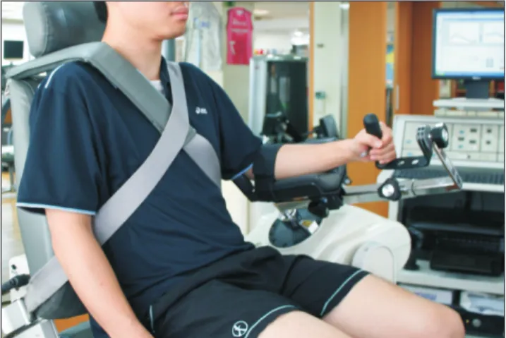
a point was awarded for every second in correct posture, and 10 points for 10 seconds and longer.13) We measured the isokinetic muscle strength of both shoulders in the following motions us- ing the Biodex System 3 (Biodex Corp., Shirley, NY, USA): the internal rotation and the external rotation at an angular velocity of 60o/s with the patient in a modified neutral sitting position 30o/30o/30o; the scapular abduction; and the scapular adduc- tion. Four measurements were taken for each motion. The peak torque deficit of the affected arm was calculated relative to the unaffected contralateral arm (Fig. 1).13-15) For the belly press test, the patient’s forearm is in internal rotation and slightly apart from the body at an angle of around 30o. The patient attempts to keep the arm in internal rotation as much as possible while
pressing their belly with his/her hand.7) For the lift off test the hand of the patient is placed behind the back and the examiner assesses how well the patient can push away a resisting force applied to the palm of the patient.9) For the bear hug test, the patient’s hand is placed flat on the contralateral clavicle and the examiner measures how well the patient maintains this posi- tion from internal rotation of the forearm as the patient’s arm is pulled.3)
Diagnostic Arthroscopy
Under general anesthesia, an arthroscopic examination was performed on the patient who was placed in a beach-chair posi- tion at a 70o angle. We examined the scapulohumeral joint first and then the subacromial space. We made a 5-mm skin incision that is 1 cm away from the acromion to create a posterolateral portal for the arthroscope insertion. We made a dissection with- in the subacromial space to ascertain a sufficient arthroscopic window.16,17) To measure subscapularis tendon tear size, we performed debridement of the region around the tear. The sub- scapularis tendon tear was classified according to Lafosse’s clas- sification (Fig. 2).6)
Statistical Analysis
Statistical analyses were conducted using PASW Statistics ver.
18.0 (IBM Co., Armonk, NY, USA). The chi-square and the lin- ear-by-linear association tests were used to analyze the correla- tion between the arthroscopic findings of the subscapularis tears (i.e., the classification of the tears) and the results of the physical examinations and of the isokinetic muscle strength tests. The correlation between the classes of subscapularis lesions and the proportion of positive outcomes of the collective and the indi- vidual physical tests and outcomes of isokinetic muscle strength tests were analyzed by independent t-tests, the Mann-Whitney U-test (nonparametric variables), and the one-way Kruskal-Wallis ANOVA test. A p-value of less than 0.05 was considered to be statistically significant.
Fig. 1. We measured isokinetic muscle strength using the Biodex System III.
Results
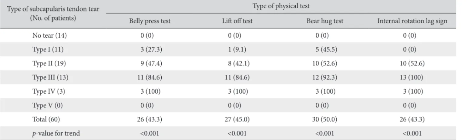
We classified the subscapularis tendon tears according to the Lafosse classification system; 14 patients (23.3%) had no tears;
11 patients (18.3%) had a Lafosse type I tear; 19 patients (31.7%) had a Lafosse type II tear; 13 patients (21.7%) had a Lafosse type III tear; and 3 patients (5.0%) had a Lafosse type IV tear. Lafosse type V tears were not observed in any. In patients without tears, when we carried out the belly press test, the lift off test, and the bear hug test and evaluated for the internal rotation lag sign, no tests returned a positive result. But as the severity of the tear
increased or as the Lafosse classification increased, we found that the proportion of the belly press test, lift off test, bear hug test, and the internal rotation lag sign that returned a positive outcome also increased with statistical significance (p<0.001).
The patients with Lafosse type I tears showed a negative inter- nal rotation lag sign (Table 1). This positive trend was significant across all the tear groups when the proportions of the cumula- tive positive outcomes of the physical tests were taken. This sta- tistical significance of the trend was lost when proportions of the positive outcomes of the tests were assessed individually rather than collectively (Table 2).
Fig. 2. (A) Lafosse type I subscapularis tear:
partial lesion of the superior 1/3 of the ten- don; (B) Lafosse type II subscapularis tear:
complete lesion of the superior 1/3 tendon;
(C) Lafosse type III subscapularis tear: com- plete lesion of the superior 2/3 of the tendon;
and (D) Lafosse type IV subscapularis tear:
complete lesion of the entire tendon.
A B
C D
Table 1. Frequency of Positive Outcomes of Physical Tests Type of subcapularis tendon tear
(No. of patients)
Type of physical test
Belly press test Lift off test Bear hug test Internal rotation lag sign
No tear (14) 0 (0) 0 (0) 0 (0) 0 (0)
Type I (11) 3 (27.3) 1 (9.1) 5 (45.5) 0 (0)
Type II (19) 9 (47.4) 8 (42.1) 10 (52.6) 10 (52.6)
Type III (13) 11 (84.6) 11 (84.6) 12 (92.3) 13 (100)
Type IV (3) 3 (100) 3 (100) 3 (100) 3 (100)
Type V (0) 0 (0) 0 (0) 0 (0) 0 (0)
Total (60) 26 (43.3) 27 (45.0) 30 (50.0) 26 (43.3)
p-value for trend <0.001 <0.001 <0.001 <0.001
Values are presented as number (%).
To compare the agreement in the severity of tear between that diagnosed preoperatively and that diagnosed during ar- throscopic examination, we carried out a correlation analysis of each patient’s KSS score and the type of Lafosse classification with which the patient’s tear was classified. We found that the KSS score and the Lafosse type did not show a statistically signifi- cant correlation.
Through isokinetic muscle strength tests, we found that La- fosse tear type did not have any statistically significant correlation with external rotation torque deficit at the 60o/s angular velocity.
But we found that with increase in Lafosse type, the internal ro- tation torque deficit at the 60o/s angular velocity also increased.
Through a trend analysis, we found that only the increase in in- ternal rotation torque deficit between patients with Lafosse type II tears and those with Lafosse type III was large enough to be statistically significant (p=0.007) (Table 2, Fig. 3).
Discussion
The prevalence of subscapularis tendon tear has been shown to range from 30% to 50%.3,18) Of all the rotator cuff tears, 5% of tears are subscsapularis tendon tears that occur independently of other tears of the rotator cuff; thus, they are less common than those of the infraspinatus tendon and the supraspinatus ten- don.2,4,6,7) Since subscapularis tendon tears usually extends to the articular side of the subscapularis tendon, its diagnosis was dif-
ficult and its function was largely overlooked despite its clinical importance.2,5,19) Yet, Barth et al.3) and Burkhart and Tehrany20) found that the subscapularis tendon plays an important role in maintaining a normal function of the shoulder. Widespread employment of arthroscopic surgery and thereby a greater dis- covery of subscapularis tears of the articular side have facilitated better diagnosis of subscapularis tendon tears.3,4) Because of the importance of the subscapularis tendon, an early diagnosis of subscapularis tendon tears would be favorable and will facilitate a better treatment and maintenance of shoulder function.3,4,20)
Studies similar to ours in the past have investigated the cor- relation between results of physical examinations and subscapu- laris tendon tears.3,4) For instance, the sensitivity and specificity of the belly press test, the bear hug test, and the lift off test have been studied to assess the diagnostic value of these physical examinations. Barth et al.3) reported that the belly press test showed a sensitivity of around 40% and a specificity of 97.9%, that the bear hug test showed a sensitivity of around 60% and a specificity of 91.7%, and that the lift off test showed a sensitivity of 17.6% and a specificity of 100%. Bartsch et al.4) reported that the belly press test showed a sensitivity of around 80% and a specificity of 88% and that the lift off test showed a sensitivity of around 40% and a specificity of around 79%.3,4) But the values between these studies do not completely agree with each other, suggesting that accurately diagnosing a subscapularis tendon tear on the basis of the results of one physical examination alone may not be feasible.
There are previous studies that have not only investigated the association between physical examination and subscapularis tendon tears but also that of isokinetic muscle strength tests and rotator cuff tears.3,4,13-15) However, studies that have investigated the association between the severity of subscapularis tendon tears and results of physical examination, of isokinetic strength tests, and of arthroscopy are few. In this study, we made a com- parative analysis of the preoperative results of physical exami- nations and of isokinetic muscle strength tests according to the severity of subscapularis tendon tears, which was verified by ar- throscopy, and evaluated whether they can be used to make an early diagnosis of a subscapularis tendon tear, as well as to gauge its size.
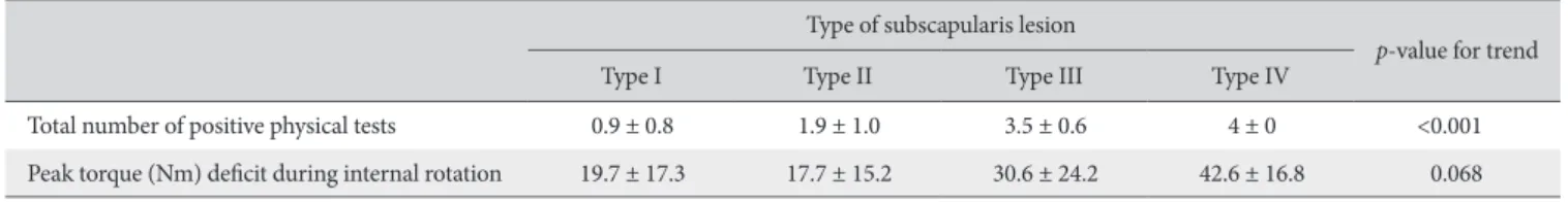
For the physical examinations we assessed (belly press test, lift off test, bear hug test, and the internal rotation lag sign), Table 2. Total Number of Positive Physical Tests and Peak Torque Deficit during Internal Rotation according to the Type of Subscapularis Lesion
Type of subscapularis lesion
p-value for trend
Type I Type II Type III Type IV
Total number of positive physical tests 0.9 ± 0.8 1.9 ± 1.0 3.5 ± 0.6 4 ± 0 <0.001
Peak torque (Nm) deficit during internal rotation 19.7 ± 17.3 17.7 ± 15.2 30.6 ± 24.2 42.6 ± 16.8 0.068 Values are presented as mean ± standard deviation.
Fig. 3. Deficit according to the severity of subscapularis tear in preoperative peak torque (Nm) during internal rotation at an angular velocity of 60o/s.
Type IV Type I
45 40 35 30 25 20 15 10 5 0
Type II Type III p=0.007
Lafosse type
Nm
we found that a greater proportion of physical examinations returned a positive sign when the patient’s severity of tear in- creased, depicted by the relationship across the Lafosse tear classes. Thus, we propose that these physical examinations as a collective unit can aid the diagnosis of subscapularis tendon tears and the prediction of their sizes, but as single tests we believe they do not have sufficient diagnostic power.
We found that the preoperative isokinetic muscle strength, specifically the internal rotation torque deficit at the 60o/s angu- lar velocity, differed between patients with Lafosse type II tears and those with Lafosse type III tears, the deficit being signifi- cantly greater in the latter. Our findings suggest that the mecha- nism of tear of the upper two thirds of the subscapularis tendon differs from that of the lower third, where in the former case more than half of the subscapularis tendon detaches from the attachment site leading to retraction of the subscapularis tendon and to collateral tears of the superior glenohumeral ligament and of the coracohumeral ligament; the resulting deterioration in the strength of the subscapularis tendon that we have seen between those with Lafosse type II tears and those with Lafosse type III tears may have manifested as the increase in internal rotation torque deficit.4,6) This finding also signifies the progression of the lesion at the later stages.
Our study is the first of its kind that investigated the correla- tion between the severity of subscapularis tendon tears and the results of preoperative evaluations. But even when the results of physical examination and the internal rotation torque deficit cannot conclusively predict the presence of a subscapularis ten- don tear nor the tear size, a subscapularis tendon tear should not be ruled out until more conclusive evidence is found. As a limitation of this study, the subjectivity of the observer who as- sessed the physical tests may have introduced unintentional bias to our interpretations.
Conclusion
In conclusion, we found that no single type of physical test or isokinetic muscle strength test can be used to diagnose sub- scapularis tendon tears at an early stage. Rather we found that the more severe a subscapularis tendon tear the greater propor- tion of physical tests, undertaken preoperatively, that returned a positive outcome. We found that severity of the subscapularis tendon tear was negatively correlated to isokinetic muscle strength; as severity of the tendon tear increased, the patient’s isokinetic muscle strength decreased. But the decrease in the isokinetic muscle strength, specifically the internal rotation peak torque deficit at the angular velocity of 60o/s, across the groups was statistically significant only between the Lafosse type II tear group and Lafosse type III tear group. Thus, taking the Lafosse type II tear as the benchmark, we propose that the internal rota- tion peak torque deficit at the angular velocity of 60o/s may be
used to differentiate subscapularis tendon tears. Further studies are needed to validate our findings.
Acknowledgements
We express our appreciation to Dr. Deok-Weon Kim from the Green Hospital for providing data relevant to this study.
References
1. Cho NS, Lee SH. Impingement syndrome & rotator cuff tear:
etiology. J Korean Arthrosc Soc. 2012;16(1):72-8.
2. Shon MS, Koh KH, Lee SS, Yoo JC. MR evaluation of tendi- nous portions in the subscapularis muscle. Clin Should Elbow.
2011;14(1):35-45.
3. Barth JR, Burkhart SS, De Beer JF. The bear-hug test: a new and sensitive test for diagnosing a subscapularis tear. Arthros- copy. 2006;22(10):1076-84.
4. Bartsch M, Greiner S, Haas NP, Scheibel M. Diagnostic values of clinical tests for subscapularis lesions. Knee Surg Sports Trau- matol Arthrosc. 2010;18(12):1712-7.
5. Keating JF, Waterworth P, Shaw-Dunn J, Crossan J. The relative strengths of the rotator cuff muscles. A cadaver study. J Bone Joint Surg Br. 1993;75(1):137-40.
6. Lafosse L, Jost B, Reiland Y, Audebert S, Toussaint B, Gobezie R. Structural integrity and clinical outcomes after arthroscopic repair of isolated subscapularis tears. J Bone Joint Surg Am.
2007;89(6):1184-93.
7. Gerber C, Hersche O, Farron A. Isolated rupture of the sub- scapularis tendon. J Bone Joint Surg Am. 1996;78(7):1015-23.
8. Adams CR, Schoolfield JD, Burkhart SS. Accuracy of pre- operative magnetic resonance imaging in predicting a sub- scapularis tendon tear based on arthroscopy. Arthroscopy.
2010;26(11):1427-33.
9. Gerber C, Krushell RJ. Isolated rupture of the tendon of the subscapularis muscle. Clinical features in 16 cases. J Bone Joint Surg Br. 1991;73(3):389-94.
10. Tung GA, Yoo DC, Levine SM, Brody JM, Green A. Subscapu- laris tendon tear: primary and associated signs on MRI. J Com- put Assist Tomogr. 2001;25(3):417-24.
11. Adams CR, Brady PC, Koo SS, et al. A systematic approach for diagnosing subscapularis tendon tears with preop- erative magnetic resonance imaging scans. Arthroscopy.
2012;28(11):1592-600.
12. Ticker JB, Burkhart SS. Why repair the subscapularis? A logical rationale. Arthroscopy. 2011;27(8):1123-8.
13. Kim DW, Sung JH, Jung JE, Ko MS. Correlation between Ko- rean Shoulder Scoring System and Isokinetic muscle strength test. J Korean Orthop Soc Sports Med. 2010;9(2):104-8.
14. Kim DW, Joo HK, Jung JE. Comparison of isokinetic strength between stage 1,2 impingement syndrome and rotator cuff
tear. J Korean Shoulder Elbow Soc. 2010;13(1):53-7.
15. Kim JO, Shim SD, Jeong MS, Chang JH, Park C. Can pre- operative isometric and Isokinetic strength predict the size of the full-thickness rotator cuff tears? Korean J Sports Med.
2009;27(1):89-94.
16. Lafosse L, Lanz U, Saintmard B, Campens C. Arthroscopic repair of subscapularis tear: Surgical technique and results. Or- thop Traumatol Surg Res. 2010;96(8 Suppl):S99-108.
17. Ko SH, Lee CC, Kim SW, Shin SM, Cho BK. Arthroscopic UU-tension band suture for rotator cuff tear above 4 cm:
comparative study with simple suture. Clin Should Elbow.
2012;15(2):99-108.
18. Yoo JC, Rhee YG, Shin SJ, et al. Subscapularis tendon tear clas- sification based on 3-dimensional anatomic footprint: a cadav- eric and prospective clinical observational study. Arthroscopy.
2015;31(1):19-28.
19. Park YB, Park YE, Koh KH, Lim TK, Shon MS, Yoo JC. Subscap- ularis tendon repair using suture bridge technique. Arthrosc Tech. 2015;4:e133-7.
20. Burkhart SS, Tehrany AM. Arthroscopic subscapularis ten- don repair: technique and preliminary results. Arthroscopy.
2002;18(5):454-63.